Rotary Instruments And Methods For Intrauterine Tissue Resection

a rotary instrument and uterine tissue technology, applied in the field of uterine tissue diagnosis and therapeutic treatment, can solve the problems of large resector or tissue removal device, large equipment, complex, etc., and achieve the effect of simple, efficient and simple resector and better control of fluid balan

Pending Publication Date: 2019-05-09
CALDERA MEDICAL
View PDF16 Cites 4 Cited by
  • Summary
  • Abstract
  • Description
  • Claims
  • Application Information

AI Technical Summary

Benefits of technology

[0007]The present invention provides a compact hysteroscope that avoids the need for use of anesthesia, have better control of fluid balance when using a hanging bag fluid source and, hence, that can be used in an office setting and simplified, efficient single-use resector that does not require special handling, cleaning, and sterilization procedures, and medical systems employing the same. These objectives are achieved, in part, by providing a single-use uterine tissue resector comprising: a handle; an elongated outer cutting tube having a proximal portion statically attached to the handle; an elongated inner cutting tube concentrically positioned and rotatable within a lumen of the outer cutting tube; and a motor positioned within the handle and coupled to a proximal end of the inner cutting tube, wherein upon activation of the motor, the motor rotates the inner cutting tube relative to the outer cutting tube. Wherein the single-use uterine tissue resector further comprises a battery positioned within the handle that is in electrical communication with the motor; wherein the handle comprises a leaf spring trigger; wherein the handle comprises a momentary switch; wherein a distal end of the inner cutting tube and a distal end of the outer cutting tube form openings into a lumen of the inner cutting tube; wherein a distal portion of the inner cutting tube and a distal portion of the outer cutting tube comprise apertures having cutting surfaces; wherein the apertures of the inner and outer cutting tubes comprise a substantially same size, shape and longitudinal placement relative to one another; wherein the single-use uterine tissue resector further comprises a fluid chamber positioned within the handle having a lateral port that protrudes out from the handle; wherein a proximal portion of the inner cutting tube passes through the fluid chamber, the proximal portion of the inner cutting tube having an aperture formed through a sidewall thereof; and wherein the handle comprises a window through which the lateral port of the fluid chamber protrudes and is rotatable relative to the handle.

Problems solved by technology

One problem associated with current hysteroscopy systems is that they are intended for use in an operating room setting with the patient being subjected to some type of anesthesia.
Anesthesia is required in particular because the size of current hysteroscopes is relatively large and, as such, they can cause discomfort and pain to the patient.
Another problem with current systems is that the resector or tissue removal devices are relatively large, complex and, hence, relatively expensive pieces of equipment.

Method used


Image

  • Rotary Instruments And Methods For Intrauterine Tissue Resection
  • Rotary Instruments And Methods For Intrauterine Tissue Resection
  • Rotary Instruments And Methods For Intrauterine Tissue Resection

Examples


Embodiment Construction

[0028]Specific embodiments of the invention will now be described with reference to the accompanying drawings. This invention may, however, be embodied in many different forms and should not be construed as limited to the embodiments set forth herein; rather, these embodiments are provided so that this disclosure will be thorough and complete, and will fully convey the scope of the invention to those skilled in the art. The terminology used in the detailed description of the embodiments illustrated in the accompanying drawings is not intended to be limiting of the invention. In the drawings, like numbers refer to like elements.

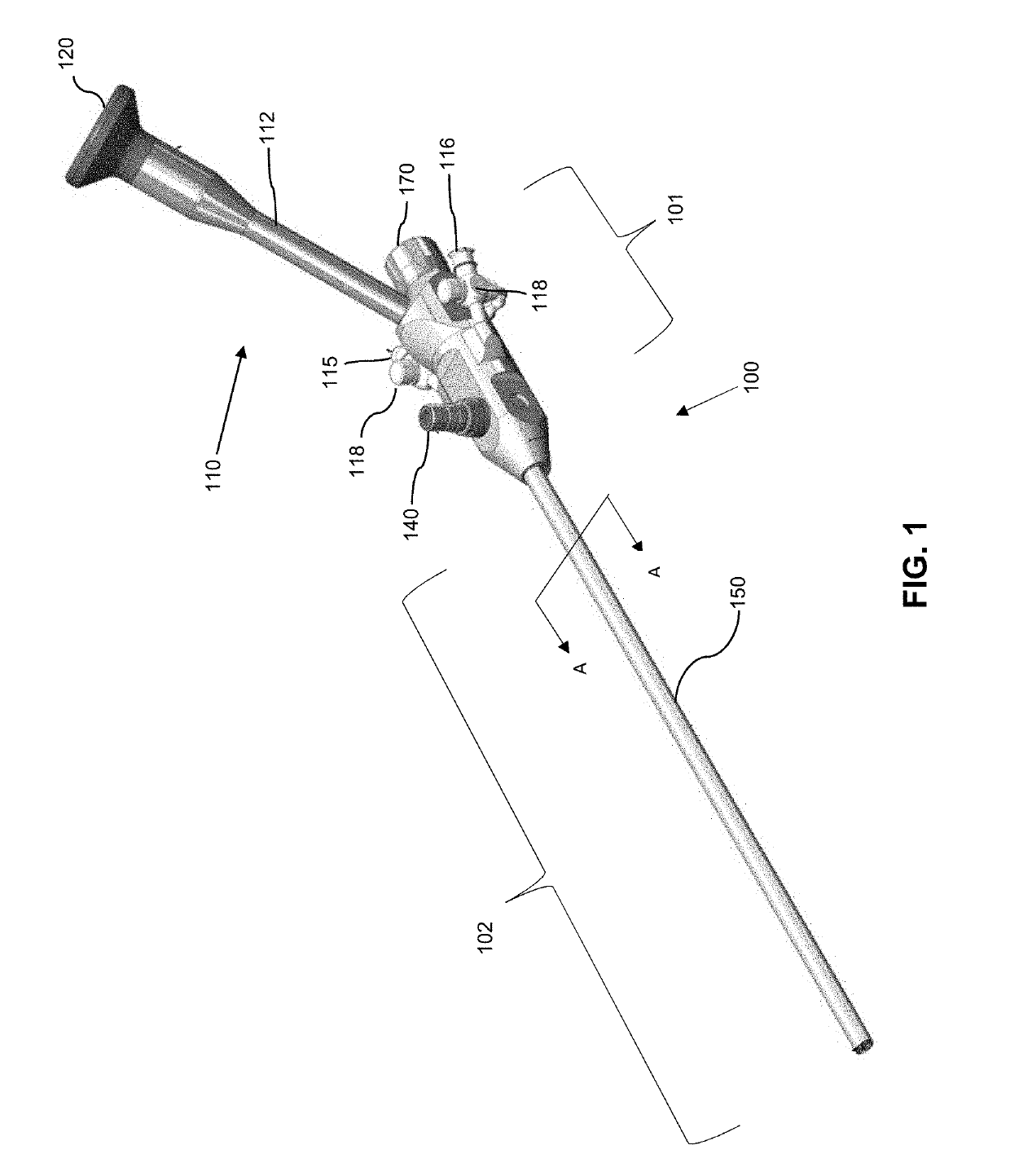
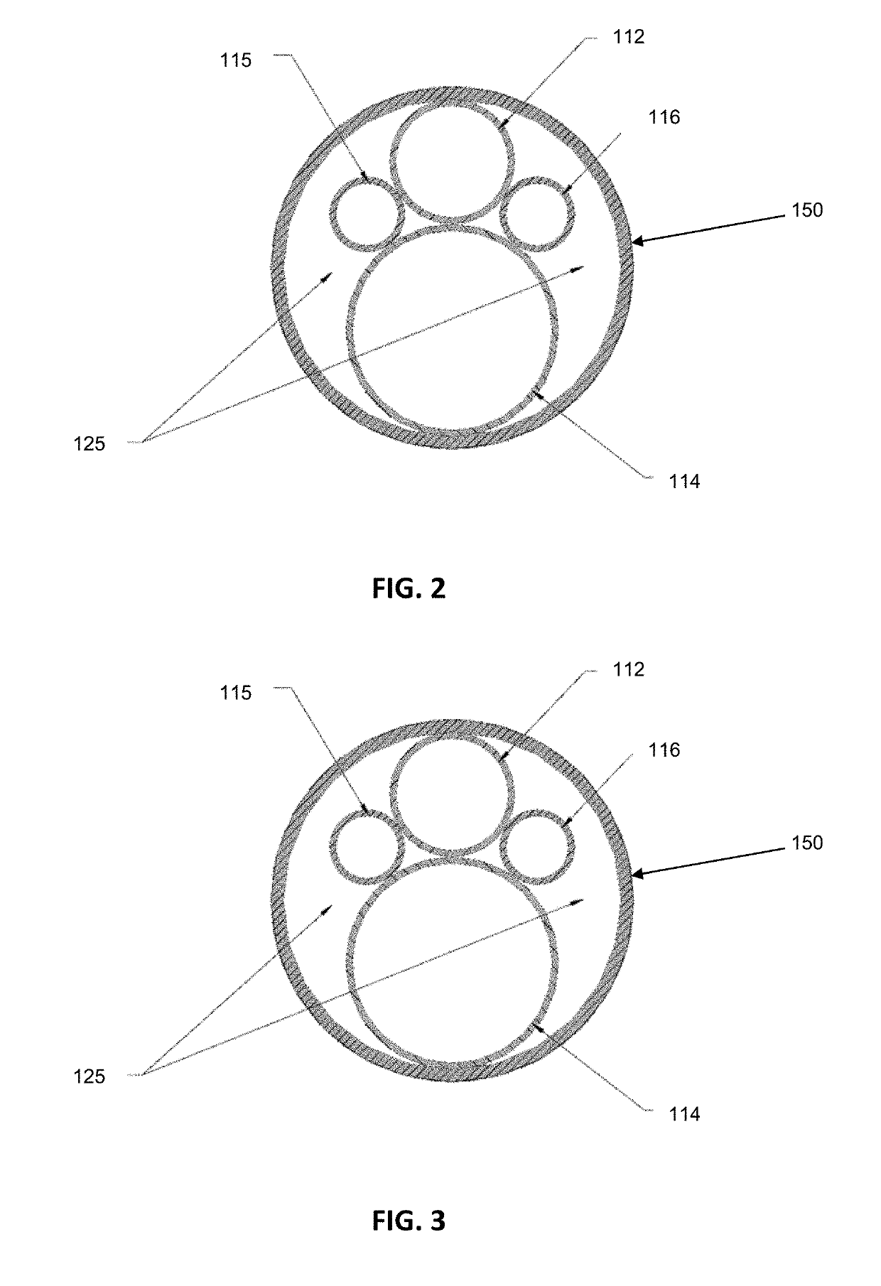
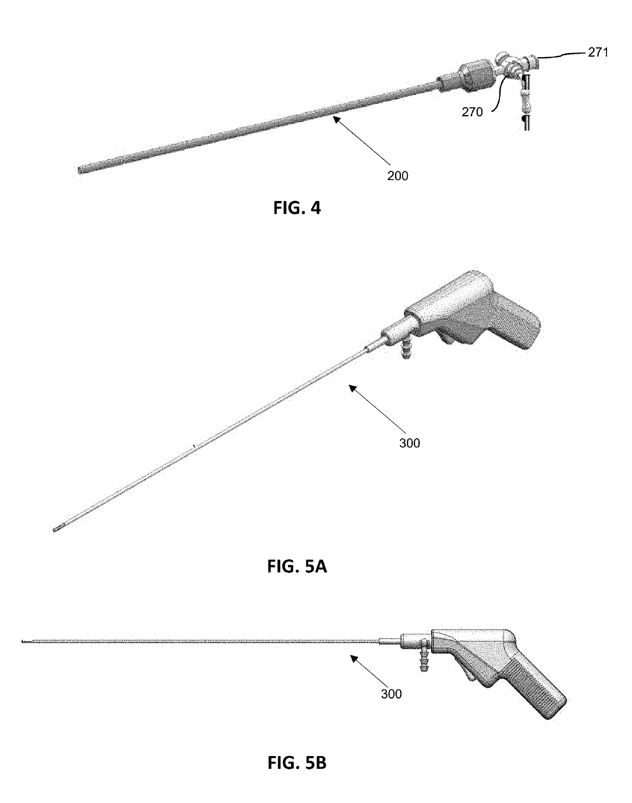
[0029]The present invention is directed to a system comprising a reusable or multi-use, multi-lumen hysteroscope for uterine distention and visualization of uterine tissue and / or tissue pathology and a single-use tissue removal device (TRD) or tissue resector to mechanically remove pathological tissues and irrigate such from the uterus.

[0030]Herein, detailed e...


PUM


Abstract

A compact multi-lumened hysteroscope, a single-use tissue resector, and a medical system employing the same for diagnosis and therapeutic treatment of uterine tissue, such as polyps and fibroids.

Description

RELATED APPLICATIONS[0001]This application claims priority to U.S. Provisional Application Ser. No. 62 / 584,059 filed Nov. 9, 2017, entitled Rotary Instruments and Methods for Intrauterine Tissue Resection and U.S. Provisional Application Ser. No. 62 / 626,431 filed Feb. 5, 2018, entitled Rotary Instruments and Methods for Intrauterine Tissue Resection, both of which are hereby incorporated herein by reference in their entireties.FIELD OF THE INVENTION[0002]The present invention relates to the field of uterine tissue diagnosis and therapeutic treatments such as resection and, more particularly, to instruments for the rotary removal of uterine tissue and methods of use thereof.BACKGROUND OF THE INVENTION[0003]Hysteroscopy broadly refers to the inspection of a uterine cavity using a hysteroscope accessed through the cervix. Hysteroscopy allows for diagnosis of intrauterine pathology and, furthermore, can be used for surgical intervention. The hysteroscope typically includes a sheath, sco...

Claims


Application Information

Patent Timeline
Patent Type & Authority Applications(United States)
IPC IPC(8): A61B17/42A61B17/32A61B1/00
CPCA61B17/42A61B17/32002A61B2017/0023A61B2017/4216A61B1/00163A61B2017/320024A61B2217/005A61B1/00071A61B1/00103A61B1/00195A61B2217/007A61B2017/00398A61B2017/00734A61B1/015A61B1/018
Inventor CHARVET, JOSE LUISLU, FELIXMUHLFELD, SANDRA
Owner CALDERA MEDICAL
Why Eureka
Try Eureka