
Fully automatic ultrasonic scanner and scan detection method

an ultrasonic scanner and fully automatic technology, applied in ultrasonic/sonic/infrasonic image/data processing, tomography, applications, etc., can solve the problems of time-consuming inspection process, incomplete, image data cannot be stored and transmitted, etc., and achieve the effect of simple structure and convenient operation

Active Publication Date: 2019-05-23
SHANGHAI SHENBO MEDICAL INSTR CO LTD
View PDF0 Cites 17 Cited by
  • Summary
  • Abstract
  • Description
  • Claims
  • Application Information

AI Technical Summary

Benefits of technology

This patent is about a device that can automatically adjust the angle of an ultrasonic probe when scanning the human body. This ensures that the probe scans against the skin surface and remains perpendicular to it. This technology improves the accuracy of ultrasonic scanning and makes it easier to detect and scan areas of the body that may be difficult to reach.

Problems solved by technology

1. the inspection must be performed by experienced professional physicians, and the inspection process is time-consuming;
2. the scan may not complete and may not cover the entire breast;
3. the detection effect depends on the physician's experience and physical condition and is instable; and
4. the image data cannot be stored and transmitted because of the non-standardized collection.
When breast cancer screening is popularized across the country, especially in rural areas or small and medium-sized cities, the expected effects for ultrasonic detection of early breast cancer cannot be achieved due to a lack of experienced professional physicians.
However, because the body shape and the size and shape of the organ of each person are not the same, the ultrasonic probe cannot make adjustment according to different curves of the human body.
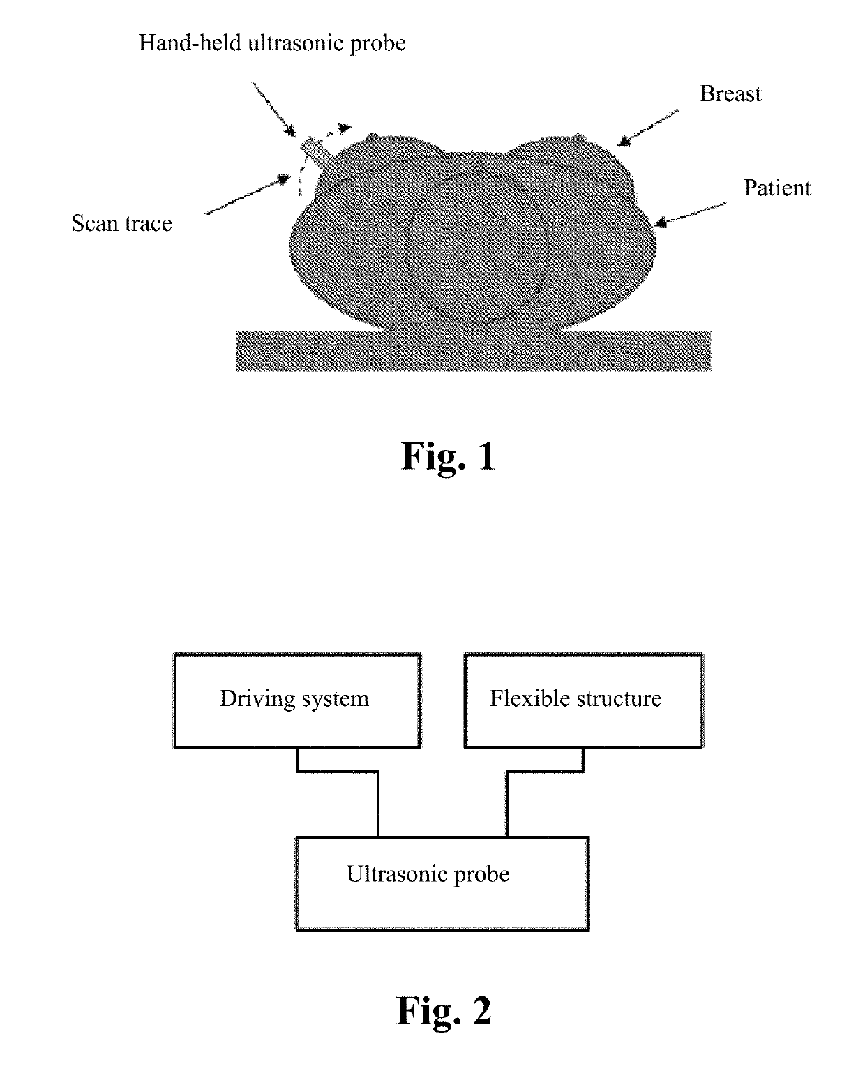
The current ultrasonic inspection is performed by a professional physician through a hand-held scanning probe or is performed according to a fixed shape, and it is difficult to adapt to various kinds of body shapes, breast sizes and compactness, thereby forming scan blind spots and resulting in missed diagnosis.
In the prior art, there is also a flexible part such as a water bag added between the skin surface of the to-be-detected organ and the ultrasonic probe; however, the ultrasonic probe is not in direct contact with the skin during detection, and the water bag between the probe and the skin causes multiple reflected interference images; and as the probe does not always keep perpendicular to the skin, the reflection angles of the collected images are different, which eventually lead to low-quality scanned images and the risk of missed diagnosis.

Method used


Image

  • Fully automatic ultrasonic scanner and scan detection method
  • Fully automatic ultrasonic scanner and scan detection method
  • Fully automatic ultrasonic scanner and scan detection method

Examples


Embodiment Construction

[0041]In the following statement, lots of technical details are proposed in order to make the readers better understand the present application. However, a person of ordinary skill in the art should understand that the technical solutions claimed by each claim of the present application can also be realized even without these technical details and various changes and modifications based on each of the following implementation manners.

[0042]In order to make the objectives, technical solutions and advantages of the present invention clearer, the implementation manners of the present invention will be further described in detail in conjunction with the accompanied drawings.

[0043]The first implementation manner of the present invention relates to a fully automatic ultrasonic scanner; and FIG. 2 is a schematic structural diagram of the fully automatic ultrasonic scanner, and the fully automatic ultrasonic scanner comprises:

[0044]an ultrasonic probe;

[0045]a driving system for driving the ...


PUM


Abstract

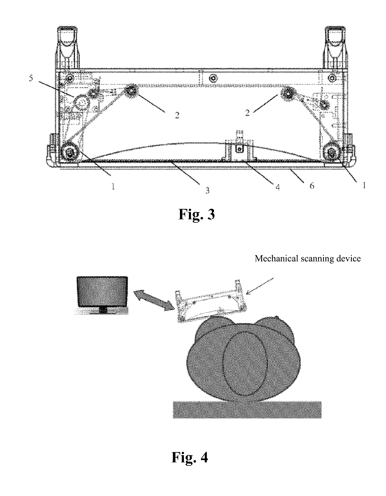
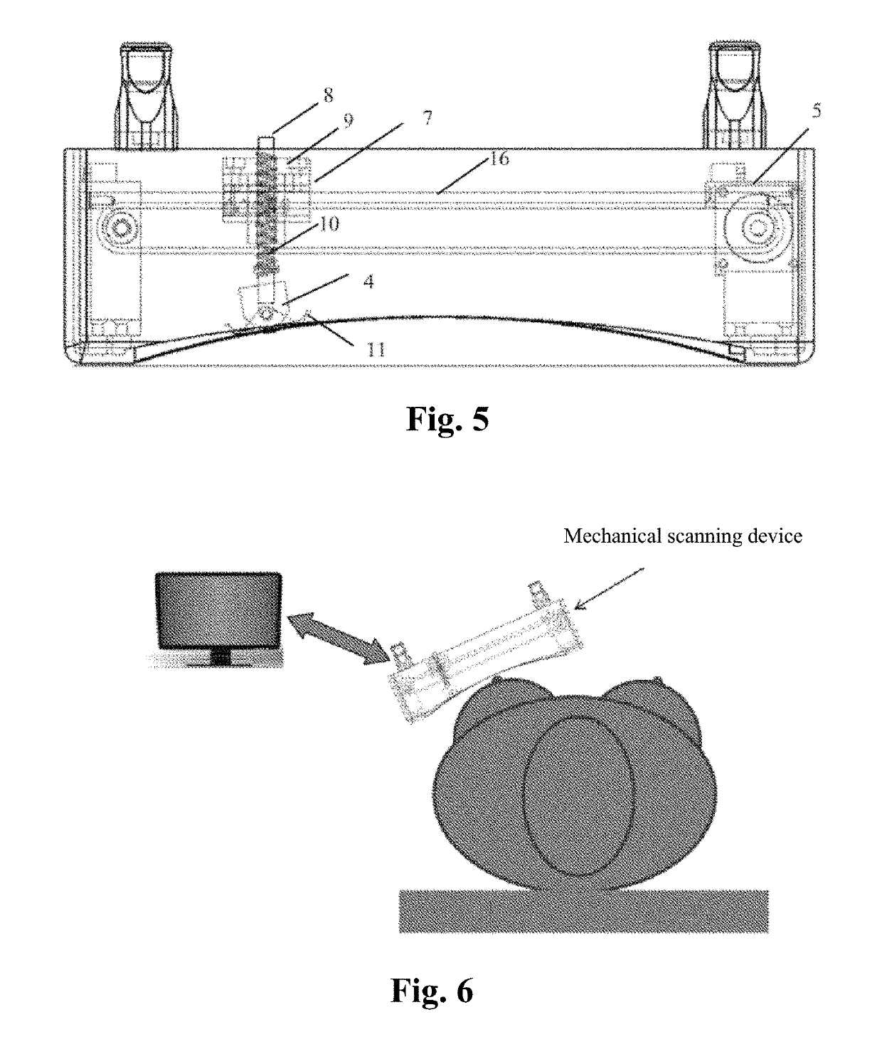
A fully automatic ultrasonic scanner and a scan detection method is provided. The fully automatic ultrasonic scanner comprises: an ultrasonic probe (4); a driving system (5) for driving the ultrasonic probe (4) to move; and a flexible structure on which the ultrasonic probe (4) is mounted, wherein the flexible structure enables the ultrasonic probe (4) to be always along a curve of a skin surface and keep perpendicular to the skin surface during scanning. The flexible structure of the fully automatic ultrasonic scanner has self-adaptive effect, can adjust the scan trace and the probe angle in real time according to different curves of the human body, and ensure that the ultrasonic probe (4) scans against the skin surface and keeps perpendicular to the skin surface, which improves the quality of the scanned images, so as to enhance the detection rate and accuracy rate of early screening, and reduces the probability of missed diagnosis.

Description

TECHNICAL FIELD[0001]The present invention relates to medical equipment, and in particular to a fully automatic ultrasonic scanner and a scan detection method.BACKGROUND ART[0002]During breast cancer screening, a physician holds a probe of an existing hand-held ultrasonic breast scanner to carry out scanning (image data collection) and on-site diagnosis (image data analysis), which has the following technical problems:[0003]1. the inspection must be performed by experienced professional physicians, and the inspection process is time-consuming;[0004]2. the scan may not complete and may not cover the entire breast;[0005]3. the detection effect depends on the physician's experience and physical condition and is instable; and[0006]4. the image data cannot be stored and transmitted because of the non-standardized collection.[0007]Therefore, hand-held breast ultrasonic methods are more suitable for treatment diagnosis instead of early screening. When breast cancer screening is popularized...

Claims


Application Information

Patent Timeline
IPC IPC(8): A61B8/00A61B5/00A61B8/08
CPCA61B8/4455A61B8/4461A61B8/4254A61B8/4416A61B5/0077A61B8/4218A61B8/0825A61B8/085A61B8/145A61B8/40A61B8/4209A61B8/4281A61B8/429A61B8/4405A61B8/466A61B8/483A61B8/5207A61B8/5253A61B8/565
Inventor TIAN, XUDONGSUN, ZHIFENG
Owner SHANGHAI SHENBO MEDICAL INSTR CO LTD
Learn More