Contactless System and Method For Assessing Tissue Viability and Other Hemodynamic Parameters

a contactless system and hemodynamic parameter technology, applied in the field of contactless system and method for assessing tissue viability and other hemodynamic parameters, can solve the problems of inflicting trauma to already vulnerable tissue, increasing the risk of wound infection, and subjective tissue excision procedures

Active Publication Date: 2019-07-25
THE BOARD OF TRUSTEES OF THE UNIV OF ARKANSAS +1
View PDF0 Cites 3 Cited by
  • Summary
  • Abstract
  • Description
  • Claims
  • Application Information

AI Technical Summary

Benefits of technology

[0009]In one aspect, a contactless system for assessing tissue viability and other hemodynamic parameters is featured. The system includes one or more light sources configured to emit lights at a predetermined wavelength sensitive to hemoglobin concentration associated with spontaneous hemodynamic oscillations at tissue in a predetermined area of a human subject. One or more polarizers each coupled to one or more of the one or more light sources are configured to polarize the light to a polarized state such that the polarized light in the polarized state diffuses into the tissue in the predetermined area at a predetermined depth and the polarized light is maintained in the polarized state at the predetermined depth. One or more detectors each including a detector polarizer coupled thereto are configured to discriminate the light maintained in the polarized state and at the predetermined depth and configured to generate a plurality of frames of the tissue in the predetermined area at the predetermined depth. A controller is coupled to the one or more light sources and the one or more detectors and is configured to: acquire the plurality of frames, select a region of interest having the same coordinates for each of the plurality of frames, average the number of pixels within each region of interest to create a raw reference signal, detrend the raw reference signal to create a detrended raw reference signal, perform frequency domain analysis of the detrended raw reference signal, identify a frequency band of interest associated with the spontaneous hemodynamic oscillations, perform an inverse fast Fourier transform within the frequency band of interest to generate a reference signal indicative of blood volume oscillations at a selected spontaneous hemodynamic oscillation, for each sample of the reference signal at a predetermined point in time, multiply the sample by each pixel of a frame at the same predetermined point in time to generate a three-dimensional coordinate matrix including a plurality of correlation matrix frames at each predetermined point in time, and add the plurality of correlation matrix frames at each predetermined point in time to generate a two-dimensional hemodynamic map indicative of the strength of the spontaneous hemodynamic oscillation to assess the viability of the tissue in the predetermined area.

Problems solved by technology

Although bleeding is typically assumed to mean the tissue is viable, this conventional tissue excision procedure is subjective and imprecise because it relies on visual inspection that does not preclude the possibility that some necrotic tissue or biofilm will be inadvertently left in the wound site.
Because flowmetry requires the probe to directly contact with the burn wound, it may increase the risk of wound infection and may inflict trauma to already vulnerable tissue.
Additionally, because LDI measures perfusion in one spot at the time, assessing a large burn wound may be a time-consuming process.
Additionally, there is some risk that LDI may not detect necrotic tissue in the wound bed.
The major drawback associated with conventional ICG video-angiography is that intravascular dye injection is required.
While conventional LDI and ICG each offer a unique approach to detecting tissue viability, both techniques are cumbersome to manipulate in a surgical setting, have a large size, and do not provide for real-time diagnosis, critical for tissue viability assessment during an excision procedure.

Method used


Image

  • Contactless System and Method For Assessing Tissue Viability and Other Hemodynamic Parameters
  • Contactless System and Method For Assessing Tissue Viability and Other Hemodynamic Parameters
  • Contactless System and Method For Assessing Tissue Viability and Other Hemodynamic Parameters

Examples


Embodiment Construction

[0021]Aside from the preferred embodiment or embodiments disclosed below, this invention is capable of other embodiments and of being practiced or being carried out in various ways. Thus, it is to be understood that the invention is not limited in its application to the details of construction and the arrangements of components set forth in the following description or illustrated in the drawings. If only one embodiment is described herein, the claims hereof are not to be limited to that embodiment. Moreover, the claims hereof are not to be read restrictively unless there is clear and convincing evidence manifesting a certain exclusion, restriction, or disclaimer.

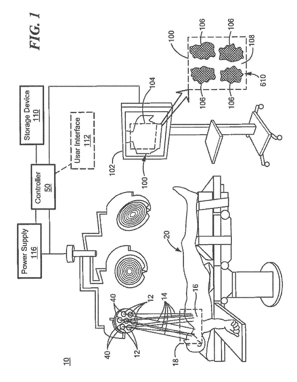
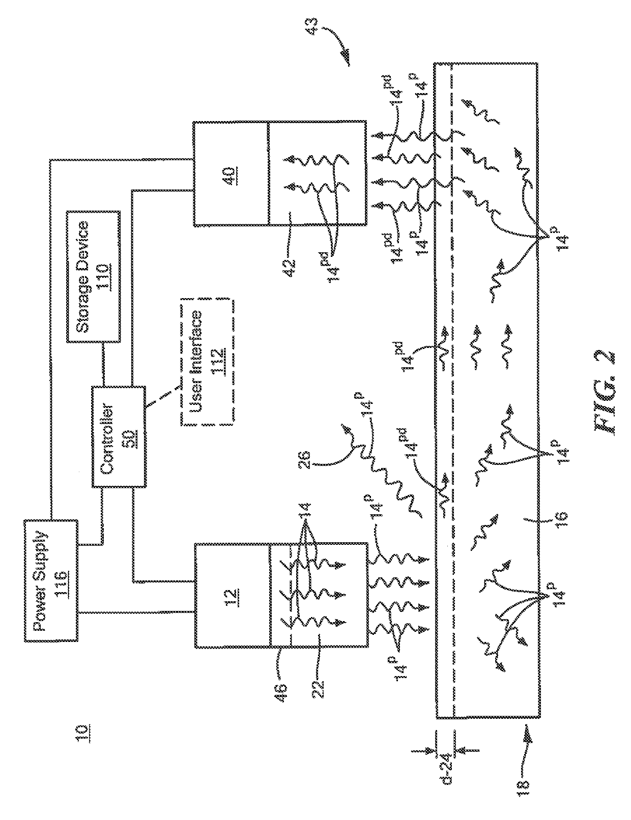
[0022]FIG. 1 shows one embodiment of contactless system 10 for assessing of tissue viability and other hemodynamic parameters. System 10 preferably includes one or more light sources 12 configured to emit light 14 at a predetermined wavelength sensitive to hemoglobin concentration associated with spontaneous hemodynamic osc...


PUM


Abstract

A contactless system for assessing tissue viability and other hemodynamic parameters includes one or more light sources configured to emit lights at a predetermined wavelength sensitive to hemoglobin concentration associated with spontaneous hemodynamic oscillations at tissue in a predetermined area of a human subject. One or more polarizers are each coupled to one or more of the one or more light sources and are configured to polarize the light to a polarized state such that the polarized light in the polarized state diffuses into the tissue in the predetermined area at a predetermined depth and the polarized light is maintained in the polarized state at the predetermined depth. One or more detectors each including a detector polarizer coupled thereto are configured to discriminate the light maintained in the polarized state and at the predetermined depth and are configured to generate a plurality of frames of the tissue in the predetermined area at the predetermined depth. A controller is coupled to the one or more light sources and the one or more detectors. The controller is configured to: acquire the plurality of frames, select a region of interest having the same coordinates for each of the plurality of frames, average the number of pixels within each region of interest to create a raw reference signal, detrend the raw reference signal to create a detrended raw reference signal, perform frequency domain analysis of the detrended raw reference signal, identify a frequency band of interest associated with the spontaneous hemodynamic oscillations, and perform an inverse fast Fourier transform within the frequency band of interest to generate a reference signal indicative of blood volume oscillations at a selected spontaneous hemodynamic oscillation. For each sample of the reference signal at a predetermined point in time, the controller multiplies the sample by each pixel of a frame at the same predetermined point in time to generate a three-dimensional coordinate matrix including a plurality of correlation matrix frames at each predetermined point in time. The controller adds the plurality of correlation matrix frames at each predetermined point in time to generate a two-dimensional hemodynamic map indicative of the strength of the spontaneous hemodynamic oscillation to assess the viability of the tissue in the predetermined area.

Description

RELATED APPLICATIONS[0001]This application claims benefit of and priority to U.S. Provisional Application Ser. No. 62 / 621,873 filed Jan. 25, 2018, under 35 U.S.C. §§ 119, 120, 363, 365, and 37 C.F.R. § 1.55 and § 1.78, which is incorporated herein by this reference.GOVERNMENT RIGHTS[0002]This invention was made with government support under contract number W81XWH-17-C-0169, awarded by the Department of Defense. The government has certain rights in the invention.FIELD OF THE INVENTION[0003]This invention relates to a contactless system and method for assessing tissue viability and other hemodynamic parameters.BACKGROUND OF THE INVENTION[0004]Burn wounds may be classified into four categories of increasing depth: superficial, intermediate partial thickness, deep partial thickness, and full thickness. The latter two classifications typically require aggressive interventions that involve the debridement of necrotic tissue and the application of split thickness skin grafts. These are mor...

Claims


Application Information

Patent Timeline
Patent Type & Authority Applications(United States)
IPC IPC(8): A61B5/00
CPCA61B5/0071A61B5/445A61B5/14551A61B5/7257A61B5/14556A61B5/02405A61B5/02028A61B5/0205A61B5/0261A61B2560/0223A61B5/1455A61B5/349
Inventor PIERRO, MICHELEQUINN, KYLEWOESSNER, ALAN
Owner THE BOARD OF TRUSTEES OF THE UNIV OF ARKANSAS
Why Eureka
Try Eureka