
Anti-nerve growth factor antibodies and methods of preparing and using the same
a growth factor and growth factor technology, applied in the field of antibodies, to prevent the upregulation of neuropeptides, reduce pain, or remove pain
- Summary
- Abstract
- Description
- Claims
- Application Information
AI Technical Summary
Benefits of technology
Problems solved by technology
Method used
Image

Examples
example 1
Production of Antibodies
[0163]Whole antibody sequences were produced by combining equinised light chain and heavy chain variable domains of SEQ ID NO:1 and SEQ ID NO:2, respectively to C-terminal equine constant heavy chain or constant light chain domains. The equinised aD11 VH domain was combined with two different equine heavy chain constant domains; HC2 (IgG2) and HC6 (IgG6) and the equinised aD11 VL domain with the equine kappa light chain constant domain. The sequences of the full-length mature antibody chains are shown in SEQ ID 4 (light chain with kappa constant domain) and 6 (heavy chain with HC2 constant domain). The combined amino acid sequences were converted to expressible form in mammalian cells by the optimal selection of codons and full chemical gene synthesis and cloning into a mammalian cell expression vector pcDNA3.1+. The resultant cDNAs were transfected into CHO cells and the supernatants analysed as detailed in Examples 2 to 5.
example 2
Determining Binding of Antibodies to Murine and Equine NGF
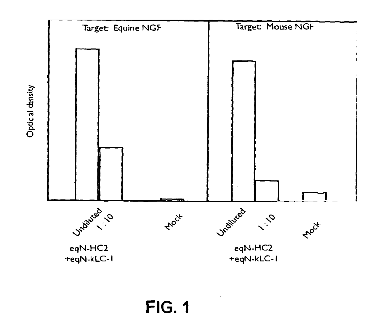
[0164]Equinised heavy and light chain cDNAs were transfected into CHO cells, the supernatants harvested and reacted in ELISA format with either equine or murine NGF. Following incubation and wash steps, the bound equine antibody was detected by reactivity with a goat-anti equine IgG specific polyclonal antibody linked to horseradish peroxidase (HRP) and developed using TMB. The optical density of the resulting product was measured at 450 nm and compared with that from mock empty vector transfected supernatant (denoted as “Mock” in FIG. 1).
[0165]The results are shown in the graph of FIG. 1. Binding to mouse NGF is shown for the HC2 (IgG2 constant domain) equinised antibody (termed eqN-HC2+eqN-kLC-1). In the second part of the graph, binding of the eqN-HC2+eqN-kLC-1 antibody comprising the eqN-kLC-1 light chain and the eqN-HC2 (IgG2) constant chain to equine NGF is shown.
example 3
Purification of Equinised Antibodies
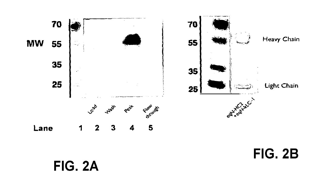
[0166]The supernatants obtained from Example 2 were purified using a Protein A column, separated by SOS-PAGE and tested for reactivity to anti-equine IgG polyclonal antibody HRP. The SOS-PAGE gel was also stained using Coomassie blue to detect heavy and light chains. The anti-equine IgG polyclonal antibody preparation predominantly recognises the equine heavy chains. The results are shown in FIGS. 2A and B. The results show purification of equine anti-NGF with type 2 heavy chain by Protein A, as illustrated by a Western blot developed with anti-equine polyclonal antibody HRP. The peak fraction was analysed by Coomassie stained SDS-PAGE. Some degradation of the heavy and light chain is apparent by SOS-PAGE. The Coomassie blue stained gel (FIG. 2B, shows presence of heavy and light chains as well as complete antibody (MW of 70).
PUM
| Property | Measurement | Unit |
|---|---|---|
| concentration | aaaaa | aaaaa |
| concentration | aaaaa | aaaaa |
| concentration | aaaaa | aaaaa |
Abstract
Description
Claims
Application Information