
Systems and methods for medical imaging

Inactive Publication Date: 2019-10-10
WADE JACK
View PDF5 Cites 3 Cited by
  • Summary
  • Abstract
  • Description
  • Claims
  • Application Information

AI Technical Summary

Benefits of technology

This patent describes a technology that allows for the capture of high-resolution images and videos using an array of image sensors. By multiplying the number of sensors and processing the gathered data in real-time, the technology can provide medical professionals with better visibility during procedures and even non-invasively identify healthy or diseased tissue. The technology can magnify images without using digital zoom, which can result in a loss of clarity. This is achieved by retrieving raw image data from only the sensors corresponding to the area of interest and adjusting compression to show the data on a display. Overall, this technology can capture and enhance images for improved medical practice.

Problems solved by technology

While this brings many benefits to patients, it presents a number of challenges for the surgeon who must work within a very confined surgical compartment.
In particular, surgeons must deal with poor visibility, limited lighting and a narrow viewing angle.
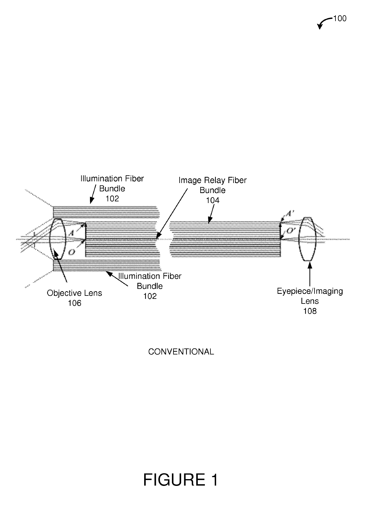
Because of their size, conventional endoscopes (and the like) tend to have limited imaging resolution and often fail to provide more than one perspective of biological tissue.
However, image resolution of the CCD remains as a limiting factor in terms of image magnification and precludes the ability to zoom in to see very fine detail, or at a cellular level.
For some embodiments, magnification is facilitated without the need for digital zoom, which uses the same number of pixels in the portion of the image being magnified as there are in that same portion in the unmagnified image and thus results in a loss of clarity in the enlarged image.

Method used


Image

  • Systems and methods for medical imaging
  • Systems and methods for medical imaging
  • Systems and methods for medical imaging

Examples


Embodiment Construction

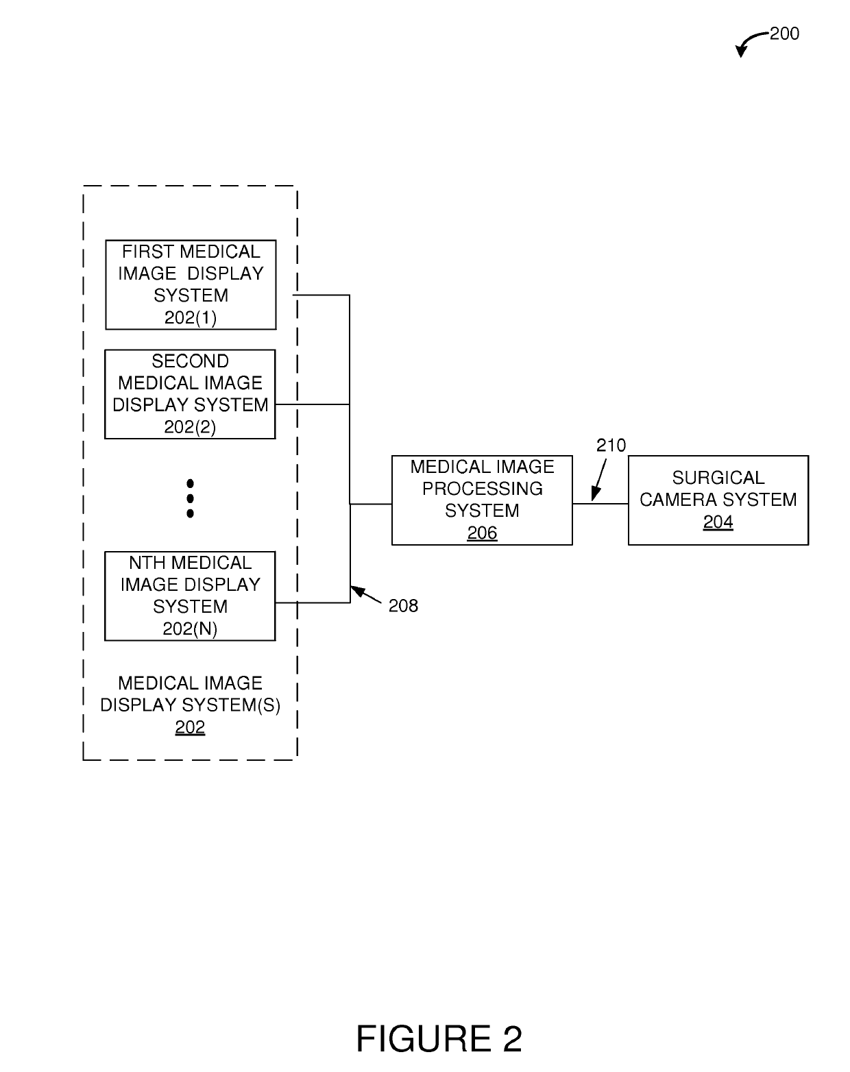
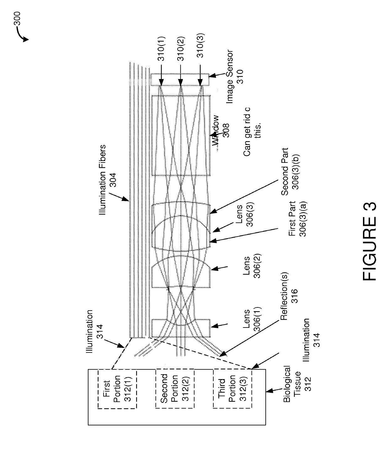
[0032]Systems and methods disclosed herein provide an imaging system such as an endoscopic imaging system for a variety of applications. Embodiments of the systems and methods disclosed herein can be configured to utilize a plurality of image sensors as an array of image sensors to capture images for display or recording. In various embodiments, algorithms or other processing techniques can be used to provide high resolution imaging and to provide real-time image magnification without the loss of resolution, or without the same amount of loss of resolution as would be experienced by typical conventional “digital zoom” techniques.

[0033]Particularly, in various embodiments, an array of image sensors is used to capture images from the endoscope. The optical fiber or lens system used to transmit the images from the objective lens to the image sensor is configured to allow sections or portions of the tissue, organ, cavity, or other sample being imaged to be mapped to corresponding image ...


PUM


Abstract

The disclosed technology relates to high resolution imagery for a medical environment. Various embodiments provide for a surgical camera, such as an endoscopic camera, comprising an array of image sensors. The array of image sensors may be configured to capture static images or video, may be configured to capture imagery of biological tissue (e.g., surfaces), and may be specifically configured to capture imagery within a surgical environment. For some embodiments, the array of image sensors multiplies the number of available pixels by the number of image sensors, thereby facilitating high resolution image data (e.g., one or more static images or video). The collected image data may be processed at or near real-time to provide very high-quality imagery (e.g., images or video) that can be enhanced or magnified for medical purposes, which can provide a medical care giver (e.g., surgeon) better visibility during medical procedures.

Description

CROSS REFERENCE TO RELATED APPLICATIONS[0001]The preset application is a continuation of U.S. patent application Ser. No. 14 / 709,241, filed May 11, 2015, which claims the benefit of claims the benefit of U.S. Provisional Patent Application Ser. No. 61 / 991,406, filed May 9, 2014, the contents of which is hereby incorporated by reference herein in its entirety.TECHNICAL FIELD[0002]The technology disclosed herein relates to imaging tools and, in particular, some embodiments relate to systems and methods for imaging a selected subject matter (e.g., tissue, organs or a body cavity) in a medical environment, such as a surgical environment.DESCRIPTION OF RELATED ART[0003]Minimally invasive medical procedures have been increasing in popularity and have become commonplace inpatient care. For instance, endoscopes and other types of medical imaging devices are indispensable tools in patient care, especially in in-patient and out-patient surgical environments. Advantages of using endoscopes inc...

Claims


Application Information

Patent Timeline
IPC IPC(8): A61B1/00A61B1/05A61B17/00A61B1/07
CPCA61B1/0002A61B1/051A61B2562/04A61B1/07A61B1/0005A61B2505/05A61B1/00009A61B1/00039A61B17/00234A61B90/361A61B2090/306A61B2576/00A61B1/05A61B1/00188A61B1/00096A61B1/00011A61B1/042G16H30/40A61B1/000095A61B1/00042
Inventor WADE, JACK
Owner WADE JACK
Learn More