
Method and system for determining virtual outputs for a multi-energy x-ray imaging apparatus

a multi-energy, x-ray imaging technology, applied in the field of x-ray imaging, can solve the problems of obscuring the tissue being imaged, affecting the image quality,

Active Publication Date: 2019-12-12
KA IMAGING INC
View PDF1 Cites 13 Cited by
  • Summary
  • Abstract
  • Description
  • Claims
  • Application Information

AI Technical Summary

Benefits of technology

The patent describes a method and system for improving the performance of a multi-energy x-ray imaging apparatus. The method involves receiving multiple outputs from the apparatus and using a general algorithm to determine parameters and generate a virtual output algorithm for the apparatus. The virtual output algorithm can then be used to generate at least one virtual output with reduced noise and object scattered radiation. The virtual output can be used for correcting faulty pixels, obtaining bone mineral density measurements, and other applications. The system includes a multi-energy x-ray imaging apparatus with multiple sensor layers, a mid-filter layer, and an anti-grid layer. The sensor layers can include a photoconductor layer, scintillator layer, or scintillator-infused glass substrate layer. The technical effects of the patent include improved image quality, reduced noise, and improved performance of the multi-energy x-ray imaging apparatus.

Problems solved by technology

In x-ray imaging, a large source of noise that often decreases the quality of an image is anatomical noise.
This noise can obscure the tissue being imaged or can be misread as anatomical abnormalities.
In this case, the ribs are a major source of anatomical noise, as they are not the anatomy of interest.
One challenge in DE imaging comes from the need to obtain two separate low and high energy images.
In practice, a total cancellation of a specific tissue type is not commonly possible.
These non-idealities also mean that the theoretical value of the weighting factors may not provide the best possible cancellation, requiring the observer to calculate their ideal value experimentally or qualitatively.
Unfortunately, the temporal separation inherent in this technique causes motion artifacts to appear in the final images, which can pose a large challenge to a radiologist or observer interpreting it.
These artifacts are noticeable distortions in the image caused by slight misalignments of the anatomies in the sequential images, and generally stem from patient or object motion that occurs during and in between image acquisitions.
Motion artifacts will appear due to these movements, which can be a particular hindrance in cardiac and pulmonary imaging due to the large presence of the heart.
Furthermore, this issue will compound as more image acquisitions are added in multi-energy imaging, since the total acquisition time will increase, allowing for more patient motion.
A practical issue that arises with the single-shot approach is that to obtain the ideal effective energy separation between the layers, the sensitive material—be it a scintillator or a direct conversion material—mass loading (or, equivalently, their thicknesses) must be tuned for specific tissue types and patient anatomy.
Since at a commercial level it is only feasible to build a few specific configurations, this leaves a compromised solution that can best fit all target applications and patient types as the only practical one.

Method used


Image

  • Method and system for determining virtual outputs for a multi-energy x-ray imaging apparatus
  • Method and system for determining virtual outputs for a multi-energy x-ray imaging apparatus
  • Method and system for determining virtual outputs for a multi-energy x-ray imaging apparatus

Examples


Embodiment Construction

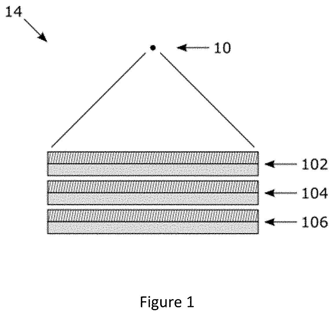
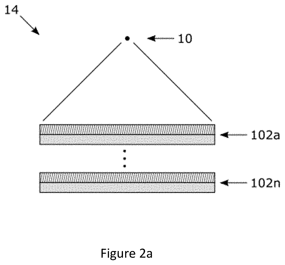
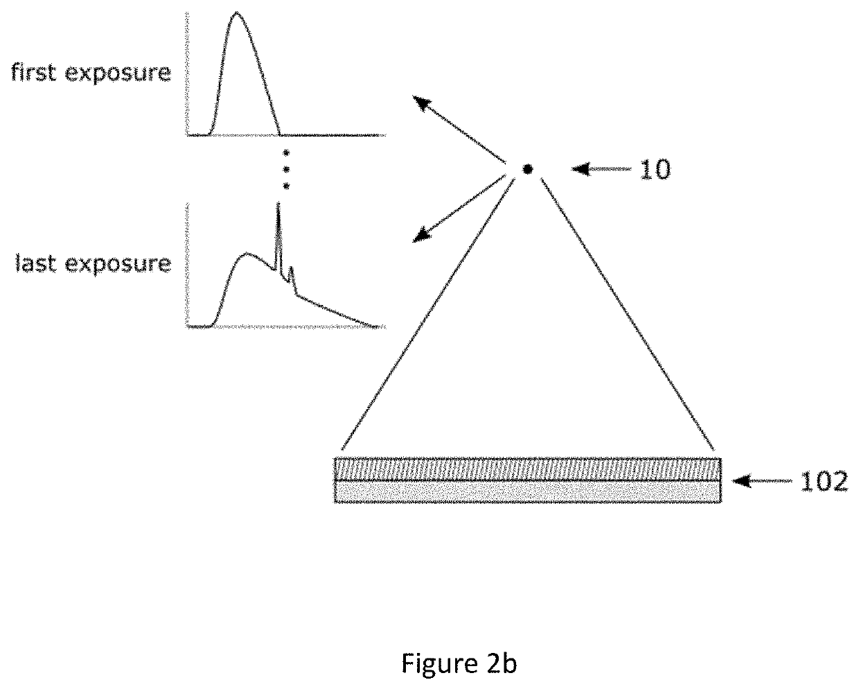
[0041]The disclosure is directed at a method and apparatus for determining virtual outputs for a multi-energy x-ray imaging apparatus. In one embodiment, the method receives actual outputs from the layers of a multi-layer x-ray imaging apparatus and then processes the outputs to determine outputs for other non-existent layers within the multi-layer x-ray imaging apparatus as if they were actual physical layers within the x-ray imaging apparatus. In another embodiment, the method receives actual outputs from different spectral / energy exposures obtained from a multi-shot imaging apparatus and then processes the outputs to determine the outputs for other non-obtained spectral / energy exposures.

[0042]FIG. 8 illustrates a general diagram of a radiographic imaging environment. As shown, an x-ray source 10 generates an x-ray beam, or x-rays, 11 that is transmitted towards an object 12, e.g. a patient's hand, for imaging by a radiography detector system (RDS) 14. The results of the x-ray exp...


PUM


Abstract

The disclosure is directed at a method and apparatus for determining virtual outputs for a multi-energy x-ray apparatus. Based on the application that the x-ray apparatus is being used for, a general algorithm can be determined or selected. Inputs received from the x-ray apparatus can be substituted into the general algorithm to generate a virtual output algorithm for the x-ray apparatus. Virtual outputs can then be calculated using the virtual output algorithm.

Description

CROSS-REFERENCE TO OTHER APPLICATIONS[0001]This application claims priority from U.S. Provisional Application No. 62 / 682,540 filed Jun. 8, 2018 the contents of which are hereby incorporated by reference.FIELD OF THE DISCLOSURE[0002]The disclosure is generally directed at x-ray imaging and, more specifically, at a method and system for determining virtual outputs for a multi-energy x-ray imaging apparatus.BACKGROUND OF THE DISCLOSURE[0003]The quality of a medical image and by extension its value as a tool depends only on how well it can convey the anatomy of the patient being imaged to the observer, such as a physician. The better the anatomy is understood, the more accurate information the physician has to make decisions.[0004]In x-ray imaging, a large source of noise that often decreases the quality of an image is anatomical noise. It is caused by a superposition of the normal anatomy that stems from the two-dimensional (2D) projection of the three-dimensional (3D) patient. This no...

Claims


Application Information

Patent Timeline
IPC IPC(8): A61B6/00
CPCA61B6/4266A61B6/5282A61B6/4208A61B6/482A61B6/5205A61B6/505A61B6/4241A61B6/483A61B6/462A61B6/4216A61B6/586G01T1/242
Inventor KARIM, KARIM S.LOPEZ MAURINO, SEBASTIANGHANBARZADEH, SINA
Owner KA IMAGING INC
Learn More