Imaging system for screening and diagnosis of breast cancer

a breast cancer and imaging system technology, applied in the field of subcutaneous cellular mass detection, can solve the problems of no device available for a woman to use, no effective preventive method, and greatly reduced survival chances, so as to improve survival rate, reduce the risk of breast cancer, and improve the effect of screening and diagnosis

Pending Publication Date: 2020-01-09
BANPIL PHOTONICS
View PDF7 Cites 1 Cited by
  • Summary
  • Abstract
  • Description
  • Claims
  • Application Information

AI Technical Summary

Benefits of technology

[0014]The present invention aims to overcome problems associated with current technologies by providing a device that is friendly to users and makes screening and diagnosis of breast cancer more sensitive, more rapid, non-invasive, and less costly, allowing for early detection of emerging tumors through routine self-examination.
[0017]It is an object of this invention to encourage routine breast cancer screening that is self-operable, more private, easier to use, yet cost-effective.
[0018]It is an object of this invention to raise the accuracy of diagnosis and reduce the rate of false positives and false negatives.

Problems solved by technology

However, if the tumor is not detected until it has grown large and spread into the lymphatic system, chances of survival are greatly decreased.
Unfortunately, there is no device available that a woman may use to regularly screen herself for breast cancer at home.
It is not an effective preventive method because finding a lump likely indicates that the tumor has already been growing for years.
There is no evidence that routine examination reduces morality rates, and it is no longer a recommended screening method.
Such genetic screening does not detect the presence of breast cancer, but it may reveal a person's susceptibility to develop it.
Having a close relative diagnosed with breast cancer increases a woman's risk of breast cancer.
Moreover, since only a small percentage of breast cancer occurrences is caused by inheriting mutations, it is not a method that would be beneficial for the general public.
However, there is continued debate about whether this method is less helpful than it is helpful.
For example, one of the drawbacks is that false positives create long-lasting psychological stress and anxiety, which can affect the patient's wellness and behavior for many years.
On the other hand, false negatives estimated up to thirty percent occur, which can lead to missed opportunities for treatment if regular checking is not done.
Mammograms do not work well in younger women because their breast tissue is denser.
Cost can further be an issue, more so because insurance policies tend not to cover mammograms for women under forty, even though women in their teens or twenties are sometimes diagnosed with breast cancer.
Other drawbacks include discomfort and limitation in detection accuracy.
Patients undergoing mammograms have their breasts compressed, which can cause pain or discomfort.
Tumors sized smaller than one millimeter are difficult, if not impossible, to detect.
While it increases the detection rate of breast cancer, it also increases the rate of false positives.
However, it also produces greater false positives and is expensive, costing thousands of dollars.
It is a time-consuming and invasive procedure requiring injection of a contrast agent that poses a risk to patients with a history of renal disease.
Removing tissue from the breast can be a physically and psychologically challenging procedure.
Currently, this technique is not used for breast cancer screening and is typically only used for large-scale imaging of the entire body to determine if the cancer has spread from the breast area.
In addition, this technique is not non-invasive because it requires the injection of the dye as well as a visit to the physician.
While the use of light in the NIR range has the advantage of being highly sensitive and specific (can also reach deep tissue), it has the same problems as CAT scans in that it requires the injection of a drug and cannot be performed without the aid of a physician.
However, this technology is limited by the difficulty in distinguishing between absorption and scattering in tissue, as well as the need to rely on secondary factors (oxygen metabolism and blood volume) to determine the presence of a tumor.
Nevertheless, it can be seen that current methods of breast cancer screening and diagnostic methods can be stress inducing, unreliable, bulky, invasive, and costly.
Moreover, some women may also feel a stigma associated with breast cancer, and going through medical procedures related to breast cancer can make them feel especially vulnerable to privacy issues.

Method used


Image

  • Imaging system for screening and diagnosis of breast cancer
  • Imaging system for screening and diagnosis of breast cancer
  • Imaging system for screening and diagnosis of breast cancer

Examples


Embodiment Construction

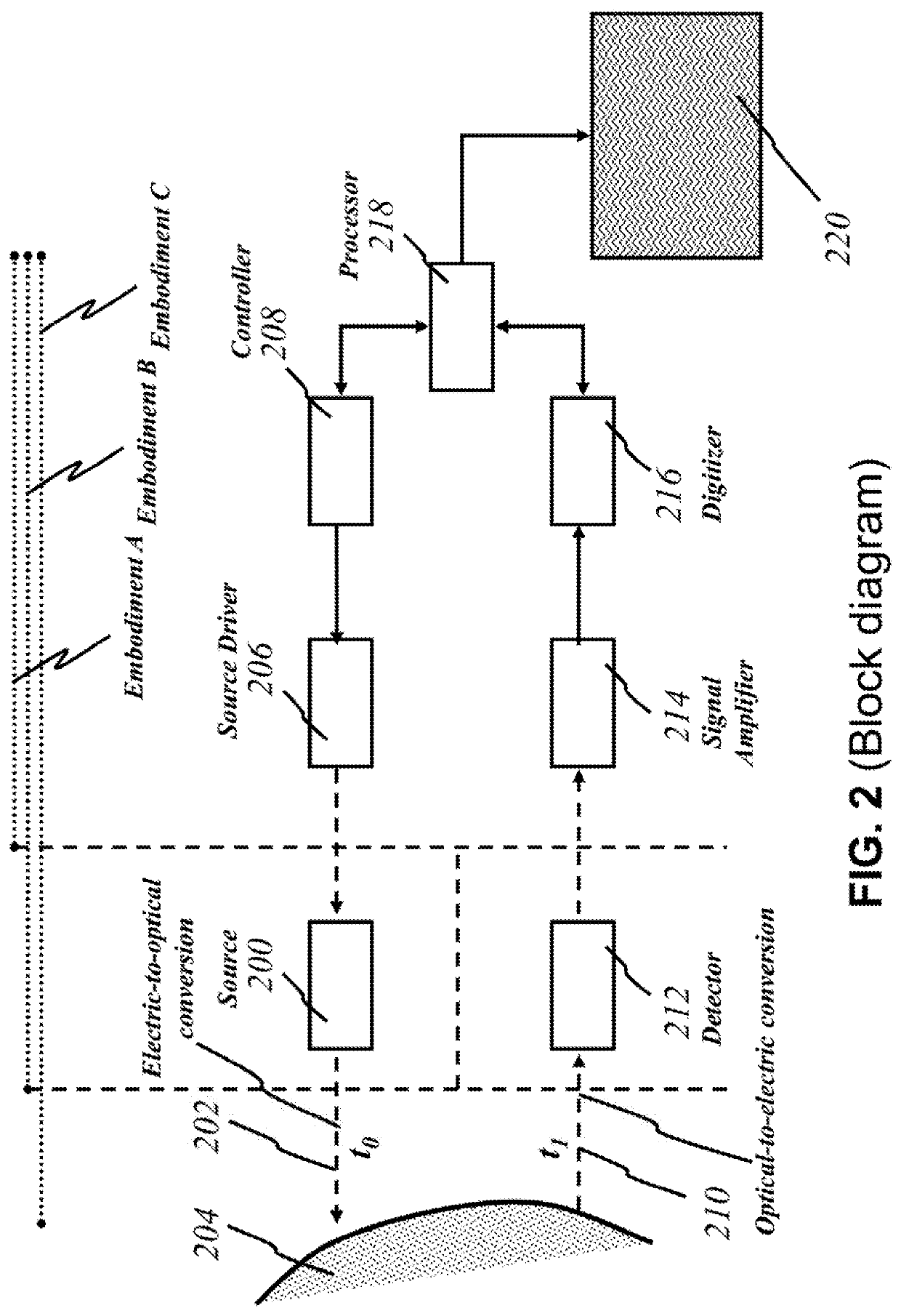
[0056]Reference numerals refer to corresponding parts labeled throughout the figures. The embodiments described herein pertain to a device that detects and images subcutaneous cellular mass through optical techniques. The embodiments pertain to methods and apparatuses for screening and diagnosis of breast cancer.

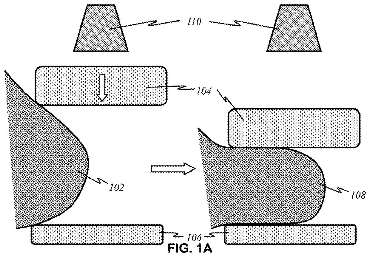
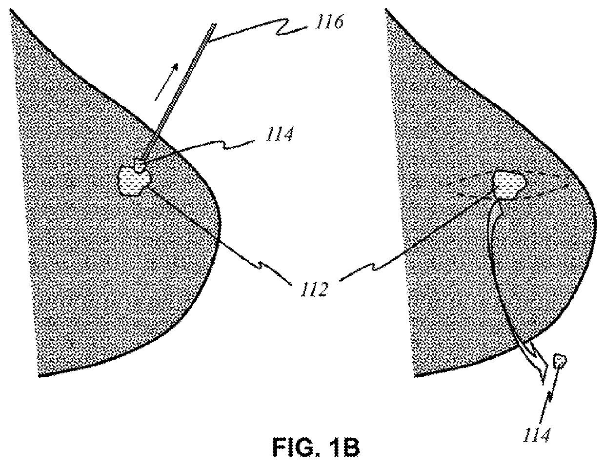
[0057]As used herein, the term “area of interest” and “area of concern” refer to parts of bodily tissue where cancer cells are suspected or known to be. For example, a woman (or man) may undergo mammography on her breasts where she felt a lump. The general area where the lump was would be an area of concern because it is suspected that cancer cells may be developing in the lump. In particular, a “tumor” is an abnormal growth of cells, especially malignant neoplasms that invade nearby cells (cancer).

[0058]As used herein, the term “biomass” refers to a total mass or volume of organic matter, typically from the human body. It could be an entire organ or portion thereof, a secti...


PUM


Abstract

This invention provides a non-invasive diagnosis system that is not only capable of producing high-resolution, three-dimensional images of abnormalities of tissue growth inside the body but, it can also detect the type of abnormalities and their location using multispectral imaging techniques. It is possible to provide a portable, non-invasive device that is handheld and with which women may use to screen themselves for early detection of breast cancer without the need to visit a physician. As the present invention uses broadband sources and / or multiple coherent sources, secondary factors such as oxygen metabolism or blood volume associated with the cancer tissues could also be detected to provide further verification of the type. This invention would raise the accuracy of diagnosis and reduce the rate of false positives and false negatives.

Description

[0001]This application is a divisional of and claims the benefit of priority to co-owned U.S. patent application Ser. No. 14 / 984,717 of the same title filed Dec. 30, 2015 issuing as U.S. Pat. No. 10,357,162 on Jul. 23, 2019, the foregoing being incorporated herein by reference in its entirety.FIELD OF THE INVENTION[0002]This invention relates to detection of subcutaneous cellular mass utilizing optics and imaging techniques. More particularly, this invention is related to detecting (a) abnormal growth of tissues inside the body, (b) their types, (c) their dimensions, and (d) their location from outside the body (through non-invasive contact or non-contact with the body). More specifically, this invention is related to the means to detect abnormalities of tissue growth inside the body, their types, dimensions, and location from outside, more particularly the early diagnosis of the cancer, especially breast cancer. This invention also relates to a medical device that emits electromagn...

Claims


Application Information

Patent Timeline
Patent Type & Authority Applications(United States)
IPC IPC(8): A61B5/00
CPCA61B5/7246A61B5/742A61B5/0091A61B5/0013A61B5/708A61B2560/0425A61B5/4312A61B2562/164A61B2562/066
Inventor DUTTA, ACHYUT KUMAR
Owner BANPIL PHOTONICS
Why Eureka
Try Eureka