[0041]The long-term or stable effectiveness of the invention methods can be assessed as described herein, for example, by the endothelial cell phenotype and density not being compromised. Another way to assess the long-term or stable effectiveness of the invention method is by determining that the density of keratocytes is not reduced after treatment, at time points greater than 4 weeks or 1 month. Yet another way to assess the long-term or stable effectiveness of the invention method is by determining that apoptosis is not induced in any cell type after treatment, at time points greater than 4 weeks or 1 month. Accordingly, in a particular embodiment, the corneal haze formation is prevented or reduced for a period greater than or equal to 4 months, and the endothelial cell phenotype and density is not compromised. In another embodiment, the corneal haze formation is prevented or reduced for a period greater than or equal to 4 months, and the density of keratocytes is not reduced. In other embodiments, the HDACi is selected from the group consisting of Entinostat (MS-275); Panobinostat (LBH589); Trichostatin A (TSA); Mocetinostat (MGCD0103); Belinostat (PXD101); Romidepsin (FK228, Depsipeptide); MC1568; Tubastatin A HCl; Givinostat (ITF2357); Dacinostat (LAQ824); CUDC-101; Quisinostat (JNJ-26481585); Pracinostat (SB939); PCI-34051; Droxinostat; Abexinostat (PCI-24781); RGFP966; AR-42; Ricolinostat (ACY-1215); Tacedinaline (CI994); CUDC-907; M344; Tubacin; RG2833 (RGFP109); Resminostat; Tubastatin A; WT161; ACY-738; Tucidinostat (Chidamide); TMP195; (ACY-241); BRD73954; BG45; 4SC-202; CAY10603; LMK-235; CHR-3996; Splitomicin; Santacruzamate A (CAY10683); Nexturastat A; TMP269; HPOB; Valproic acid sodium salt (Sodium valproate), and derivatives of any of these members, or a physiologically acceptable salt of any of these members.
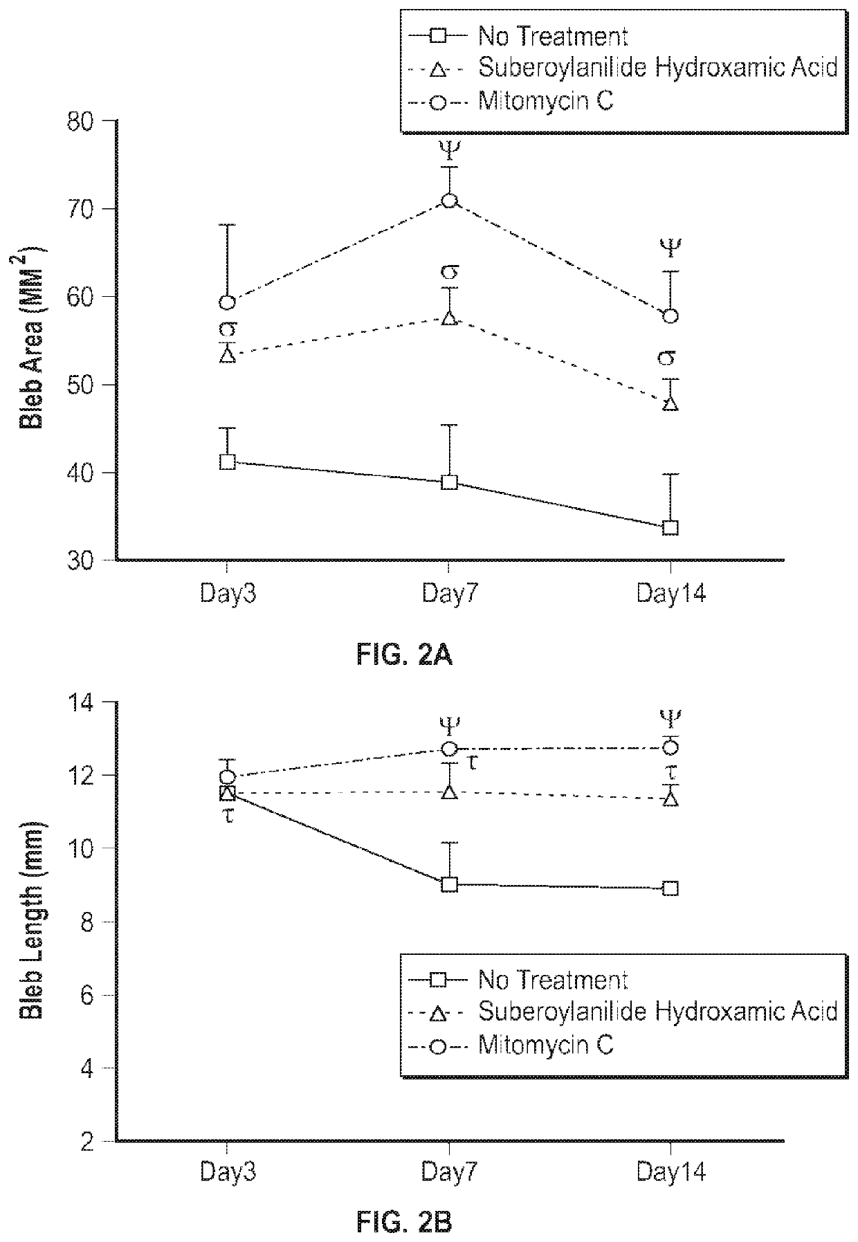
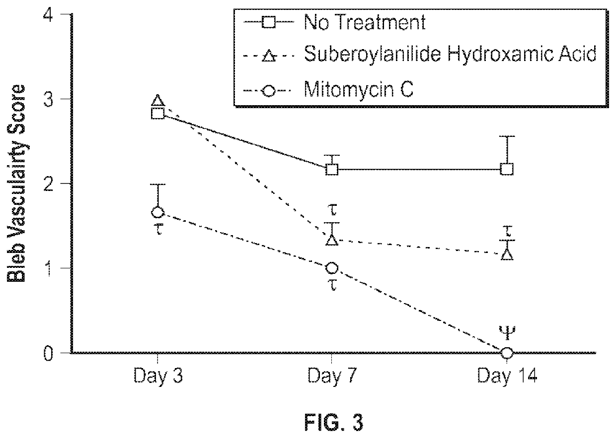
[0042]Adjunct therapy is needed to reduce corneal haze after PRK, especially with higher diopter treatments. Our group has previously demonstrated that HDAC inhibitors effectively reduce corneal haze and scarring in vivo in the rabbit cornea without causing significant acute side effects. See, e.g., Sharma A, et al., Trichostatin A inhibits corneal haze in vitro and in vivo. Invest Ophthalmol Vis Sci. 2009; 50(6):2695-2701; Tandon A et al., Vorinostat: a potent agent to prevent and treat laser-induced corneal haze. J Refract Surg. 2012; 28(4):285-90; Donnelly K S et al., Suberoylanilide hydroxamic acid (vorinostat): its role on equine corneal fibrosis and matrix metalloproteinase activity. Vet Ophthalmol. 2014; 17 Suppl 1:61-68; and Gronkiewicz K M et al., Molecular mechanisms of suberoylanilide hydroxamic acid in the inhibition of TGF-β1-mediated canine corneal fibrosis. Vet Ophthalmol. 2016; 19(6):480-487; each of which are incorporated herein by reference in their entirety. In accordance with the present invention, we found adjunct topical SAHA and MMC application after PRK significantly prevented corneal haze and decreased the pro-fibrotic biomarkers in vivo in rabbits. Although haze inhibition by SAHA was less than MMC, this difference was statistically insignificant and appeared clinically irrelevant based on the slit-lamp subjective analysis. The most remarkable findings of the present study were the detection of significantly reduced cytotoxicity and enhanced safety profile by SAHA compared to MMC. SAHA application demonstrated markedly improved keratocyte viability and phenotype, reduced keratocyte and endothelial apoptosis and strikingly better endothelial cellular morphology. These results indicate that topical adjunct SAHA application after PRK would be a safer alternative to MMC in preventing post-PRK corneal haze.
[0043]The corneal wound healing response plays a central role in the outcome of refractive surgery. Pharmacologically broad acting agents, specifically steroids and MMC, are most commonly used to control post-PRK scarring. Increasingly precise targeted control of the corneal wound healing response will lead to faster recovery times, more accurate refractive outcomes and decreased complication rates. In vitro analysis reveals that expression of α-SMA in stress fibers confers to the differentiated myofibroblast at least a two-fold stronger contractile activity compared with α-SMA-negative fibroblasts. We found many α-SMA expressing myofibroblasts after PRK in the anterior stroma of rabbit cornea. It is likely that these cells contribute to corneal scar formation and refractive outcome of the procedure. The clearance of α-SMA expressing cells from the anterior stroma of MMC treated corneas suggest the decrease in myofibroblasts due to MMC toxicity. This toxicity is also responsible for a diminished keratocyte population available for conversion to myofibroblasts at the site of injury.
[0044]The literature suggests that during laser photo disruption some cells are vaporized instantly, while cells in close proximity went into a slow involution form of cell death, known as apoptosis. Myofibroblasts may undergo apoptosis or undergo transdifferentiation back to a progenitor cell. The level of keratocyte apoptosis distribution, along with activated stromal keratocytes repopulation, are likely contributors of corneal wound healing associated with variability and regression after PRK.
[0045]Previous studies demonstrated that topical application of MMC after PRK in rabbits not only decreased keratocyte density due to apoptosis at the wound site, but it significantly delayed keratocyte repopulation and activation in the anterior stroma with normal epithelial cell differentiation. In accordance with the present invention, we observed a similar cytotoxicity pattern in MMC treated rabbit corneas in which several TUNEL+ cells at shorter times and low DAPI-stained nuclei at longer times in the anterior stroma were observed. Contrary to this, SAHA treatment did not show such damage to the anterior stroma. These findings indicate that SAHA has a superior safety profile than MMC in the treatment of corneal haze after PRK.
[0046]The corneal endothelial cells do not replicate in humans and therefore their preservation is important for corneal transparency and normal functioning. In accordance with the present invention, topical application of SAHA did not cause apoptosis in endothelial cells, and in addition showed a typical polygonal morphology and cellular density similar to untreated control corneas up to 4-months, the longest tested time point. The conflicting literature on the effects of MMC on human corneal endothelial cells exist. A nonrandomized controlled trial showed that the prophylactic use of MMC (0.02%; 10-50 seconds) inhibited haze formation but caused significant loss of corneal endothelial cells. In contrast, other studies have shown that the administration of 0.02% MMC topically applied to the cornea for 12 seconds and 40 seconds following PRK did not have a significant effect on qualitative morphometric parameters or quantitative endothelial cell density.

