Methods and compositions for promoting wound healing with decreased scar formation after glaucoma filtration surgery

a technology of scar formation and wound healing, applied in the field of ophthalmic diseases, can solve the problems of fibrosis over filtering bleb, excessive ecm synthesis, large patient population that does not respond adequately to topical drug therapy to reduce iop, etc., and achieves the effect of assessing the long-term or stable effectiveness of the invention method, and reducing the density of keratocytes

Inactive Publication Date: 2018-12-27
U S GOVERNMENT REPRESENTED BY THE DEPT OF VETERANS AFFAIRS
View PDF0 Cites 4 Cited by
  • Summary
  • Abstract
  • Description
  • Claims
  • Application Information

AI Technical Summary

Benefits of technology

[0041]The long-term or stable effectiveness of the invention methods can be assessed as described herein, for example, by the endothelial cell phenotype and density not being compromised. Another way to assess the long-term or stable effectiveness of the invention method is by determining that the density of keratocytes is not reduced after treatment, at time points greater than 4 weeks or 1 month. Yet another way to assess the long-term or stable effectiveness of the invention method is by determining that apoptosis is not induced in any cell type after treatment, at time points greater than 4 weeks or 1 month. Accordingly, in a particular embodiment, the corneal haze formation is prevented or reduced for a period greater than or equal to 4 months, and the endothelial cell phenotype and density is not compromised. In another embodiment, the corneal haze formation is prevented or reduced for a period greater than or equal to 4 months, and the density of keratocytes is not reduced. In other embodiments, the HDACi is selected from the group consisting of Entinostat (MS-275); Panobinostat (LBH589); Trichostatin A (TSA); Mocetinostat (MGCD0103); Belinostat (PXD101); Romidepsin (FK228, Depsipeptide); MC1568; Tubastatin A HCl; Givinostat (ITF2357); Dacinostat (LAQ824); CUDC-101; Quisinostat (JNJ-26481585); Pracinostat (SB939); PCI-34051; Droxinostat; Abexinostat (PCI-24781); RGFP966; AR-42; Ricolinostat (ACY-1215); Tacedinaline (CI994); CUDC-907; M344; Tubacin; RG2833 (RGFP109); Resminostat; Tubastatin A; WT161; ACY-738; Tucidinostat (Chidamide); TMP195; (ACY-241); BRD73954; BG45; 4SC-202; CAY10603; LMK-235; CHR-3996; Splitomicin; Santacruzamate A (CAY10683); Nexturastat A; TMP269; HPOB; Valproic acid sodium salt (Sodium valproate), and derivatives of any of these members, or a physiologically acceptable salt of any of these members.
[0042]Adjunct therapy is needed to reduce corneal haze after PRK, especially with higher diopter treatments. Our group has previously demonstrated that HDAC inhibitors effectively reduce corneal haze and scarring in vivo in the rabbit cornea without causing significant acute side effects. See, e.g., Sharma A, et al., Trichostatin A inhibits corneal haze in vitro and in vivo. Invest Ophthalmol Vis Sci. 2009; 50(6):2695-2701; Tandon A et al., Vorinostat: a potent agent to prevent and treat laser-induced corneal haze. J Refract Surg. 2012; 28(4):285-90; Donnelly K S et al., Suberoylanilide hydroxamic acid (vorinostat): its role on equine corneal fibrosis and matrix metalloproteinase activity. Vet Ophthalmol. 2014; 17 Suppl 1:61-68; and Gronkiewicz K M et al., Molecular mechanisms of suberoylanilide hydroxamic acid in the inhibition of TGF-β1-mediated canine corneal fibrosis. Vet Ophthalmol. 2016; 19(6):480-487; each of which are incorporated herein by reference in their entirety. In accordance with the present invention, we found adjunct topical SAHA and MMC application after PRK significantly prevented corneal haze and decreased the pro-fibrotic biomarkers in vivo in rabbits. Although haze inhibition by SAHA was less than MMC, this difference was statistically insignificant and appeared clinically irrelevant based on the slit-lamp subjective analysis. The most remarkable findings of the present study were the detection of significantly reduced cytotoxicity and enhanced safety profile by SAHA compared to MMC. SAHA application demonstrated markedly improved keratocyte viability and phenotype, reduced keratocyte and endothelial apoptosis and strikingly better endothelial cellular morphology. These results indicate that topical adjunct SAHA application after PRK would be a safer alternative to MMC in preventing post-PRK corneal haze.
[0043]The corneal wound healing response plays a central role in the outcome of refractive surgery. Pharmacologically broad acting agents, specifically steroids and MMC, are most commonly used to control post-PRK scarring. Increasingly precise targeted control of the corneal wound healing response will lead to faster recovery times, more accurate refractive outcomes and decreased complication rates. In vitro analysis reveals that expression of α-SMA in stress fibers confers to the differentiated myofibroblast at least a two-fold stronger contractile activity compared with α-SMA-negative fibroblasts. We found many α-SMA expressing myofibroblasts after PRK in the anterior stroma of rabbit cornea. It is likely that these cells contribute to corneal scar formation and refractive outcome of the procedure. The clearance of α-SMA expressing cells from the anterior stroma of MMC treated corneas suggest the decrease in myofibroblasts due to MMC toxicity. This toxicity is also responsible for a diminished keratocyte population available for conversion to myofibroblasts at the site of injury.
[0044]The literature suggests that during laser photo disruption some cells are vaporized instantly, while cells in close proximity went into a slow involution form of cell death, known as apoptosis. Myofibroblasts may undergo apoptosis or undergo transdifferentiation back to a progenitor cell. The level of keratocyte apoptosis distribution, along with activated stromal keratocytes repopulation, are likely contributors of corneal wound healing associated with variability and regression after PRK.
[0045]Previous studies demonstrated that topical application of MMC after PRK in rabbits not only decreased keratocyte density due to apoptosis at the wound site, but it significantly delayed keratocyte repopulation and activation in the anterior stroma with normal epithelial cell differentiation. In accordance with the present invention, we observed a similar cytotoxicity pattern in MMC treated rabbit corneas in which several TUNEL+ cells at shorter times and low DAPI-stained nuclei at longer times in the anterior stroma were observed. Contrary to this, SAHA treatment did not show such damage to the anterior stroma. These findings indicate that SAHA has a superior safety profile than MMC in the treatment of corneal haze after PRK.
[0046]The corneal endothelial cells do not replicate in humans and therefore their preservation is important for corneal transparency and normal functioning. In accordance with the present invention, topical application of SAHA did not cause apoptosis in endothelial cells, and in addition showed a typical polygonal morphology and cellular density similar to untreated control corneas up to 4-months, the longest tested time point. The conflicting literature on the effects of MMC on human corneal endothelial cells exist. A nonrandomized controlled trial showed that the prophylactic use of MMC (0.02%; 10-50 seconds) inhibited haze formation but caused significant loss of corneal endothelial cells. In contrast, other studies have shown that the administration of 0.02% MMC topically applied to the cornea for 12 seconds and 40 seconds following PRK did not have a significant effect on qualitative morphometric parameters or quantitative endothelial cell density.

Problems solved by technology

However, a large number of patients do not respond adequately to topical drug therapy to reduce IOP.
A major deterrent to the success of GFS is caused by aberrant post-operative wound healing resulting in excessive ECM synthesis leading to fibrosis over filtering bleb.
Development of fibrosis and collagen deposit at sclerotomy site compromises bleb's proper functioning and disable its ability to maintain non-pathologic reduced IOP.
Though these drugs are effective in preventing ocular fibrosis and improving the outcome of filtration surgery, they are known to cause sight-threatening complications including wide spread cell death, bleb leak, hypotony, and / or endophthalmitis.
The released cytokines alter the gene expression of proliferative, cytoskeletal and matrix proteins, thus leading to fibrosis.

Method used


Image

  • Methods and compositions for promoting wound healing with decreased scar formation after glaucoma filtration surgery
  • Methods and compositions for promoting wound healing with decreased scar formation after glaucoma filtration surgery
  • Methods and compositions for promoting wound healing with decreased scar formation after glaucoma filtration surgery

Examples


example 1

bitor (HDACi) Prevents Excessive Wound Healing and Scar Formation in Rabbit Model of Glaucoma Filtration Surgery

[0080]Material and Methods

[0081]Background.

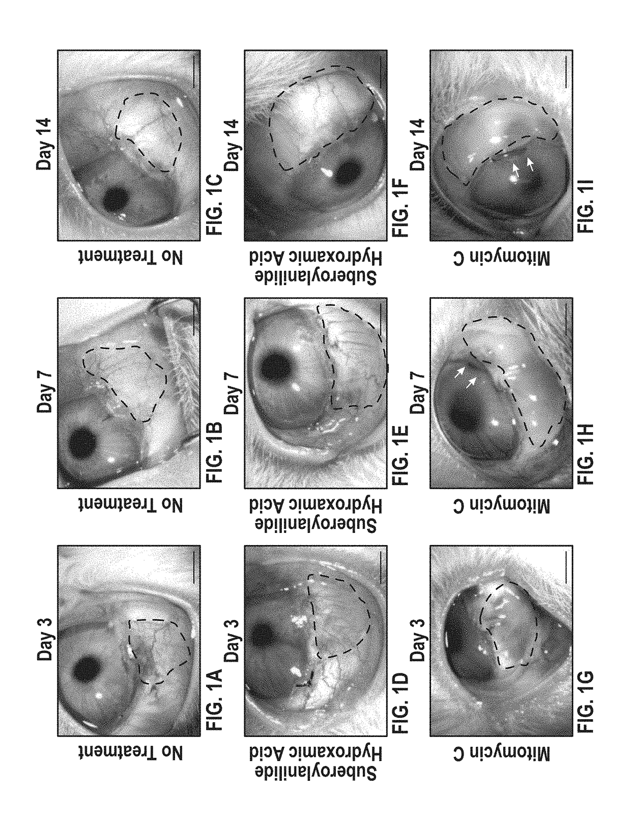
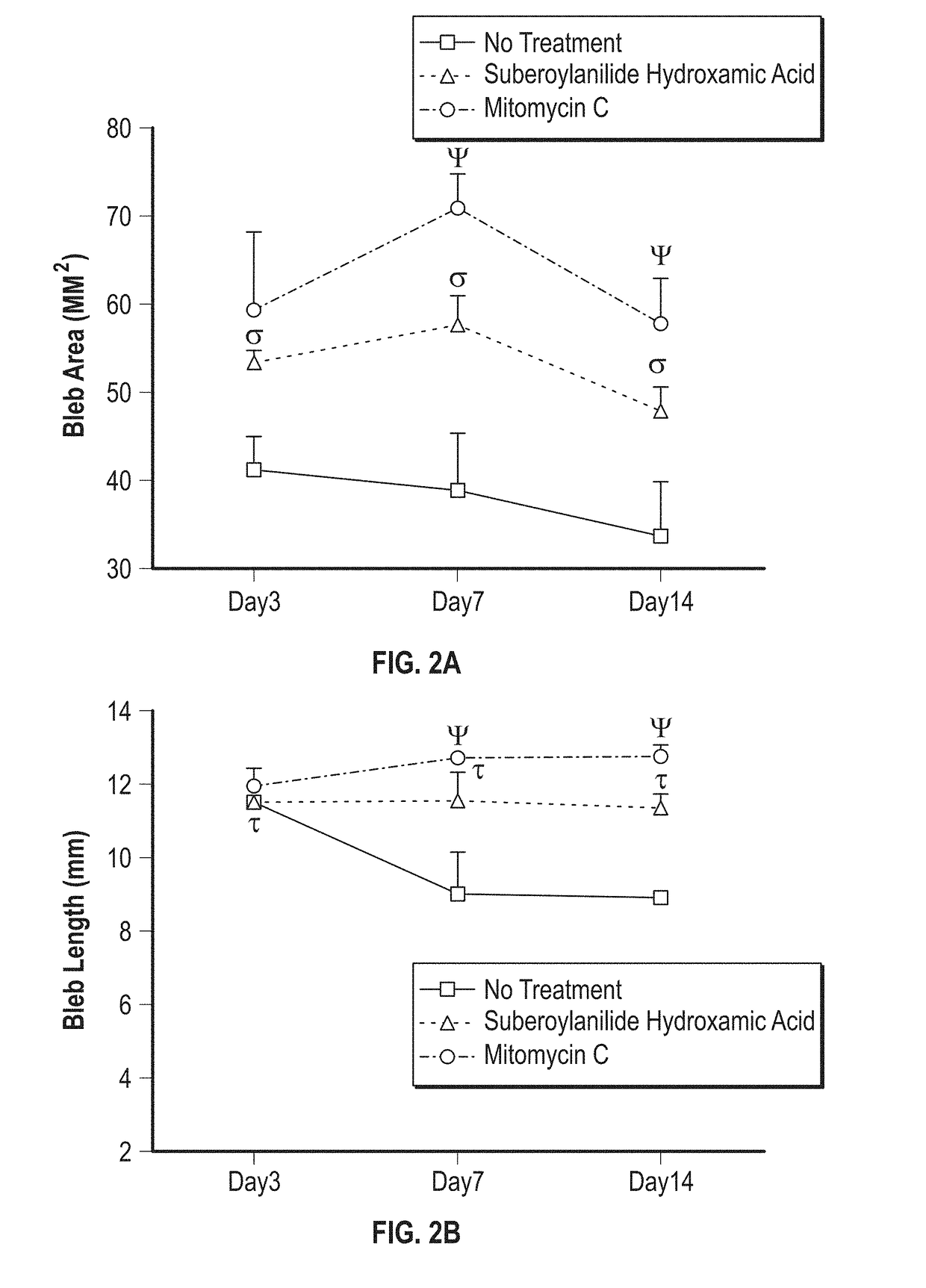
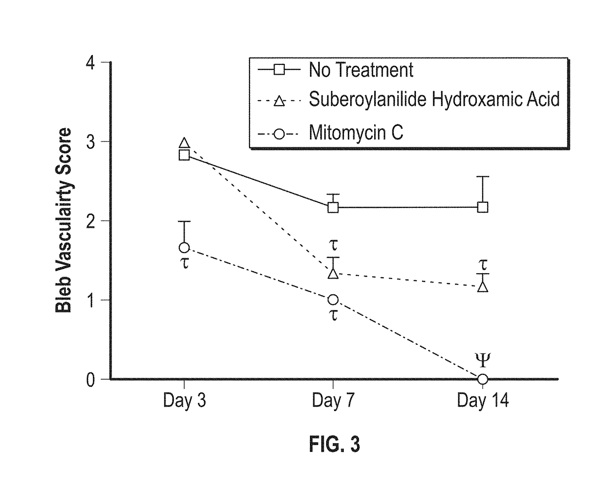
[0082]Briefly, a rabbit model of glaucoma filtration surgery (GFS) was used. Rabbits underwent GFS received Balanced Salt Solution (BSS) or SAHA (50 μM) or mitomycin C (MMC; 0.04% (w / v)). Clinical scores of intraocular pressure (IOP), bleb vascularity and slit lamp examination were performed. On postoperative day 14, rabbits were sacrificed and the bleb tissues were collected for evaluation of tissue fibrosis with H&E, Masson trichrome (MT), α-smooth muscle actin (α-SMA), and F-actin staining. Further, SAHA-mediated acetylation of histones in corneal fibroblasts and conjunctiva were determined by western blot analysis.

[0083]Preparation of SAHA Solution and Treatment Regimen.

[0084]A 10 mM stock solution of SAHA (Cayman Chemical Company, Ann Arbor, Mich., USA) was prepared by dissolving in dimethyl sulfoxide (DMSO) and then further ...

example 2

bitor (HDACi) Prevents Corneal Haze Formation Long-Term, after Photorefractive Keratectomy (PRK) Surgery

[0107]Material and Methods

[0108]Background.

[0109]Briefly, corneal haze in rabbits was produced with −9.0 diopter PRK. A single application of SAHA (25 μM) or MMC (0.02%) was applied topically immediately after PRK. Effects of the two drugs were analyzed by slit-lamp microscope, specular microscope, TUNEL assay, and immunofluorescence.

[0110]Corneal Haze Production in Rabbit Eyes.

[0111]Photorefractive keratectomy was used to produce corneal haze in rabbits by performing −9.0 diopter ablation with the Summit Apex excimer laser (Model: SVS APEX Plus ER; Alcon, Ft. Worth, Tex.) as reported previously. Briefly, the rabbits were anesthetized and local anesthesia of the cornea was achieved through the application of topical ophthalmic 0.5% proparacaine hydrochloride (Alcon, Fort Worth, Tex.). A wire lid speculum was placed and corneal epithelium was removed by gentle scraping with surgica...


PUM

PropertyMeasurementUnit
timeaaaaaaaaaa
timeaaaaaaaaaa
timeaaaaaaaaaa

Abstract

Disclosed is a method of promoting wound healing with reduced scarring after glaucoma filtration surgery in a mammalian subject in need thereof, which involve the use of a HDAC inhibitor (HDACi), such as, but not limited to, suberoylanilide hydroxamic acid (SAHA).

Description

CROSS REFERENCE TO RELATED APPLICATIONS[0001]This application claims the benefit of U.S. Provisional Patent Application No. 62 / 523,673, filed Jun. 22, 2017, which is incorporated herein by reference in its entirety.[0002]This invention was made with Government Support from the Department of Veterans Affairs and the National Eye Institute (Grant No. 2RO1EY017294-06). The Government has certain rights in the invention.[0003]Throughout the present specification certain references are referred to by superscript citation numbers, which numbers refer to the references enumerated in the list included herein before the claims. These enumerated references, and others which may be cited herein, are intended to describe the state of the art and are incorporated herein by reference in their entireties.BACKGROUND OF THE INVENTION1. Field of the Invention[0004]The present invention is related to the treatment of ophthalmic diseases in mammalian subjects, including humans.2. Related Art[0005]Glauc...

Claims


Application Information

Patent Timeline
Patent Type & Authority Applications(United States)
IPC IPC(8): A61K31/165A61K31/506A61K31/4045A61K31/4406A61K31/437A61K9/00A61P17/02A61P27/02
CPCA61K31/165A61K31/506A61K31/4045A61K31/4406A61K31/437A61K9/0048A61P17/02A61P27/02A61K31/167
Inventor MOHAN, RAJIV R.
Owner U S GOVERNMENT REPRESENTED BY THE DEPT OF VETERANS AFFAIRS
Why Eureka
Try Eureka