
System and method for generating selective stain segmentation images for cell types of interest

a cell type and image technology, applied in image analysis, image enhancement, instruments, etc., can solve the problems of incomplete or incorrect quantification of staining using these methods, and the membrane around the stain cannot be detected, so as to facilitate visualization of image analysis results.

Active Publication Date: 2020-10-08
VENTANA MEDICAL SYST INC
View PDF0 Cites 2 Cited by
  • Summary
  • Abstract
  • Description
  • Claims
  • Application Information

AI Technical Summary

Benefits of technology

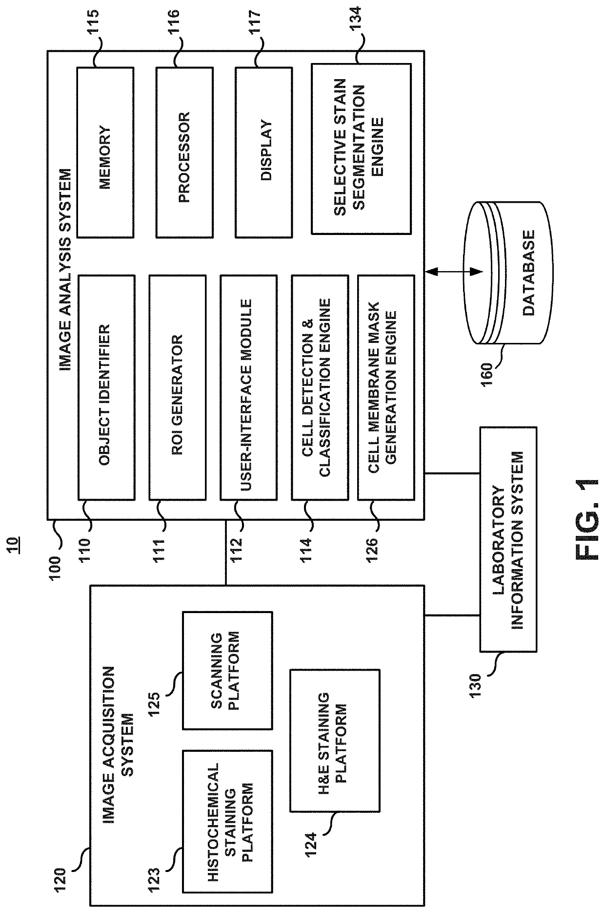
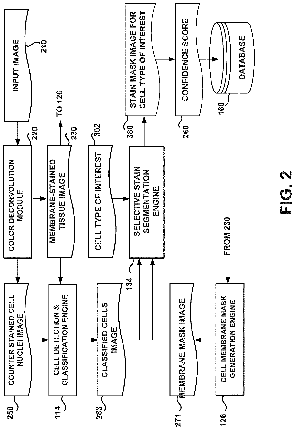
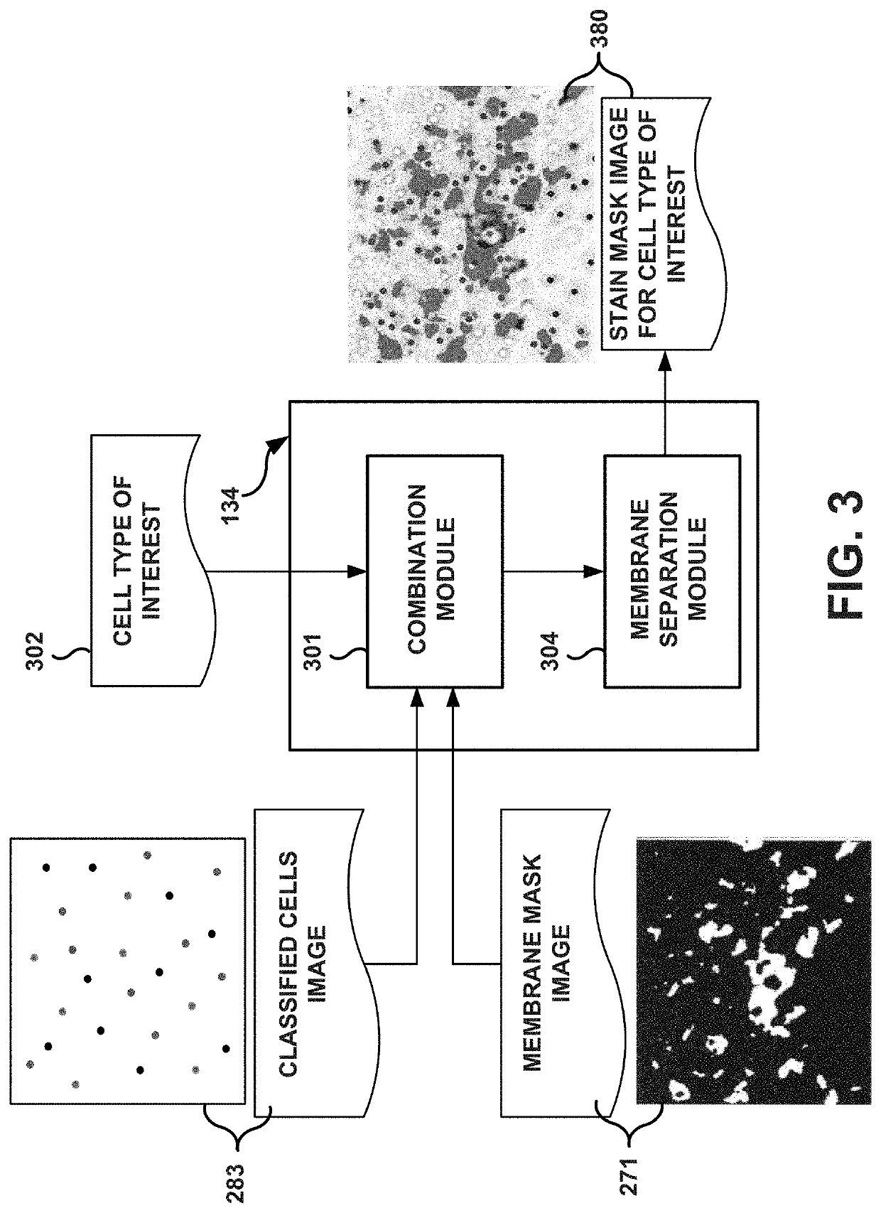
The patent describes an image analysis system that can help pathologists analyze tissue images more accurately. The system can segment the membranes of different types of cells in order to improve the scoring of whole slide results. This helps in identifying specific types of cells and their location in the tissue image. In addition, the system can also separate multiple biomarker stains in digital pathology tissue slides, making it easier for pathologists to analyze the samples.

Problems solved by technology

Hence, if these algorithms miss the nuclei or if the membrane lies outside the predefined neighborhood radius, stained membrane around them might not be detected.
Additionally, the algorithms might ignore regions that contain membrane staining in combination with other staining compartments (such as cytoplasmic staining) Thus, quantification of staining using these methods could be incomplete or incorrect.

Method used


Image

  • System and method for generating selective stain segmentation images for cell types of interest
  • System and method for generating selective stain segmentation images for cell types of interest
  • System and method for generating selective stain segmentation images for cell types of interest

Examples


Embodiment Construction

I-Definitions

[0028]Unless defined otherwise, technical and scientific terms used herein have the same meaning as commonly understood by a person of ordinary skill in the art. See, e.g., Lackie, DICTIONARY OF CELL AND MOLECULAR BIOLOGY, Elsevier (4th ed. 2007); Sambrook et al., MOLECULAR CLONING, A LABORATORY MANUAL, Cold Springs Harbor Press (Cold Springs Harbor, N.Y. 1989).

[0029]Antibody: The term “antibody” herein is used in the broadest sense and encompasses various antibody structures, including but not limited to monoclonal antibodies, polyclonal antibodies, multispecific antibodies (e.g., bispecific antibodies), and antibody fragments so long as they exhibit the desired antigen-binding activity.

[0030]Biomarker: As used herein, the term “biomarker” shall refer to any molecule or group of molecules found in a biological sample that can be used to characterize the biological sample or a subject from which the biological sample is obtained. For example, a biomarker may be a molecu...


PUM


Abstract

An image analysis system and method to generate selective stain segmentation images for at least one cell type of interest within a stained tissue image are presented. Cell membranes in the tissue image are detected to generate a corresponding membrane mask image. Cell nuclei are classified to generate a classified cells image. Selective stain segmentation images are generated for the at least one cell type of interest based on the membrane mask image and the classified cells image.

Description

CROSS REFERENCE TO RELATED APPLICATIONS[0001]The present application is a continuation of International Application PCT / EP2018 / 086040, entitled “System And Method For Generating Selective Stain Segmentation Images For Cell Types Of Interest” and filed Dec. 20, 2018, which claims priority to U.S. Provisional Patent Application No. 62 / 609,748, filed on Dec. 22, 2017. Each of these applications is hereby incorporated by reference in its entirety for all purposes.BACKGROUNDField of the Disclosure[0002]The present disclosure generally relates to digital image processing methods and systems for medical diagnosis. Particularly, the present disclosure relates to the preparation of selective stain segmentation images for specific cell types of interest.Description of Related Art[0003]In the analysis of biological specimens such as tissue sections, blood, cell cultures and the like, biological specimens are mounted on slides and stained with one or more combinations of stain and biomarkers, a...

Claims


Application Information

Patent Timeline
IPC IPC(8): G06T7/00G06T7/12G06T7/162G06K9/62
CPCG06T7/12G06T2207/10056G06T7/162G06T7/0012G06K9/6215G06T2207/30024G06T2207/10024G06T7/11G06F18/22
Inventor GUETTER, CHRISTOPHNGUYEN, KIEN
Owner VENTANA MEDICAL SYST INC
Learn More