Method for detection of cells in a cytological sample having at least one anomaly

a cytological sample and cell technology, applied in the direction of material analysis, instrumentation, computer peripheral equipment, etc., can solve the problems of low sensitivity for certain cancers, limited method of diagnosis by cytological sample analysis, and low sensitivity for low-grade bladder cancer diagnosis

Active Publication Date: 2021-01-14
OFFICE NAT DETUD & DE RECH AEROSPATIALES
View PDF3 Cites 2 Cited by
  • Summary
  • Abstract
  • Description
  • Claims
  • Application Information

AI Technical Summary

Benefits of technology

[0006]Based on this situation, one of the goals of the invention consists of providing a method for detection of cells in a cytological sample having at least one cellular anomaly so as to subsequently help in cancer detection, in particular bladder cancer, based on digitized slide images. Advantageously the invention provides, by means of a computer-implemented processing module, automated and exhaustive analysis of cells from the sample present on the slide. Thus, the analysis of the cytological sample and of all the potentially abnormal cells is complete, quick, more precise and serves to overcome the common analysis errors of cytopathologists and cytotechnicians. Further, this method assures the standardization of the cytological analysis result for the sample. But again, this method further serves to help early diagnosis of cancer called low-grade, in particular low-grade bladder cancer, by detecting cells having minor anomalies.
[0021]The solution makes it possible to benefit from the classification of the sample according to two classes and to diagnose whether the sample has a zero or a proven cellular anomaly level. Subsequently, this result will significantly help the diagnosis by a specialist of a cancer or not.

Problems solved by technology

Early diagnosis of a cancer is obviously a major issue with significant repercussions related to the survival rate of affected patients and also on the corresponding costs for caring for them.
However, this method of diagnosis by the analysis of cytological samples is limited both from a human aspect—meaning the experience and the decreasing number of cytopathologists and cytotechnologists—and also by a low sensitivity for certain cancers.
Typically, there is a very low sensitivity for diagnosis of low-grade bladder cancer because atypical or abnormal cells have not yet undergone pronounced morphological deformations and are therefore hard to detect by human eye.

Method used


Image

  • Method for detection of cells in a cytological sample having at least one anomaly
  • Method for detection of cells in a cytological sample having at least one anomaly
  • Method for detection of cells in a cytological sample having at least one anomaly

Examples


Embodiment Construction

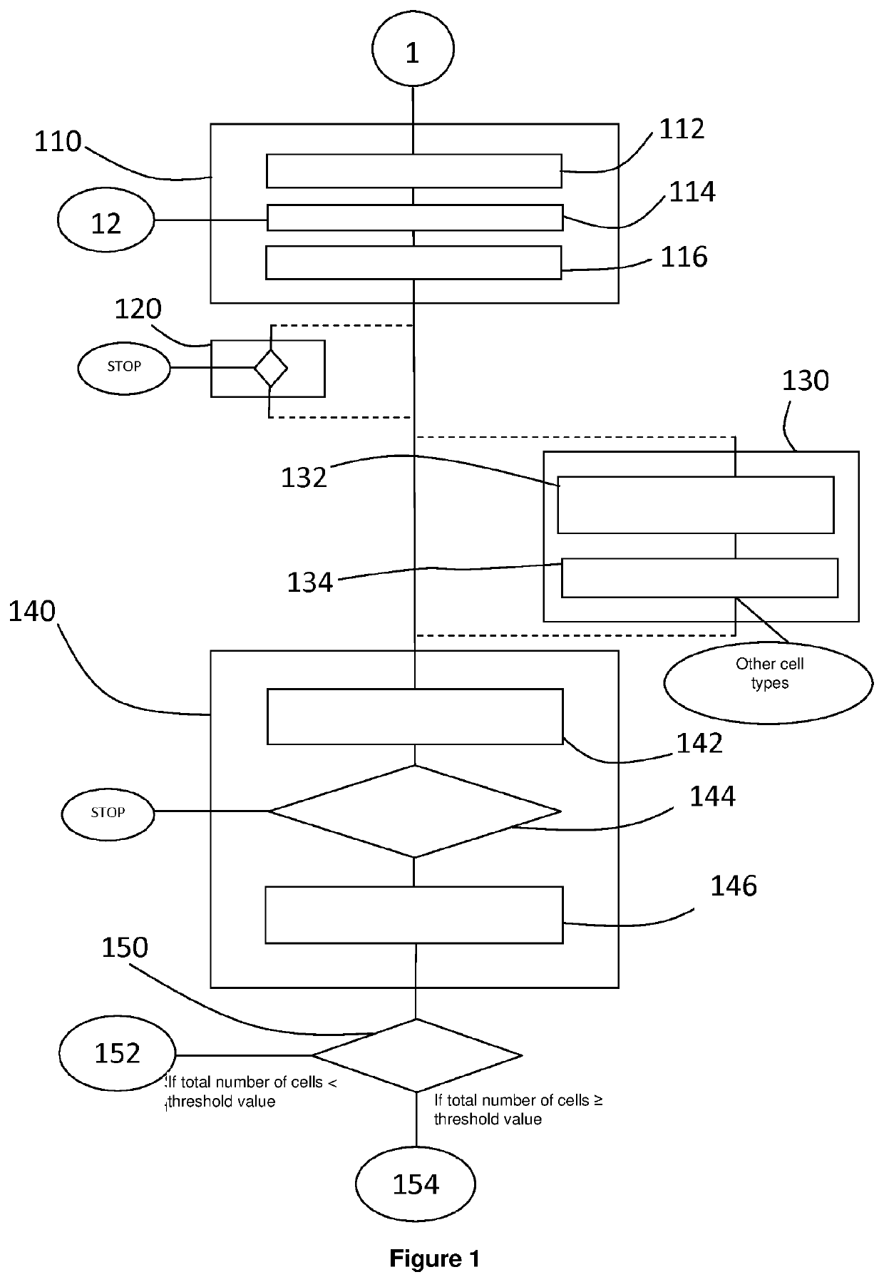
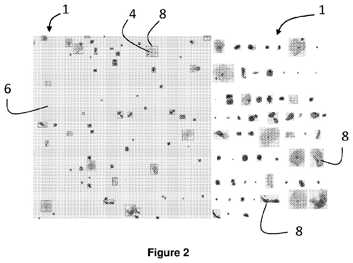
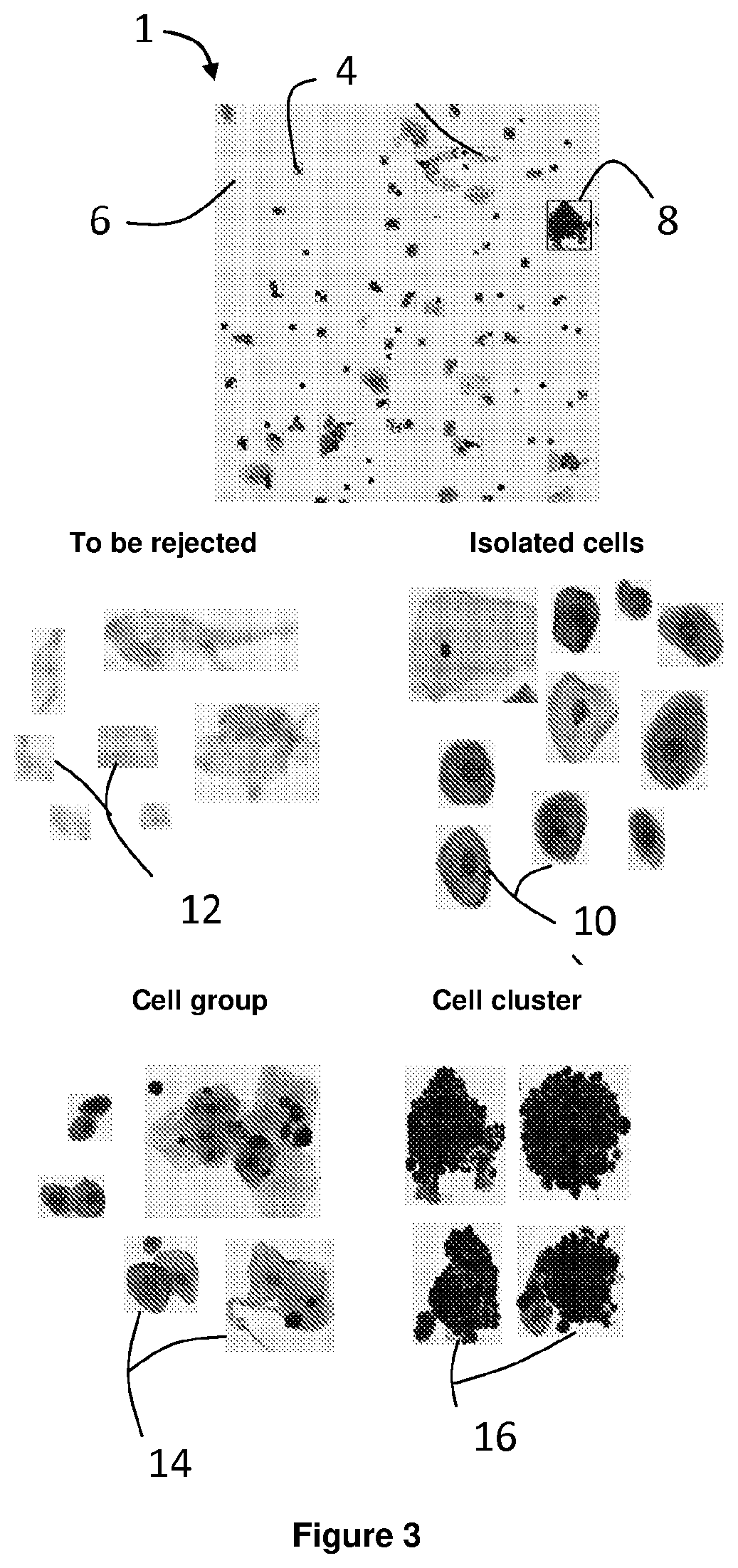
[0058]It must first be noted that the figures disclose the invention schematically. These figures represent implementation examples given without limitation and not necessarily adhering to a specific chronology.

[0059]The present invention falls in the general category of the application of computer vision of digitized images of complex and numerous objects. The invention deals with an original method for detecting cells in a cytological sample having at least one anomaly based on a first digitized image and preferably using a second digitized image of the same sample. The first images obtained with a transmission electron microscope and the second image is obtained with a fluorescence microscope. The images used come from the specific digitization of a cytological sample having undergone a preparation suited for the method, which was then spread on a cytology slide. The method according to the invention can use a cytological sample coming without distinction from fluid or from colle...


PUM

PropertyMeasurementUnit
electron microscopeaaaaaaaaaa
colorimetric detectionaaaaaaaaaa
thresholdaaaaaaaaaa

Abstract

Disclosed is a method for detecting cells having at least one anomaly in a cytological sample on the basis of at least one first digitised digitised-electron-microscopy image of the sample.

Description

CROSS-REFERENCE TO RELATED APPLICATIONS[0001]This application is the U.S. national phase of International Application No. PCT / FR2019 / 050718 filed Mar. 28, 2019 which designated the U.S. and claims priority to French Application No. 18 00251 filed Mar. 29, 2018, the entire contents of each of which are hereby incorporated by reference.BACKGROUND OF THE INVENTIONField of the Invention[0002]The present invention relates to a method for detection of cells in a cytological sample having at least one anomaly, based on one digitized image or several digitized images of the same sample. The invention also relates to a device implementing this method.Description of the Related Art[0003]The invention relates to the field of cytology which is the microscopic study of cells from a cytological sample after fixing on a slide. More precisely, it involves the study of morphological, radiometric or biochemical aspects of normal or atypical cells, meaning cells having at least one anomaly.[0004]Cance...

Claims


Application Information

Patent Timeline
Patent Type & Authority Applications(United States)
IPC IPC(8): G06K9/00G01N15/14G01N21/25G01N21/29
CPCG06K9/00147G01N15/1475G01N2015/1006G01N21/29G01N21/251G01N2015/1402G06V20/695G06V20/698
Inventor HADDAD, ZEHIRAHERBIN, STÉPHANECHAN-HON-TONG, ADRIEN
Owner OFFICE NAT DETUD & DE RECH AEROSPATIALES
Why Eureka
Try Eureka