
Generation of three-dimensional scans for intraoperative imaging

a three-dimensional scan and imaging technology, applied in the field of three-dimensional scan generation for intraoperative imaging, can solve the problems that current surgical imaging and navigation hardware and software, such as those used in the spine and orthopedic fields, still fail to deliver robust procedure guidance, as desired by surgeons

Active Publication Date: 2021-06-24
UNIFY MEDICAL
View PDF1 Cites 7 Cited by
  • Summary
  • Abstract
  • Description
  • Claims
  • Application Information

AI Technical Summary

Benefits of technology

The present invention is a surgical imaging and navigation system that includes a display, controller, 3D scanning controller, and other components to provide accurate guidance for surgical procedures. The system utilizes 3D scanning, intraoperative imaging, and tracking for precise navigation. It combines augmented reality, real-time imaging, and computer vision algorithms to provide decision support for surgeons. The system can be used in various surgical applications and is designed to improve the guidance and monitoring of procedures for hard tissues and soft tissues alike.

Problems solved by technology

Current surgical imaging and navigation hardware and software, such as those used in the spine and orthopedic fields, still fail to deliver robust procedure guidance, as desired by surgeons.

Method used


Image

  • Generation of three-dimensional scans for intraoperative imaging
  • Generation of three-dimensional scans for intraoperative imaging
  • Generation of three-dimensional scans for intraoperative imaging

Examples


Embodiment Construction

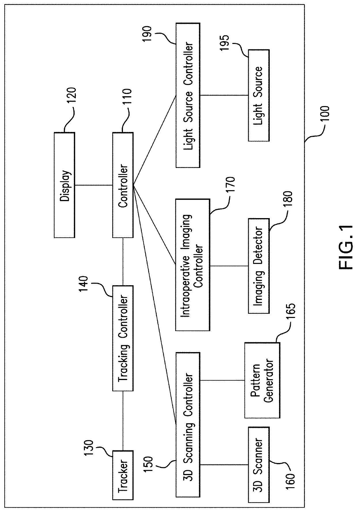
[0016]A surgical imaging and navigation system 100 of the present invention is shown in FIG. 1. In one embodiment, the surgical imaging and navigation system 100 includes a display 120, a controller 110, a 3D scanning controller 150, a 3D scanner 160, an intraoperative imaging controller 170, an imaging detector 180, a light source controller 190, a light source 195, a tracking controller 140, and a tracker 130. The 3D scanning controller 150 controls the modes and properties of the 3D scanner 160. For instance, the size of the area of 3D scanning, the resolution of 3D scanning, the speed of 3D scanning, the timing of 3D scanning may be controlled by the 3D scanning controller 150. The intraoperative imaging controller 170 controls the modes and properties of imaging detector 180. For instance, the size of the area of intraoperative imaging, the resolution of intraoperative imaging, the speed of intraoperative imaging, the timing of intraoperative imaging, and the mode of intraopera...


PUM


Abstract

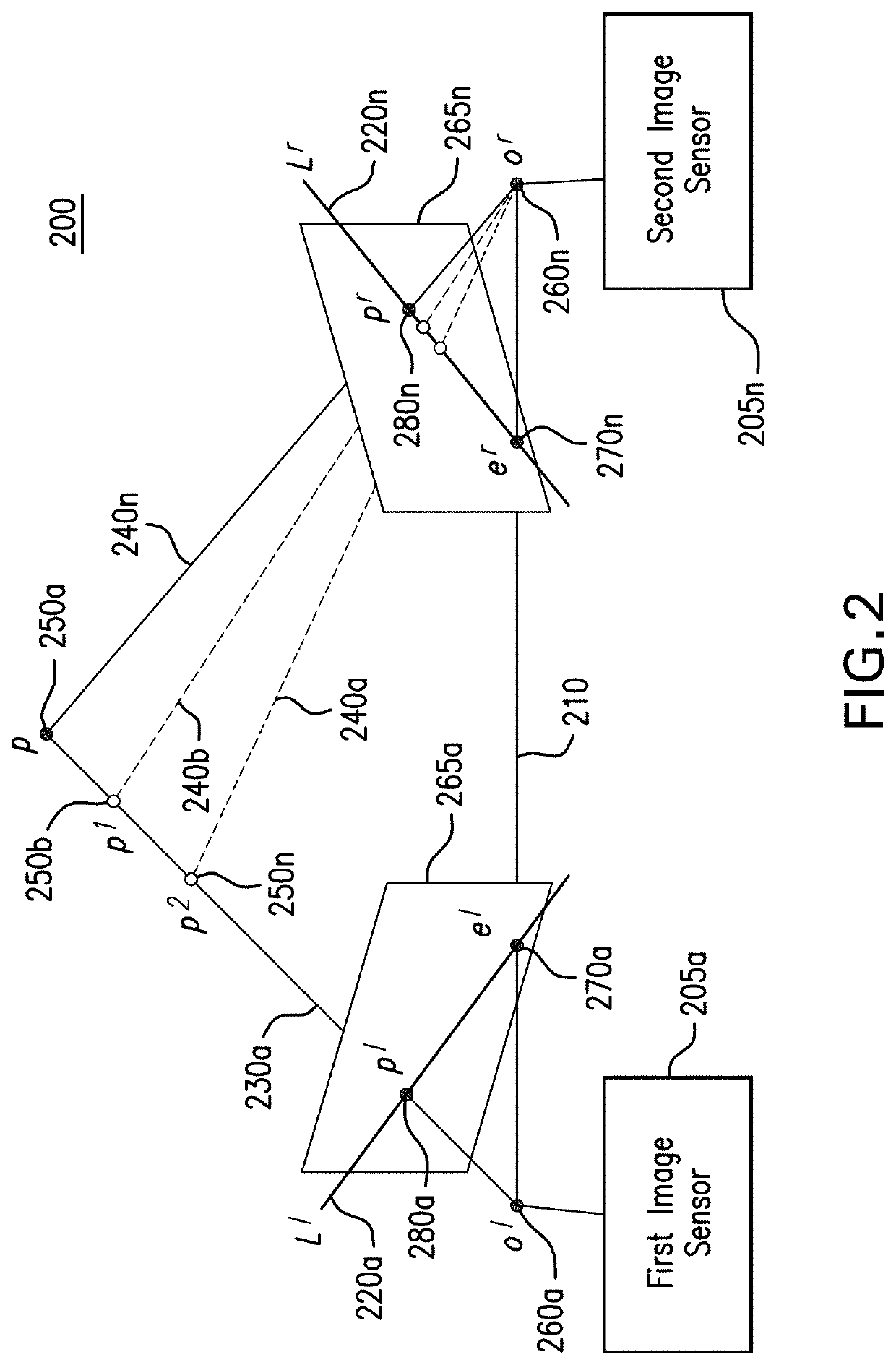
A system for executing a three-dimensional (3D) intraoperative scan of a patient is disclosed. A 3D scanner controller projects the object points included onto a first image plane and the object points onto a second image plane. The 3D scanner controller determines first epipolar lines associated with the first image plane and second epipolar lines associated with the second image plane based on an epipolar plane that triangulates the object points included in the first 2D intraoperative image to the object points included in the second 2D intraoperative image. Each epipolar lines provides a depth of each object as projected onto the first image plane and the second image plane. The 3D scanner controller converts the first 2D intraoperative image and the second 2D intraoperative image to the 3D intraoperative scan of the patient based on the depth of each object point provided by each corresponding epipolar line.

Description

CROSS-REFERENCE TO RELATED APPLICATION[0001]This application claims the benefit of U.S. Provisional Application No. 62 / 951,480 filed on Dec. 20, 2019 and U.S. Provisional Application No. 63,040,816 filed on Jun. 18, 2020, which are incorporated herein by reference in their entirety.TECHNICAL FIELD[0002]Generally, the present invention relates to apparatuses, systems, and methods of surgical imaging, navigation and tracking. In particular, the present invention relates to systems and methods for performing 3D scanning, surgical imaging, tracking, image processing, computer vision, image registration, and display. More particularly, the present invention relates to an apparatus, system, and method for utilizing 3D scanning, intraoperative imaging, light source, tracking hardware, in combination with image processing and computer vision algorithms, to perform procedural guidance.BACKGROUND OF THE INVENTION[0003]Current surgical imaging and navigation hardware and software, such as thos...

Claims


Application Information

Patent Timeline
Patent Type & Authority Applications(United States)
IPC IPC(8): G06T7/593H04N13/239G06T7/33G06T7/73A61B34/20
CPCG06T7/593H04N13/239G06T7/33H04N2013/0081A61B34/20G06T2207/10012G06T7/73G06T2207/30196G06T7/521G06T2207/30012G06T2207/20084G06T7/30H04N13/302H04N13/332H04N13/106A61B2034/2051A61B2034/2055A61B2034/2065A61B90/37A61B2090/373G06T2207/30004
Inventor LIU, YANGASKARI KARCHEGANI, MAZIYAR
Owner UNIFY MEDICAL
Learn More