Eureka AIR delivers breakthrough ideas for toughest innovation challenges, trusted by R&D personnel around the world.

Image correction of a surgical endoscope video stream

a technology of endoscopy and video stream, which is applied in image enhancement, image analysis, medical science and other directions, can solve the problems of degrading the visibility of those darker areas, affecting the visibility of surgical sites, and affecting the visibility of high contrast regions in the central area or the periphery

Active Publication Date: 2021-10-21
CMR SURGICAL LTD
View PDF6 Cites 0 Cited by
  • Summary
  • Abstract
  • Description
  • Claims
  • Application Information

AI Technical Summary

Benefits of technology

The patent describes a method for real-time image correction of a video stream from a surgical endoscope of a surgical robotic system. The method involves identifying regions of the image with different ranges of values for certain image properties, applying a mask to each region with a specific modification that modifies the image property, and deriving data from the surgical robotic system. The method may also predict a relationship between features in the image and modify the mask accordingly. The result is an improved video stream that enhances the visibility of the surgical site, reducing the need for manual adjustments and improving the overall surgical experience.

Problems solved by technology

Consequently, the surgeon is left with a poor view of the surgical site with limited visibility.
However, because other areas of the image are much darker than the central area, when the transform is applied uniformly across the image, it further degrades the visibility of those darker areas.
Whilst an improvement to uniformly applying a transform across the whole image, this approach suffers from the problem that it does not improve the visibility of high contrast regions within the central area or within the periphery.
Techniques involving manual manipulation are not suitable for real-time correction of a video stream from an endoscope as is required in this field to enable the surgeon to better view the surgical site he is operating in.

Method used


Image

  • Image correction of a surgical endoscope video stream
  • Image correction of a surgical endoscope video stream
  • Image correction of a surgical endoscope video stream

Examples


Embodiment Construction

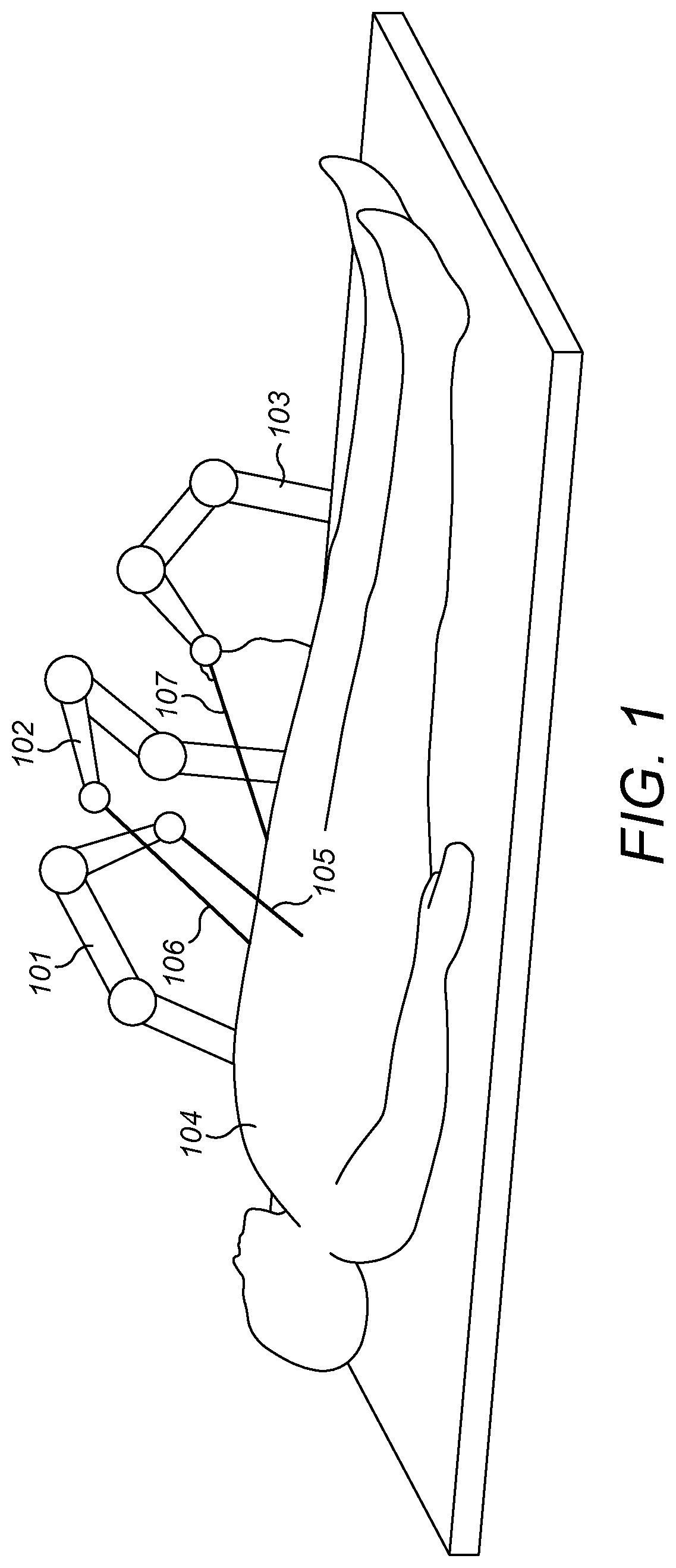
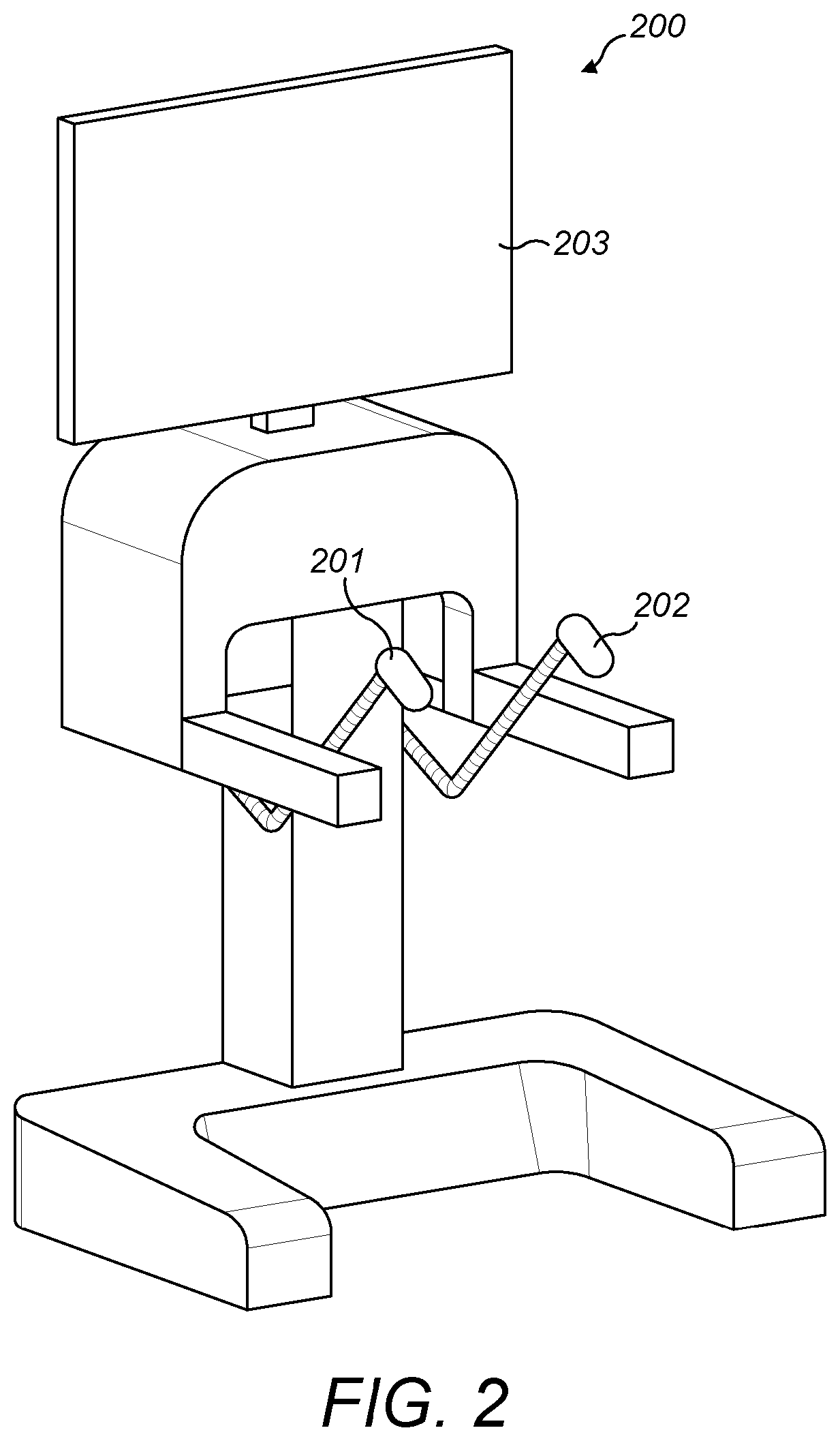
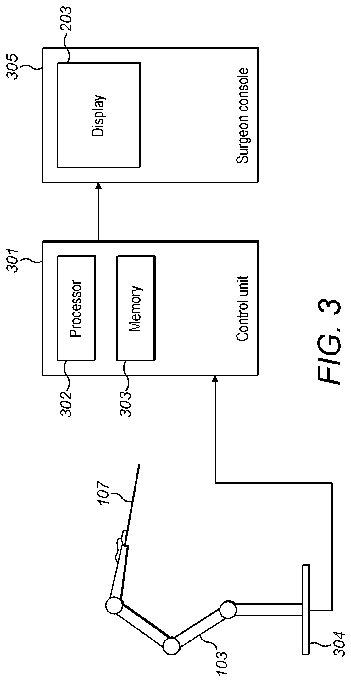
[0025]FIG. 1 illustrates surgical robots 101, 102, 103 performing an operation on a person 104. Each robot comprises an articulated arm connected to a base. Each of robots 101 and 102 has a surgical instrument 105, 106 connected to the end of its arm. A surgical endoscope 107 is connected to the arm of robot 103. The surgical instruments 105, 106 and surgical endoscope 107 each access the surgical site through an incision in the body 104. A surgeon controls the surgical instruments, and optionally the surgical endoscope, from a surgeon console 200, shown in FIG. 2. The surgeon manipulates hand controllers 201, 202. A control system converts the movement of the hand controllers into control signals to move the arm joints and / or instrument end effector of one of the surgical robots. The video feed from the surgical endoscope 107 at the surgical site is displayed on display 203. The surgeon is thereby able to view the surgical site including the instrument end effector that he is manip...


PUM


Abstract

A method of real-time image correction of a video stream from a surgical endoscope of a surgical robotic system. The video stream comprises a sequence of images. The method comprises: for an image in the sequence of images, identifying a plurality of regions, each region having a different range of values of at least one image property to another region; applying a mask to the image which applies a region-specific modification to each region, each region-specific modification modifying the at least one image property of that region; deriving data from the surgical robotic system; determining a relationship between features in the image from the derived data; modifying the mask for a predicted relationship between the features at the time of a subsequent image in the video stream; and applying the modified mask to the subsequent image in the video stream.

Description

BACKGROUND[0001]This disclosure relates to image correction of the video stream from a surgical endoscope of a surgical robotic system.[0002]In minimally invasive surgery, the surgical site is imaged via a surgical endoscope. The endoscope is a camera which typically has a tip comprising a light source for illuminating the site and one or more lenses for acquiring the video stream. The video stream is displayed in real time on a display enabling the surgeon to view the surgical site and the surgical instruments he is manipulating.[0003]In order to sufficiently illuminate the surgical site, the light source on the endoscope is very intense. In the resulting video stream, the illuminated area is in high contrast to the remainder of the image which is in shadow. In minimally invasive surgery the endoscope has a very narrow diameter, thus the light source is very close to the optical axis of the endoscope. Tissue is wet and shiny, and hence causes a strong reflection back into the lens ...

Claims


Application Information

Patent Timeline
Patent Type & Authority Applications(United States)
IPC IPC(8): G06T5/00G06T7/11G06T7/174G06T5/50G06T7/579A61B34/30
CPCG06T5/009G06T7/11G06T7/174G06T5/50G06T7/579G06T5/008A61B2034/301G06T2207/10016G06T2207/10024G06T2207/10028G06T2207/10068G06T2207/20208G06T2207/30004A61B34/30A61B1/00149A61B1/00193A61B34/37A61B90/361A61B1/000095G06T5/94G06T5/20G06T5/92
Inventor HARES, LUKE DAVID RONALD
Owner CMR SURGICAL LTD
Why Eureka
Eureka Blog
Learn More