Supercharge Your Innovation With Domain-Expert AI Agents!

Glaucoma implant device

a technology of glaucoma and implant device, applied in the field of medical devices, can solve the problems of insufficient control of intraocular pressure, further symptom progression, and drug treatment needs to be much improved, and achieve the effect of maximizing the rate of aqueous humor resorption and better intraocular pressure control

Inactive Publication Date: 2022-03-03
AJL OPHTHALMIC
View PDF4 Cites 0 Cited by
  • Summary
  • Abstract
  • Description
  • Claims
  • Application Information

AI Technical Summary

Benefits of technology

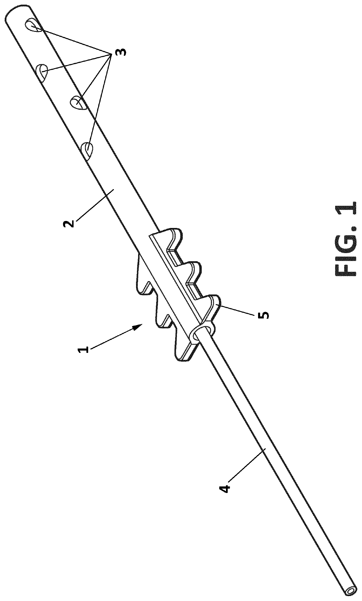
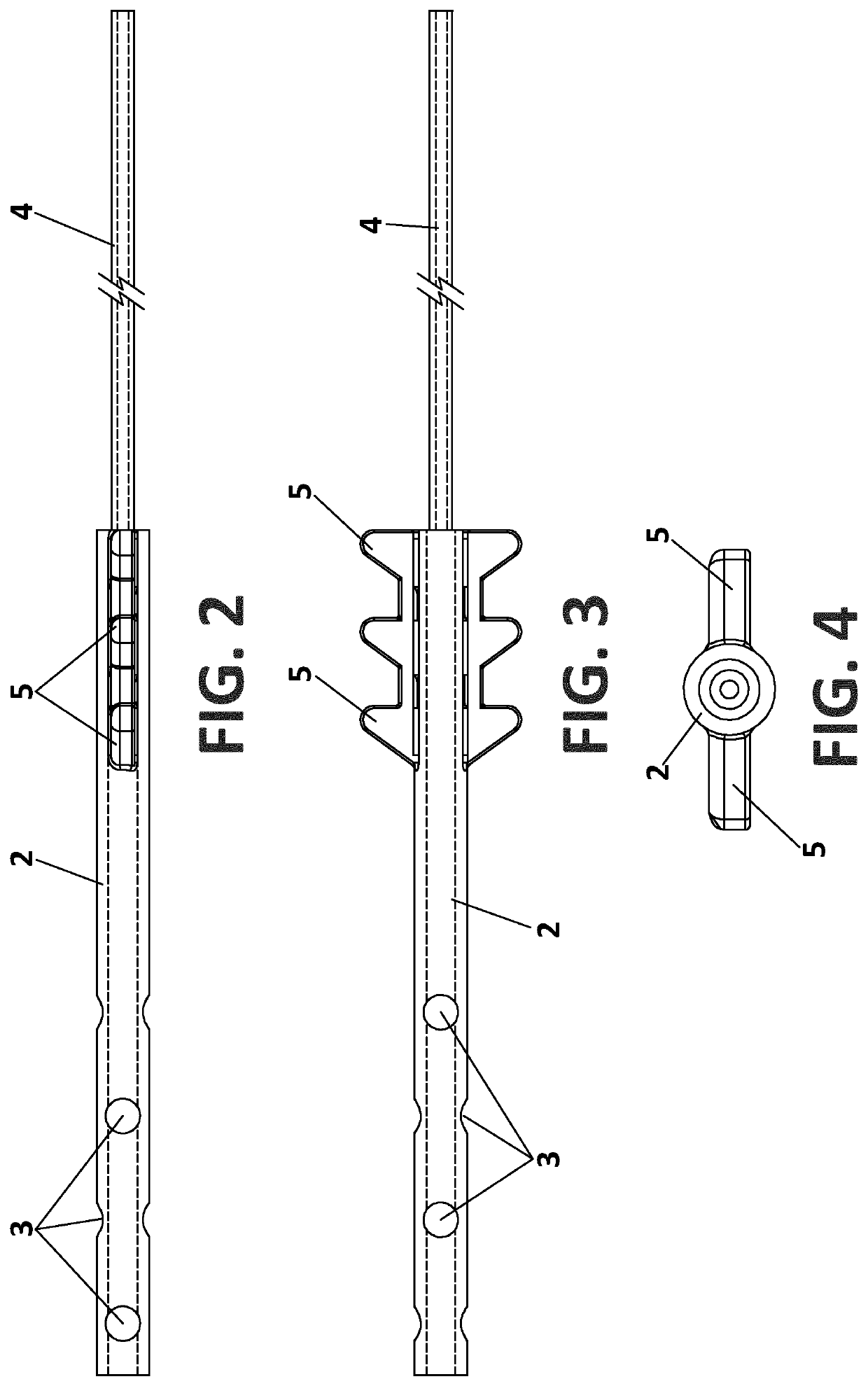
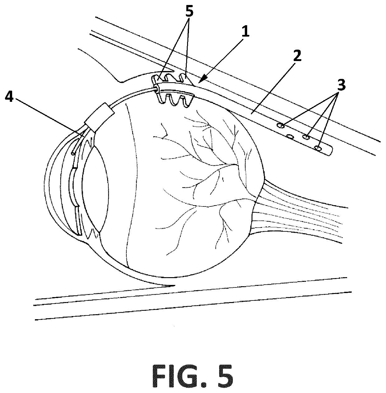
The present invention is a glaucoma implant device that can better control intraocular pressure. It consists of an elongated member with two tube-like sections of different diameters. The larger diameter tube has a fixation flange and is placed in the anterior chamber of the eye. The smaller diameter tube is cut to length during the operation and introduced directly into the anterior chamber. This device diverts aqueous humor from the anterior chamber to the retrobulbar space, which avoids the need for blebs or shunts on the cornea. The holes in the distal tube allow intercellular canaliculi to form between fat cells, which maximizes the rate of aqueous humor resorption throughout the eye.

Problems solved by technology

Increased pressure due to glaucoma can cause damage to the optic nerve, which can lead to blindness.
Unfortunately, drug treatments need much improvement, as they can cause adverse side effects and often fail to adequately control intraocular pressure.
Moreover, lack of compliance leads to further symptom progression.
Such a procedure can be quite traumatic as the implants are large and can result in various adverse events such as infections and scarring, leading to the need to re-operate.

Method used


Image

  • Glaucoma implant device
  • Glaucoma implant device
  • Glaucoma implant device

Examples


Embodiment Construction

[0012]As used herein, the term “distal” is defined as in the direction of the eye of the patient and away from the surgeon that will implant the device. The term “proximal” is defined as in the direction of the surgeon.

[0013]The glaucoma implant device of the invention comprises an elongate member 1 made of surgical silicone or any of various other biocompatible polymers or gels. It is provided with two tube-like sections, both having an interior flow pathway. The flow pathway provides a fluid pathway between the eye's anterior chamber and a suprachoroidal space when the elongate member is implanted in the eye.

[0014]The proximal tube 4 is of the same diameter as that of a standard tube shunt, its lumen diameter lies between 0.1 and 0.25 mm and is longer than the distal tube 2. The distal tube 2, of a length between 10 to 20 mm, has a greater diameter, between 1 and 3 mm, is provided with holes (at least 2 and up to 30 in all directions) and presents optionally one or more fixation f...


PUM


Abstract

Glaucoma implant device with an elongate member with two tube-like sections of different diameter, the tube of larger diameter being provided with holes and at least one fixation flange. The smaller diameter tube can be cut at an appropriate length during the operation, and is introduced directly into the anterior chamber and fixed with surgical tape.

Description

BACKGROUND OF THE INVENTIONField of the Invention[0001]The present invention relates to medical devices, in particular to devices for treating glaucoma by diverting aqueous humour out of the eye's anterior chamber through a surgically implanted duct passageway or shunt.Description of the Related Art[0002]Increased pressure due to glaucoma can cause damage to the optic nerve, which can lead to blindness. Treatment strategies have focused on keeping the intraocular pressure down in order to preserve as much vision as possible and have included the use of drugs that lower intraocular pressure through various mechanisms. Unfortunately, drug treatments need much improvement, as they can cause adverse side effects and often fail to adequately control intraocular pressure. Moreover, lack of compliance leads to further symptom progression.[0003]With respect to surgical procedures, one way to treat glaucoma is to implant a drainage device, or shunt, in the eye. The drainage device functions ...

Claims


Application Information

Patent Timeline
Patent Type & Authority Applications(United States)
IPC IPC(8): A61F9/007
CPCA61F9/00781
Inventor SPONSEL, WILLIAM ERICLARRA MATEOS, EVAMARTINEZ EZKURRA, SERGIOÁLVAREZ PÉREZ, IVAN
Owner AJL OPHTHALMIC
Features
  • R&D
  • Intellectual Property
  • Life Sciences
  • Materials
  • Tech Scout
  • Unparalleled Data Quality
  • Higher Quality Content
  • 60% Fewer Hallucinations
Learn More