Apparatus and method for detecting bone fracture

a technology of apparatus and bone fracture, applied in the field of ultrasonography, can solve the problems of more computational time or effort, and achieve the effect of reducing the cost of computational time or effor

Pending Publication Date: 2022-03-31
KONINKLIJKE PHILIPS ELECTRONICS NV
View PDF2 Cites 0 Cited by
  • Summary
  • Abstract
  • Description
  • Claims
  • Application Information

AI Technical Summary

Benefits of technology

[0006]It would be advantageous to provide an improved approa

Problems solved by technology

Moreover, the more large area to be processed, th

Method used


Image

  • Apparatus and method for detecting bone fracture
  • Apparatus and method for detecting bone fracture
  • Apparatus and method for detecting bone fracture

Examples


Embodiment Construction

[0034]The present invention will be described with respect to particular embodiments and with reference to certain drawings but the invention is not limited thereto but only by the claims. The drawings described are only schematic and are non-limiting. In the drawings, the size of some of the elements may be exaggerated and not drawn to scale for illustrative purposes.

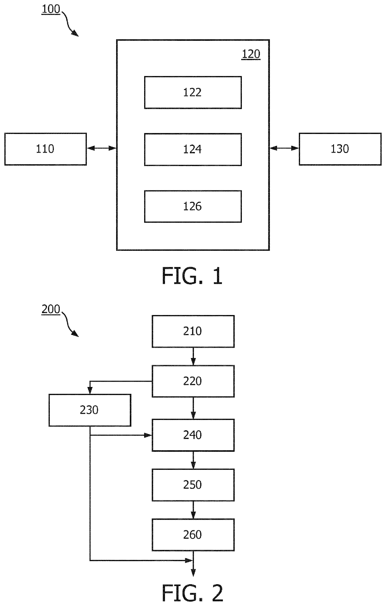
[0035]FIG. 1 illustrates an ultrasound system 100 for detecting bone fracture of a subject in accordance with some embodiments of the present invention. FIG. 2 illustrates a method 200 of detecting bone fracture of a subject on basis of ultrasound images in accordance with some embodiments of the present invention.

[0036]The ultrasound system 100 comprises an apparatus 110 for detecting bone fracture of a subject, an ultrasound image acquisition unit 110, and a user interface 130. The ultrasound image acquisition unit 110 can be an ultrasound probe. The apparatus 120 is communicatively connected to the ultrasound image ...


PUM


Abstract

The present invention proposes an apparatus (120) and method for detecting bone fracture of a subject on basis of ultrasound images. The apparatus (120) comprises a first fracture detector (122) and a second fracture detector (124). The first fracture detector (122) is configured to receive a first ultrasound image of a region of the subject, to identify a bone in the first ultrasound image, to identify at least one focus area within the region on basis of the identified bone, to generate focus area information indicating position of the at least one focus area, and to instruct an acquisition of a second ultrasound image of the region acquired based on the generated focus area information. The second fracture detector (124) is configured to receive the second ultrasound image, and to detect bone fracture on the basis of the second ultrasound image. The second ultrasound image has a higher resolution in the at least one focus area than in the rest of the region, and the resolution of the at least one focus area in the second ultrasound image is higher than the resolution of the at least one focus area in the first ultrasound image.

Description

FIELD OF THE INVENTION[0001]The present invention relates to ultrasound imaging, and more particularly to an apparatus and method for detecting bone fracture on basis of ultrasound images.BACKGROUND OF THE INVENTION[0002]Bone fractures are quite common from traumatic injury (injuries from trauma). For example, rib fractures are the most common injuries resulting from blunt check(chest) trauma. There are several different approaches for diagnosis of bone fractures, including physical examination, conventional radiography such as diagnostic x-ray, advanced medical imaging modalities such as CT, MRI or SPECT(not often recommend for this task), and ultrasound imaging. Currently, physical examination and conventional radiography are the main diagnostic tools used in daily clinical practice, but cannot detect all bone fractures. For example, only around 49% of rib fractures are detected upon precise physical examination and conventional radiographs. CT and MRI can provide most detailed in...

Claims


Application Information

Patent Timeline
IPC IPC(8): A61B8/08A61B8/00
CPCA61B8/0875A61B8/469A61B8/54A61B8/085A61B8/08
Inventor XU, JING PINGRAJU, BALASUNDAR IYYAVUGADES, ANTHONY M.
Owner KONINKLIJKE PHILIPS ELECTRONICS NV
Why Eureka
Try Eureka