Medical image analysis using machine learning and an anatomical vector

a machine learning and anatomical technology, applied in image enhancement, medical/anatomical pattern recognition, instruments, etc., can solve problems such as considerable computational effort, and achieve the effect of improving the computational efficiency of the network

Pending Publication Date: 2022-04-21
BRAINLAB
View PDF6 Cites 1 Cited by
  • Summary
  • Abstract
  • Description
  • Claims
  • Application Information

AI Technical Summary

Benefits of technology

[0006]The disclosed methods encompass registering a tracked imaging device such as a microscope having a known viewing direction and an atlas to a patient space so that a transformation can be established between the atlas space and the reference system for defining positions in images of an anatomical structure of the patient. Labels are associated with certain constituents of the images and are input into a learning algorithm such as a machine learning algorithm, for example a convolutional neural network, together with the medical images and an anatomical vector and for example also the atlas to train the learning algorithm for automatic segmentation of patient images generated with the tracked imaging device. The trained learning algorithm then allows for efficient segmentation and / or labelling of patient images without having to segment the patient images using a registered atlas each time, thereby saving on computational effort.GENERAL DESCRIPTION OF THE INVENTION
[0053]A random decision forest is an ensemble of randomly trained decisions trees. The key aspect of the forest model is the fact that its component trees are all randomly different from one another. This leads to decorrelation between the individual tree predictions and, in turn, results in improved generalization and robustness.
[0072]Because pooling summarizes the responses over a whole neighbourhood, it is possible to use fewer pooling units than detector units, by reporting summary statistics for pooling regions spaced k pixels apart rather than one pixel apart. This improves the computational efficiency of the network because the next layer has roughly k times fewer inputs to process.
[0081]In fact, for every layer, we compute two sets of gradients: the partial derivatives of z with respect to the layer parameters wi, and that layer's input xi. The termδ⁢⁢zδ⁢⁢wi,as seen in Equation 4, can be used to update the current (i-th) layer's parameters. The termδ⁢⁢zδ⁢⁢xican be used to update parameters backwards, e.g., to the (i−1)-th layer. An intuitive explanation is: xi is the output of the (i−1)-th layer andδ⁢⁢zδ⁢⁢xiis how xi should be changed to reduce the loss function. Hence, we could viewδ⁢⁢zδ⁢⁢xias the part of the “error” supervision information propagated from z backward till the current layer, in a layer by layer fashion. Thus, we can continue the back propagation process, and useδ⁢⁢zδ⁢⁢xito propagate the errors backward to the (i−1)-th layer. This layer-by-layer backward updating procedure makes learning a CNN much easier.
[0083]Sinceδ⁢⁢zδ⁢⁢xi+1is already computed and stored in memory, it requires just a matrix reshaping operation (vec) and an additional transpose operation to get∂z∂vec⁡(xi+1),which is the first term in the right hand side (RHS) of both equations. So long as we can compute∂vec⁡(xi+1)∂(vec⁡(wi)T)⁢⁢and⁢⁢∂vec⁡(xi+1)∂(vec⁡(xi)T),we can easily get what we want (the left hand side of both equations).∂vec⁡(xi+1)∂(vec⁡(wi)T)⁢⁢and⁢⁢∂vec⁡(xi+1)∂(vec⁡(xi)T)are much easier to compute than directly computing∂z∂(vec⁡(wi)T)⁢⁢and⁢⁢∂z∂(vec⁡(xi)T),because xi is directly related to xi+1, through a function with parameters wi.

Problems solved by technology

This requires registering the patient images to the atlas which is associated with considerable computational effort.

Method used


Image

  • Medical image analysis using machine learning and an anatomical vector
  • Medical image analysis using machine learning and an anatomical vector
  • Medical image analysis using machine learning and an anatomical vector

Examples


Embodiment Construction

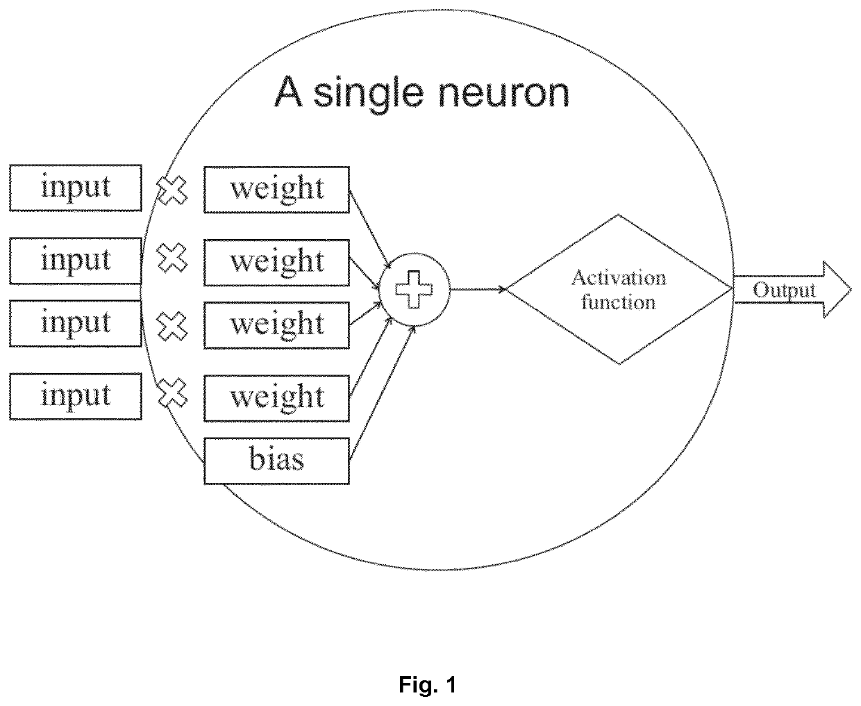
[0130]FIG. 1 illustrates the structure of a neuron as part of a neural network, for example a convolutional neural network, in which input is assigned certain weights for processing by an activation function which generates the output of the neuron.

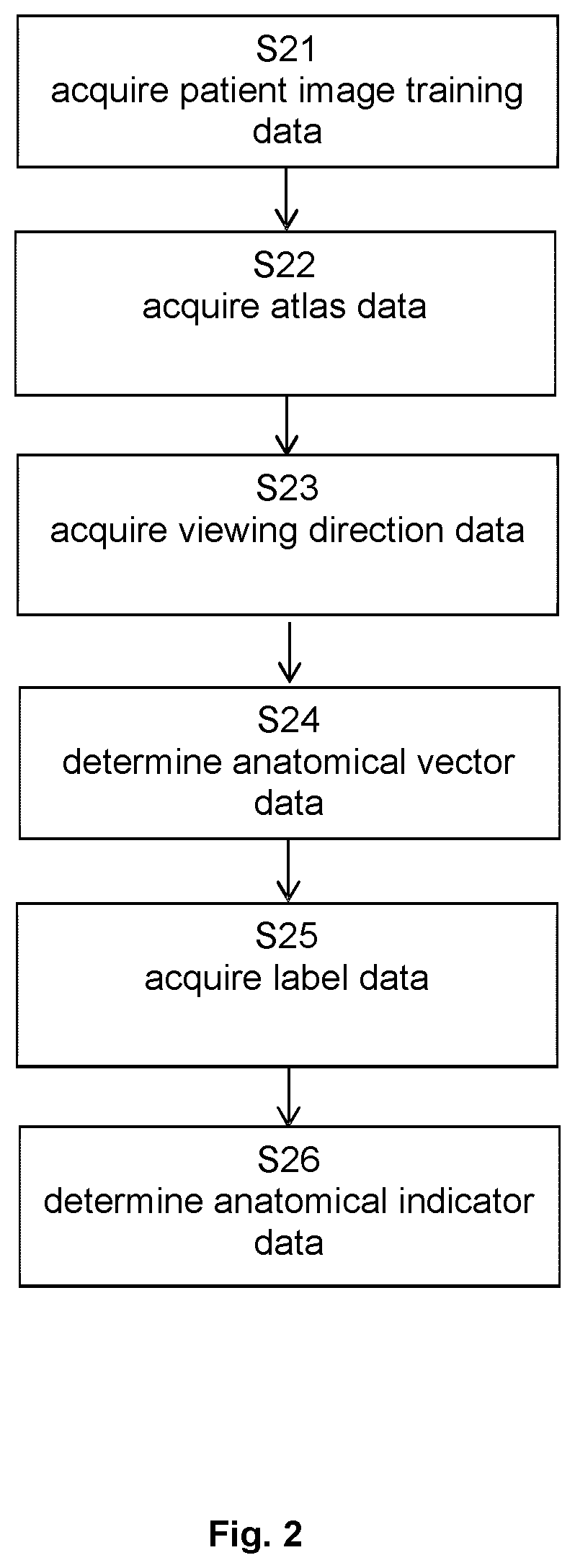
[0131]FIG. 2 describes the basic flow of the method according to the first aspect, which starts in step S21 with acquiring the patient training image data, continues to step S22 which encompasses acquisition of the atlas data, and then proceeds to acquiring the viewing direction data in step S23. On that basis, step S24 calculates the anatomical vector data, which is followed by acquisition of the label data in step S25. Finally, the anatomical indicator data is determined in step S26.

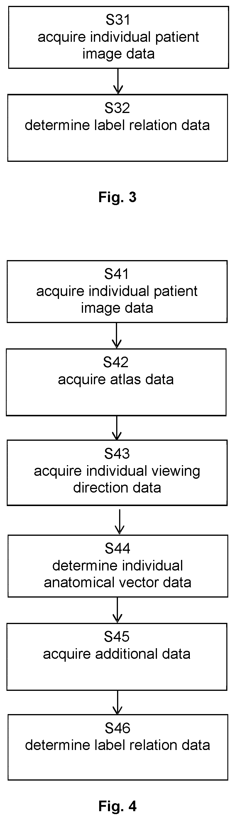
[0132]FIG. 3 illustrates the basic steps of the method according to the second aspect, in which step S31 encompasses acquisition of the individual patient image data and step 32 determines the label relation data.

[0133]FIG. 4 illustrates the basic steps of t...


PUM


Abstract

Disclosed is a computer-implemented method which encompasses registering a tracked imaging device such as a microscope having a known viewing direction and an atlas to a patient space so that a transformation can be established between the atlas space and the reference system for defining positions in images of an anatomical structure of the patient. Labels are associated with certain constituents of the images and are input into a learning algorithm such as a machine learning algorithm, for example a convolutional neural network, together with the medical images and an anatomical vector and for example also the atlas to train the learning algorithm for automatic segmentation of patient images generated with the tracked imaging device. The trained learning algorithm then allows for efficient segmentation and / or labelling of patient images without having to register the patient images to the atlas each time, thereby saving on computational effort.

Description

FIELD OF THE INVENTION[0001]The present invention relates to a computer-implemented method of training a learning algorithm for determining a relation between a label for indicating a position or type of an anatomical structure in a medical image on the one hand and the position or type of the anatomical structure in the medical image on the other hand as wells as methods of using the trained learning algorithm for segmenting and / or labelling medical patient images, a corresponding computer program, a computer-readable storage medium storing such a program and a computer executing the program, as well as a system comprising an electronic data storage device and the aforementioned computer.TECHNICAL BACKGROUND[0002]Medical patient images can be segmented or labelled using an anatomical atlas. This requires registering the patient images to the atlas which is associated with considerable computational effort.[0003]The present invention has the object of providing methods for more effi...

Claims


Application Information

Patent Timeline
Patent Type & Authority Applications(United States)
IPC IPC(8): G06T7/00G06V10/82G06V20/69G06T7/136
CPCG06T7/0014G06V10/82G06V20/695G06V2201/03G06T7/136G06T2207/30004G06T2207/10061G06V20/698G06T7/0012G06N20/00G06F18/214G06F18/24323G06V10/764G06T7/70G06V20/70G06V10/26G06V10/774G06T2207/10056G06T2207/20081G06T2207/20084
Inventor VILSMEIER, STEFANSCHMALER, JENS
Owner BRAINLAB
Why Eureka
Try Eureka