
Fundus oculi imaging device and fundus oculi imaging method using same

a technology of fundus oculi and imaging device, which is applied in the field of fundus oculi imaging device and fundus oculi imaging method, can solve the problems of difficult to have the fundus oculi examination in an area lacking medical institutions, and achieve the effect of clear and bright retinal image, rapid and accurate alignment of the first imaging module, and improved alignment accuracy

Pending Publication Date: 2022-05-26
ROOTEEHEALTH INC
View PDF0 Cites 0 Cited by
  • Summary
  • Abstract
  • Description
  • Claims
  • Application Information

AI Technical Summary

Benefits of technology

The fundus oculi imaging device and method described in this patent make it possible to quickly and accurately align the device for capturing retinal images. The device uses a corneal image to extract information about the pupil and the light needed for retinal imaging, which helps to accurately position the device. The device can move in three directions, allowing for better alignment. The second light source used for alignment is more sensitive to the retinal image, allowing for faster and more accurate alignment of the first light source. Overall, this invention allows for better quality and accuracy in fundus oculi imaging.

Problems solved by technology

Because a fundus oculi camera according to the related art has to examine the fundus oculi of an examinee while being fixed to a certain position, the examinee has to visit medical facilities such as a hospital in order to get the fundus oculi examination, and it may be difficult to have the fundus oculi examination in an area lacking medical institutions.

Method used


Image

  • Fundus oculi imaging device and fundus oculi imaging method using same
  • Fundus oculi imaging device and fundus oculi imaging method using same
  • Fundus oculi imaging device and fundus oculi imaging method using same

Examples


Embodiment Construction

Technical Problem

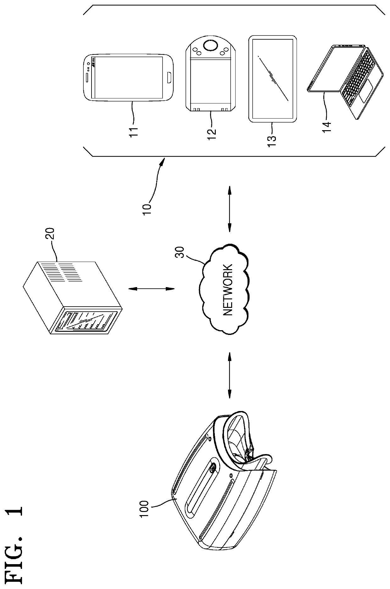
[0005]The present disclosure provides a fundus oculi imaging device capable of aligning a position of an imaging module rapidly and precisely, and obtaining a clear and exact retinal image, and a fundus oculi imaging method using the fundus oculi imaging device.

Technical Solution to Problem

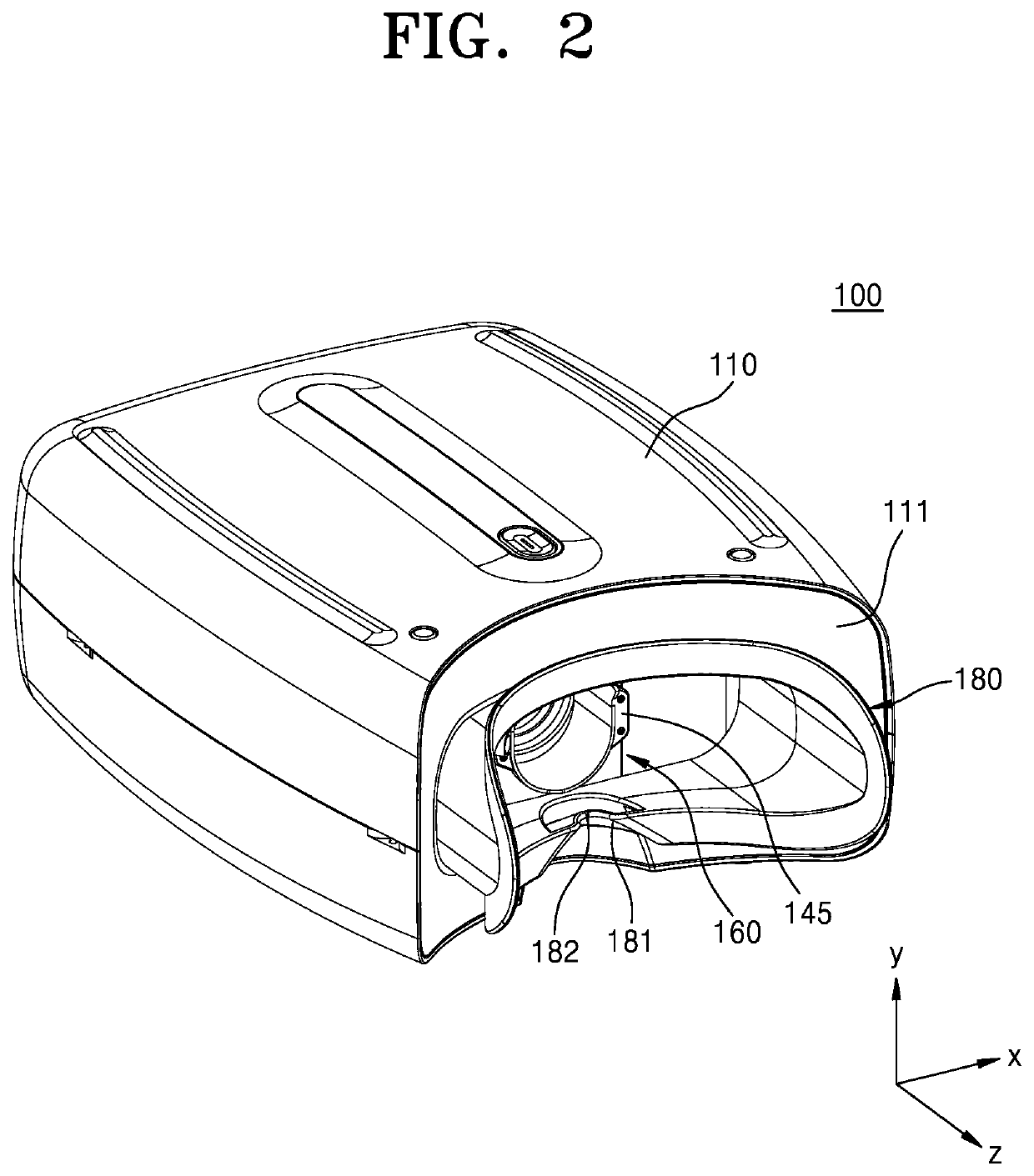
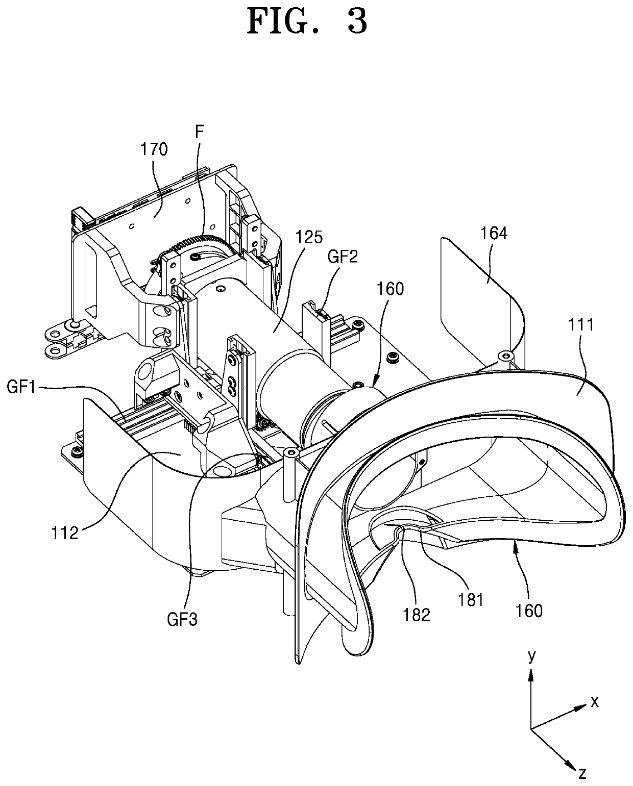
[0006]According to an aspect of the present disclosure, a fundus oculi imaging device includes: a housing; a first imaging module that is installed to be movable in the housing and captures a retinal image of an examinee; a light irradiation module moving along with the first imaging module in the housing and irradiating light to an eye of the examinee; and a second imaging module installed on a side of the housing and capturing an image of a cornea or a pupil, to which light is irradiated from the light irradiation module, of the examinee.

Advantageous Effects of Disclosure

[0007]A fundus oculi imaging device and method according to one or more embodiments of the present disclosure...


PUM


Abstract

This application relates to a fundus oculi imaging device and a fundus oculi imaging method including the same. In one aspect, the fundus oculi imaging device includes a housing and a first imaging module that is installed to be movable in the housing and captures a retinal image of an examinee. The fundus oculi imaging device may also include a light irradiation module moving along with the first imaging module in the housing and irradiating light to an eye of the examinee. The fundus oculi imaging device may further include a second imaging module installed on a side of the housing and capturing an image of a cornea or a pupil, to which light is irradiated from the light irradiation module, of the examinee.

Description

TECHNICAL FIELD[0001]One or more embodiments of the disclosure relate to an apparatus and a method, and more particularly, to a fundus oculi imaging device and a fundus oculi imaging method.BACKGROUND ART[0002]Fundus oculi refers to a posterior part of a retina in an eyeball, and through a fundus oculi examination, a central portion of the retina, e.g., a macula, an optic disc, and retinal blood vessels, etc. may be observed. In addition, severity of hypertension may be determined and diabetic complication related to eyes may be examined through the fundus oculi examination. A shape of the optic disc is used to diagnose various optic neuropathy such as glaucoma, increased intracranial pressure, optic neuritis, ischemic optic neuropathy, etc., and is essential for diagnosing retinal diseases such as macular degeneration, retinopathy of prematurity, etc. In particular, an early diagnosis of glaucoma and macular degeneration, which are two of three major causes of blindness, may be pos...

Claims


Application Information

Patent Timeline
Patent Type & Authority Applications(United States)
IPC IPC(8): A61B3/12A61B3/00A61B3/15
CPCA61B3/12A61B3/152A61B3/0008A61B3/0016A61B3/145
Inventor KOOK, KYUNG MIN
Owner ROOTEEHEALTH INC
Learn More