Real-time correction of regional tissue deformation during endoscopy procedure

a tissue deformation and endoscopy technology, applied in the field of real-time correction of regional tissue deformation during endoscopy procedures, can solve the problems of compromising the guiding capability of pre-procedural image models, mismatch between device space and image space,

Pending Publication Date: 2022-06-30
CANON USA
View PDF4 Cites 0 Cited by
  • Summary
  • Abstract
  • Description
  • Claims
  • Application Information

AI Technical Summary

Problems solved by technology

However, a number of factors may contribute to mismatch between the device space and image space throughout the procedure including, for example, breathing and bulk motions, as well as the pulling and stretching interaction of the catheter with the interior sidewall, particularly wh

Method used


Image

  • Real-time correction of regional tissue deformation during endoscopy procedure
  • Real-time correction of regional tissue deformation during endoscopy procedure
  • Real-time correction of regional tissue deformation during endoscopy procedure

Examples


Embodiment Construction

[0021]Various exemplary embodiments, features, and aspects of the disclosure will be described below with reference to the drawings.

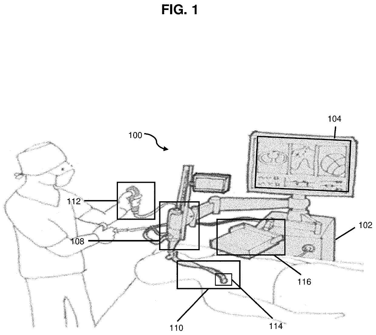
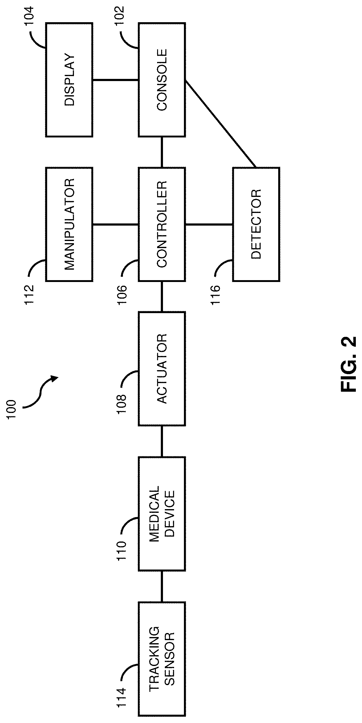
[0022]In the present disclosure, one or more configurations are described that functionally implement real-time correction of regional tissue deformation during an endoscopy procedure with imaging modalities including, for example, CT (computed tomography), MRI (magnetic resonance imaging), IVUS (intravascular ultrasound), PET (positron emission tomography), X-ray imaging, combinations or hybrids thereof, or the like. Configurations may be configured to facilitate placement of medical tools, catheters, needles or the like, and may be free standing, patient mounted, or the like. The present disclosure is not limited to any particular configuration.

[0023]According to some embodiments, the present disclosure may be configured to obtain medical image data from one or more imaging arrangements configured to implement image processing for visual guidance of d...


PUM


Abstract

A method for visualization guidance of device-to-image registration includes acquiring image data of a patient, generating a 3D model of the anatomy of the patient based on the image data, performing initial registration between image space and device space, advancing a device into a structure, tracking trajectory of the device in relation to the model, determining divergence between the device trajectory and the model, and effecting correction to the model or the device trajectory to minimize the divergence.

Description

CROSS REFERENCE TO RELATED APPLICATIONS[0001]The present application claims the benefit of priority from U.S. Provisional Application No. 63 / 132,163 filed Dec. 30, 2020, which is hereby incorporated by reference herein in its entirety.BACKGROUNDField of the Disclosure[0002]The present disclosure generally relates to medical imaging and, more particularly, to a method, apparatus, and medium for visualization guidance of device-to-image registration during an endoscopy procedure.Description of the Related Art[0003]During an image guided surgical procedure, an instrument such as an endoscopic catheter device glides into a patient. Medical images such as CT, MRI, or the like, can be used to capture patient anatomy a priori, and serve as a source to generate a three-dimensional guidance map. A catheter device may have an electromagnetic sensor on the tip and a position of the catheter device tip in the patient can be detected by a detector which may be provided outside of the patient's b...

Claims


Application Information

Patent Timeline
IPC IPC(8): A61B34/20G06T19/00G06T19/20A61B34/10A61B5/00
CPCA61B34/20G06T19/003G06T19/20A61B34/10A61B2034/107G06T2219/2004G06T2210/41A61B2034/2055A61B2034/105A61B5/7264A61B5/061A61B2505/05G06T2219/2021A61B2034/2051
Inventor ZHANG, HUALEI SHELLEYNINNI, BRIAN
Owner CANON USA
Why Eureka
Try Eureka