

Protease resistant compositions for wound healing
a technology of compositions and proteases, applied in the direction of peptide sources, instruments, peptide/protein ingredients, etc., can solve the problems that their forms or specific distributions in the wound may not support their normal activities, and achieve the effect of accelerating the rate of wound closur
- Summary
- Abstract
- Description
- Claims
- Application Information
AI Technical Summary
Problems solved by technology
Method used
Image

Examples
example 1
Production of Fibronectin-free Substrates
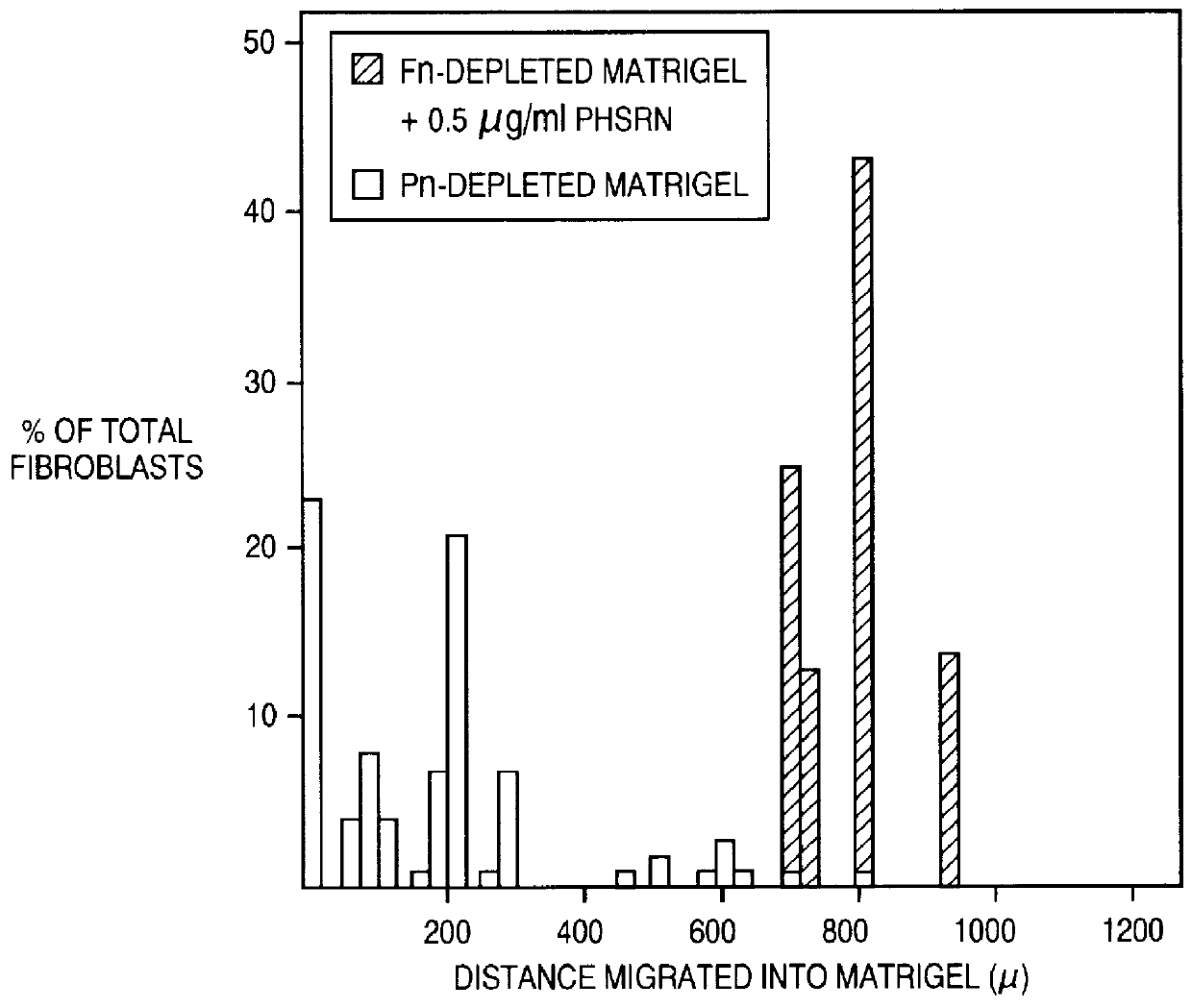
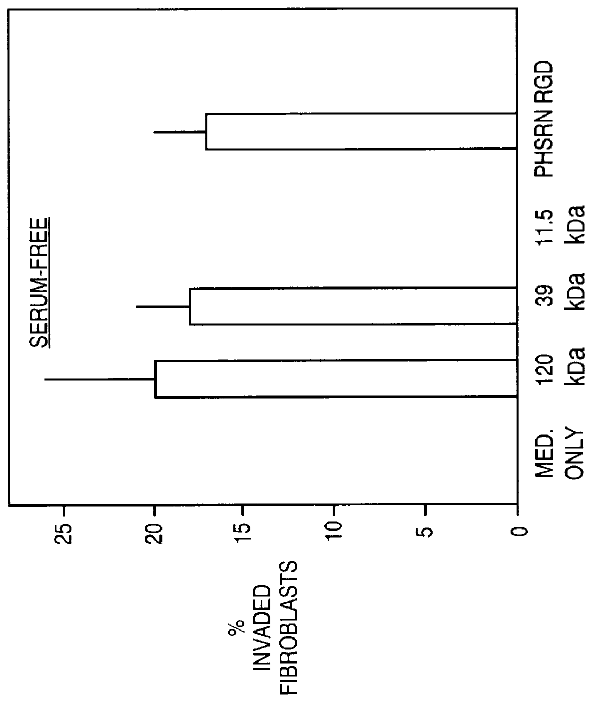
This example describes a purification approach for removal of plasma fibronectin (and / or cellular fibronectin) from a substrate (Matrigel). In this example, removal was attempted by affinity chromatography over Gelatin-Sepharose (a technique which can be used to remove plasma fibronectin from fetal calf serum).
The Gelatin-Sepharose beads were obtained from Pharmacia (Catalog# 17-0956-01). Two Kontes columns were set up with about 2 mls of Gelatin-Sepharose beads at 4 C. to prevent gelling of the Matrigel. The columns were then rinsed with about 10 column volumes of PBS to remove the preservative from the beads. The columns were drained to the top of the beads; then Matrigel was carefully added to the column. Once the Matrigel had entered the column, PBS was added to the top of the column. The Matrigel which was passed over the first column was collected and passed over the second column. The fibronectin-depleted Matrigel collected from the se...
example 2
Production of Fibronectin-free Substrates
This example describes a purification approach for removal of plasma fibronectin (and / or cellular fibronectin) from a substrate (Matrigel). In this example, removal was attempted by successive panning on gelatin. Eight wells of 24-well plate were coated with a 2% gelatin solution (the gelatin was obtained from Becton Dickinson Labware, Catalog #11868). The wells were filled with the gelatin solution which had been heated to 50 C. and incubated for 3 minutes. Then the solution was removed and the wells were allowed to air dry. Following drying, the wells were thoroughly rinsed with H.sub.2 O followed by two rinses with PBS. The plates were again allowed to dry; thereafter they were stored at -20 C. until use. Matrigel was thawed on ice and then added to one of the wells of a gelatin-coated plate (between 800 .mu.l and 1 ml of Matrigel was added to a well of a 24-well plate). The plate was placed in a bucket of ice in a 4 C. room on an orbital ...
example 3
Production of Fibronectin-free Substrates
This example describes a purification approach for removal of plasma fibronectin (and / or cellular fibronectin) from a substrate (Matrigel). In this example, removal was attempted by gelatin panning followed by antibody panning.
Anti-fibronectin Antibody-coated Wells
Wells of a 24-well plate were coated with an anti-fibronectin antibody. A mouse monoclonal antibody to human fibronectin was obtained from Oncogene Science (Catalog #CP13). Each well was incubated with 1 ml of antibody at a concentration of 30 .mu.l / ml for 2 hours at room temperature. Each well was then incubated with a solution of 3% BSA in PBS for 2 hours at room temperature. Following the two incubation periods, the wells were thoroughly washed with PBS and stored at -20 C. until use.
Depleting Matrigel of Fibronectin
Matrigel was panned over eight gelatin-coated wells (as described above in Example 2) to remove most of the fibronectin and its fragments. Thereafter, the Matrigel wa...
PUM
| Property | Measurement | Unit |
|---|---|---|
| concentration | aaaaa | aaaaa |
| concentration | aaaaa | aaaaa |
| pH | aaaaa | aaaaa |
Abstract
Description
Claims
Application Information