Method for enhancing myoblast migration and invasion in the context of gene therapy

a technology of myoblast migration and invasion, applied in the field of gene therapy, can solve the problems of unclear ability of bfgf to be of any use in modulating the migration and transplantation of myoblasts, limited knowledge available on the basic biology underlying the fate of implanted myoblasts, etc., and achieve the effect of enhancing the migration and invasion of myoblasts and greater consistency

Inactive Publication Date: 2001-09-04
RGT UNIV OF MICHIGAN
View PDF1 Cites 14 Cited by
  • Summary
  • Abstract
  • Description
  • Claims
  • Application Information

AI Technical Summary

Benefits of technology

Human myoblasts responded to growth factor treatment differently from mouse myoblasts. Treatment of human myoblasts with any growth factors, including TNF-.alpha. and bFGF, had no appreciative effects on MMP-1, MMP-2 or MMP-9 expression. The high basal level of MMP-2 expression of human cells may make them less sensitive to additional treatment with these growth factors, which primarily function to increase MMP-2 expression. This is further supported by the observation that fibronectin induces significant activation of MMP-2, and increases its effects on migration of human myoblasts (FIG. 7a). Interestingly, the effect of fibronectin alone on human myoblast invasion is small, but together with bFGF, its effect is synergistic increasing to several fold higher than the BSA control level. These results indicate that the factors and conditions which affect migration and invasion of human myoblasts are somewhat different from those of mouse cells. Human myoblasts have a great potential for migration, presumably due to the high constitutive MMP-2 expression, though an appropriate priming stimulus by treatment with growth factors or fibronectin is still needed for cells to initiate migration (FIG. 7a). Invasion of human myoblasts across MATRIGEL.RTM. is less than mouse cells (FIG. 7b). The differences observed between mouse and human cells appear not to be dependent on the age of the individual from which cells were isolated, as myoblasts from individuals 44 and 8 years old behaved similarly (data not shown).
Transfection assays allow for a great deal of flexibility in assay development. The wide range of commercially available transfection vectors will permit the expression of the MMPs. In one embodiment, cells are transiently transfected with an expression construct comprising nucleic acid encoding MMP-1 or MMP-2 that may include (in operable combination) an inducible promotor allowing for the expression of a metalloprotease to levels higher than in the untransfected cells. Increased expression of these metalloproteases may enhance migration and invasion of myoblasts in combination with the compounds to be screened. Cells can be exposed to the agent suspected of modulating myoblast migration and invasion, MMP expression would be turned on, if necessary, and migration and invasion can be measured by techniques known to those practiced in the art. The compositions of the present invention would be used as positive controls. Rates of migration and invasion of cells exposed to the compounds to be screened are compared to rates of migration and invasion of the cells exposed to the compounds of the invention. Transfection with a control expression vector (e.g. an empty expression vector) would serve to compare the effect of MMP over expression on migration and invasion. Rates of migration and invasion can be quantitated by any of a number of ways reported in the literature and known to those practiced in the art.
In another embodiment, stably transfected cell lines expressing MMP-1 or MMP-2 are produced as stocks for further assays. The use of an inducible promoter may be utilized in these systems. Screening assays for compounds suspected of modulating myoblast migration and invasion would be conducted in the same manner as with the transient transfection assays. Using stably transfected cell lines would allow for greater consistency between experiments and allow for inter-experimental comparisons.
The present invention demonstrates that cultured myoblasts can be induced to express peptides from transfected expression constructs. Additionally, the present invention demonstrates that cultured myoblasts can be induced to migrate and invade muscle tissue. This technology may be used in the delivery of therapeutic gene products thereby allowing for an effective method of gene therapy. In one embodiment, the myoblasts are obtained from an immunocompatable donor or from the patient. The myoblasts are transfected with the construct able to express the protein of interest. The myoblasts are then cultured with bFGF and FN, as described herein. Thereafter, at least a portion of the myoblasts would be introduced into the patient. In another embodiment myoblasts would be transfected with a marker protein (e.g. GFP, luciferase or .beta.-gal), prior to culturing with bFGF and FN, to permit the monitoring of the migration and invasion of the myoblasts. In yet another embodiment the myoblasts would be transfected with MMP-1 or MMP-2, prior to culturing with bFGF and FN, to permit.enhanced migration and invasion. The latter embodiment, therefore, comprises myoblasts transfected with a first vector encoding a therapeutic gene product and a second vector encoding a metalloprotease gene.
Gelatin zymography for assaying MMPs was carried out as previously described (Guerin and Holland "Synthesis and secretion of matrix-degrading metalloproteinases by human skeletal muscle satellite cells" Devel. Dynamics 202:91-99, 1995) with minor modifications. Briefly, myoblasts were grown in 6 cm tissue culture plates to approximately 70% confluence, then rinsed three times with serum-free DMEM and incubated for 3 hours in DMEM containing 0.2% BSA to eliminate the effects of serum. Growth factors were added and cells were incubated for 24 hours. Culture medium was then collected, centrifuged to pellet detached cells, and concentrated ten- to twenty-fold using the Centricon-10 (Amicon, Beverly, Mass.) system. The protein concentration of the supernatants was determined using the Bio-Rad protein microassay system with BSA used as the standard. Samples were stored at -70.degree. C. until use. For gelatin zymography, aliquots (10 .mu.g as the total protein per sample) were electrophoresed at constant voltage on a 10% polyacrylamide gel containing 2 mg / ml gelatin. The gel was rinsed three times for 15 min in 2.5% Triton-X 100 to remove SDS and renature the proteins, then incubated in MMP activation buffer (0.05 M Tris-HCl, pH 7.5 with 5 mM CaCl.sub.2) for 24 hours at 37.degree. C. with constant shaking. Gels were stained overnight in 0.5% Coomassie blue R-250, and destained for 1 hour in 40% methanol:10% acetic acid. Proteinase activity was quantified by densitometric scanning of bands using a Bio-Rad Gel Doc 1000 video camera imaging system (Bio Rad, Hercules, Calif.).

Problems solved by technology

Dynamics 202:91-99, 1995), but currently there is only limited knowledge available on the basic biology underlying the fate of implanted myoblasts, the importance of MMPs and their relationship to physiological stimuli in myoblast migration and invasion in vitro and in vivo.
However, the ability of bFGF to be of any use in modulating the migration and transplantation of myoblasts has remained unclear.

Method used


Image

  • Method for enhancing myoblast migration and invasion in the context of gene therapy
  • Method for enhancing myoblast migration and invasion in the context of gene therapy
  • Method for enhancing myoblast migration and invasion in the context of gene therapy

Examples


example ii

Zymography Analysis of Mouse Myoblasts

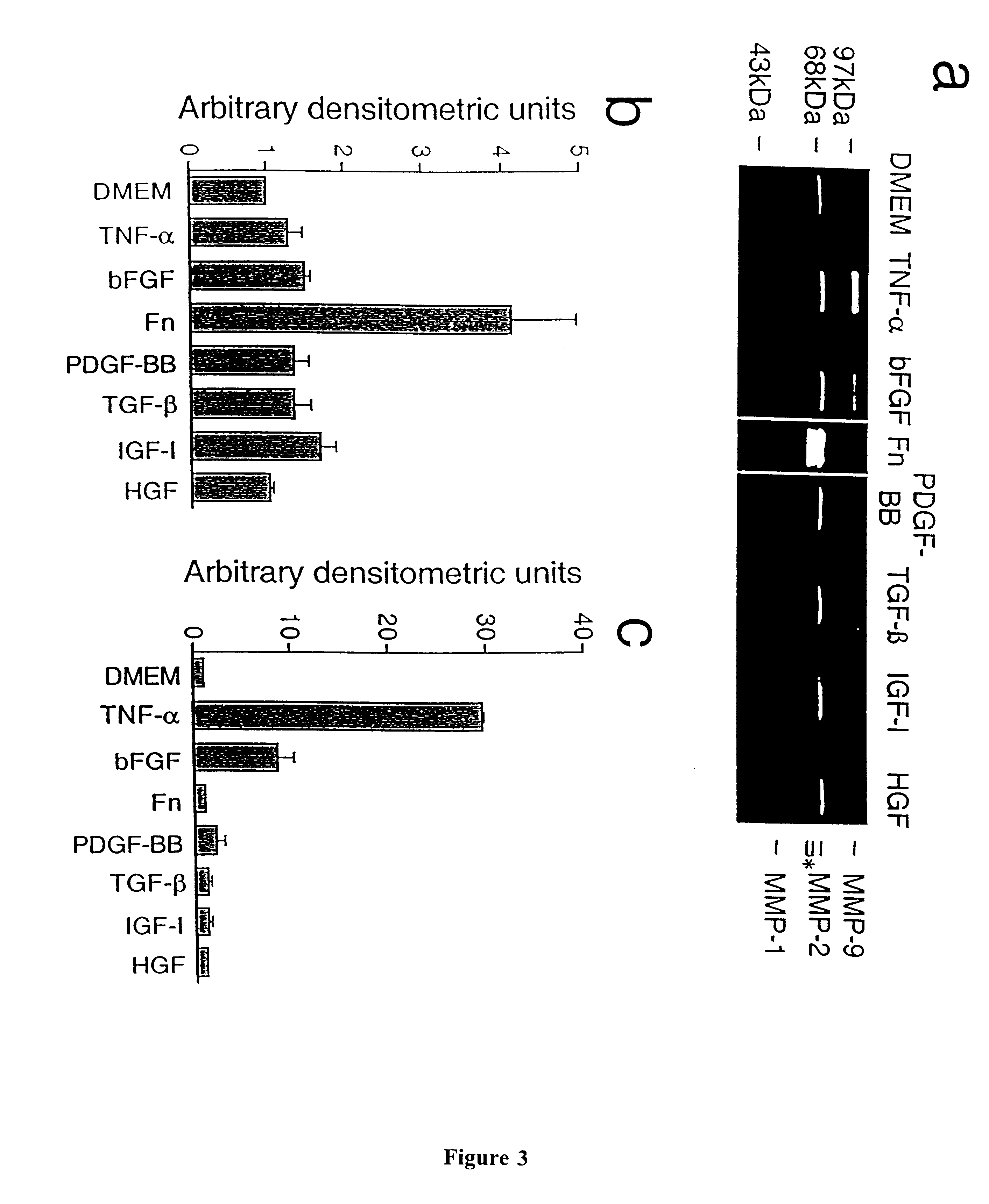
The effects of growth factors and fibronectin on MMP expression by mouse myoblasts are shown in FIG. 3a. Mouse myoblasts grown in serum-free medium constitutively expressed MMP-2 (zymogen form, 72kDa), which still appears as a zymogram band due to its inherent gelatinase activity (Reich et al. "Effects of inhibitors of plasminogen activator, serine proteinases and collagenase IV on the invasion of basement membranes by metastatic cells" Cancer Research 48:3307-3312, 1998) (FIG. 3). Proteolytic degradation of gelatin due to MMPs appears as clear bands against the dark background. Bands marked with an asterisk (64 and 62 kDa) indicate the activated forms of MMP-2. The lane for fibronectin treatment was run simultaneously on a separate gel, and the scanned picture is placed in the order for comparison. Treatment of mouse myoblasts with bFGF, PDGF-BB, TGF-.beta. and IGF-I had modest but consistent effects on total MMP-2 expression, increasing its ex...

example iii

MMP Over-expression and Mouse Myoblast Migration and Invasion

Transient over-expression of MMP-1, MMP-2, and MMP-9 was tested in myoblasts to determine whether expression of individual MMPs was sufficient to produce increased migration and / or invasion. Transient transfection rather than stable transduction was used, because secretion of over-expressed MMPs by transfected cells (approximately 20-25% of the cells) should be sufficient to allow most, if not all, cells access to increased levels of secreted MMPs and avoid prolonged exposure of cells to over-expressed MMPs. Successful transfection of MMP-1, MMP-2 and MMP-9 were confirmed by gelatin zymography, showing dramatically increased intensity of bands of approximately 57, 72 and 92 kDa (human MMP-9 is smaller than the mouse counterpart), respectively (FIG. 5a), and by Northern blot analyses (FIG. 5b, c, and d) of the transfected cells. The high molecular weight bands within the bracketed region marked with + presumably represent c...

example iv

Human Myoblast Migration, Invasion and MMP Expression

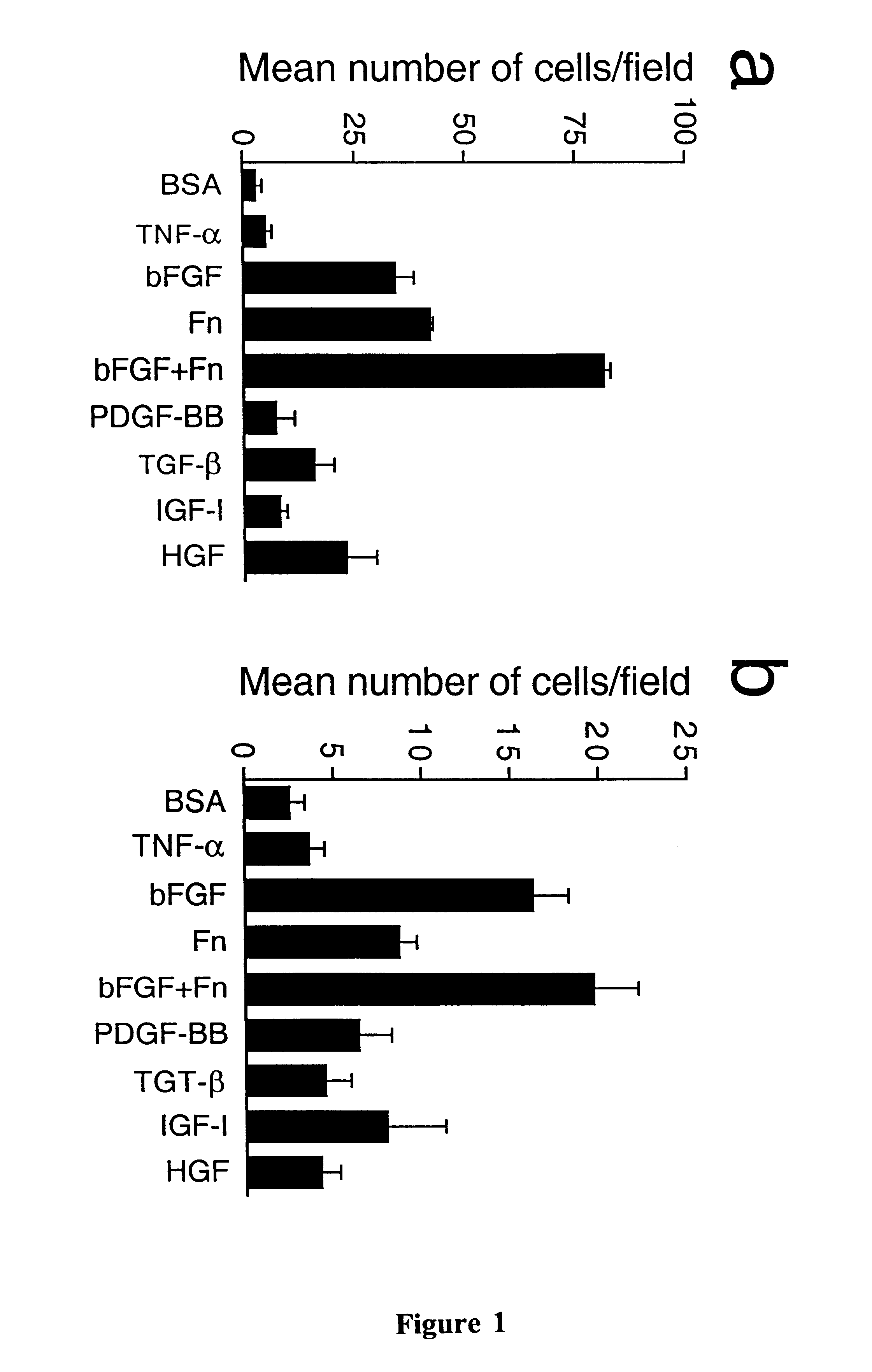
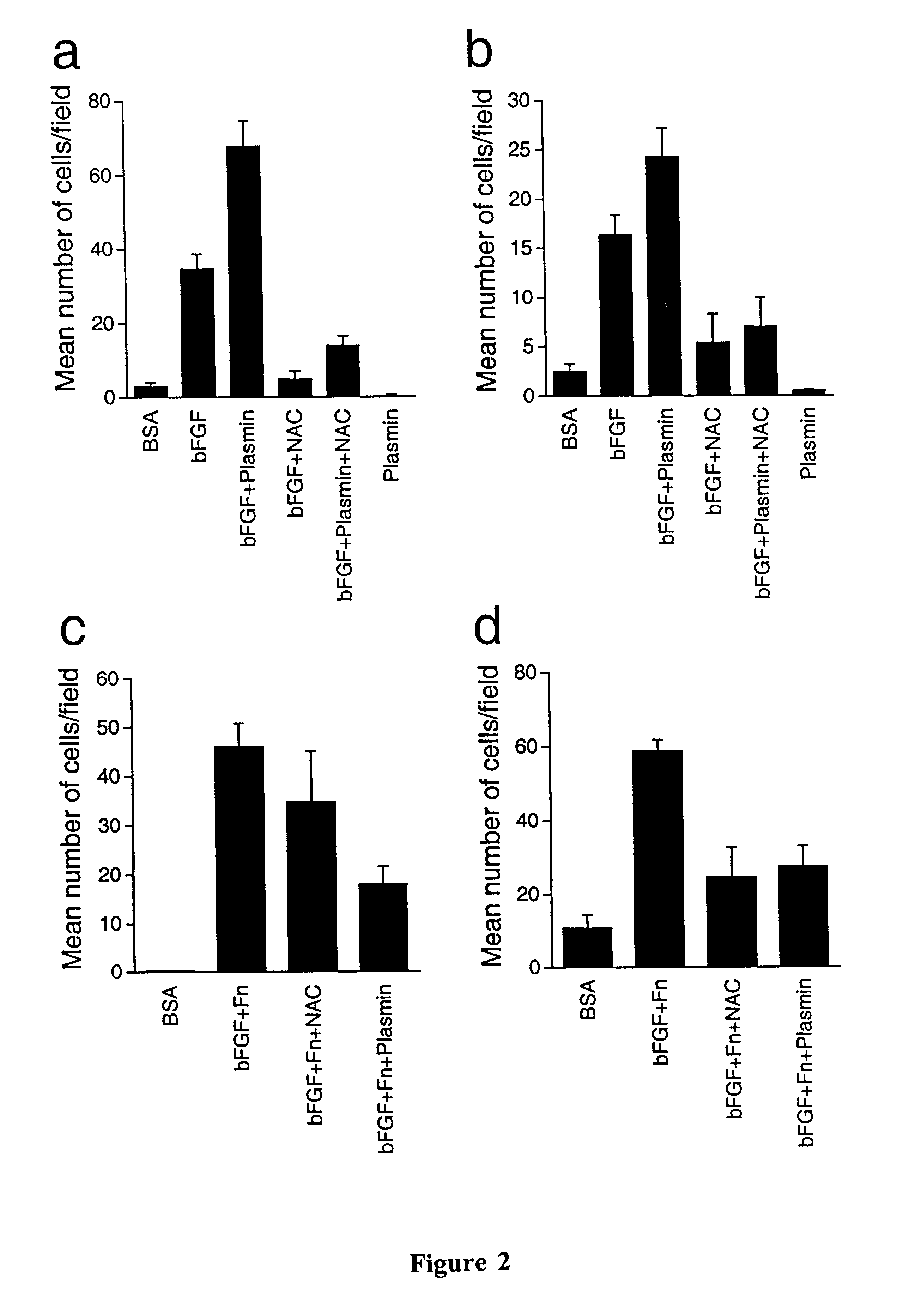
The effects of growth factors on human myoblast migration in vitro (12 hour time point) were somewhat different from those observed with mouse myoblasts. All the growth factors tested showed substantial stimulatory effects over the BSA control, ranging from 20-100-fold. The greatly elevated level of migration of human myoblasts was due in part to the extremely low migration in the BSA control (basal level) of human cells compared to mouse cells. The largest effects on human myoblast migration were produced by fibronectin (100-fold), PDGF (about 62-fold), TGF-.beta. (about 54-fold) and HGF (46-fold) over the control level, while bFGF produced only a 37-fold stimulation (FIG. 7a). Moreover, unlike mouse myoblasts, the combination of fibronectin and bFGF produced approximately the same effects as fibronectin alone. These effects were significantly increased by plasmin treatment, and greatly reduced by NAC (FIG. 7c), indicating the cr...


PUM

PropertyMeasurementUnit
concentrationsaaaaaaaaaa
pHaaaaaaaaaa

Abstract

A novel, empirically derived composition of cytokines and myoblasts is described, that allows for the migration of myoblasts through connective barriers, along with methods employing the composition in the in vivo migration of myoblasts for therapeutic purposes and gene therapy, as well as methods for the identification of agents that are agonistic or antagonistic to myoblast migration in vitro or in vivo.

Description

This invention generally relates to a novel composition comprising myoblasts and growth factors. The growth factors may include, for example, basic fibroblast growth factor (bFGF) and fibronectin (FN). The invention also relates to a novel composition comprising myoblasts transfected with vectors expressing metalloproteases and growth factors. Additionally, the invention relates to the use of these compositions in assays for the identification of agents which are antagonistic or agonistic for myoblast migration either in vivo or in vitro. Furthermore, the invention relates to the use of these compositions for the treatment of degenerative muscle diseases. Further still, the invention relates to the therapeutic use of these compositions and methods in gene therapy.The ability of myoblasts to migrate through connective tissue barriers has important implications for muscle development, muscle regeneration, and myoblast-mediated gene transfer. During embryonic development, myogenic prec...

Claims


Application Information

Patent Timeline
Patent Type & Authority Patents(United States)
IPC IPC(8): C12N5/06A61K35/12A61K48/00A61K35/34A61P21/00C12N15/09A61P21/04A61P43/00C12N5/02C12N5/077C12N5/10C12N15/50
CPCC12N5/0658A61K35/12C12N2501/60A61K48/00A61P21/00A61P21/04A61P43/00
Inventor KURACHI, KOTOKU
Owner RGT UNIV OF MICHIGAN
Why Eureka
Try Eureka