Method and apparatus for measurement of microtome performance

a microtome and performance technology, applied in the field of microtome sectioning histology tissue samples, can solve the problems of loss of precision, degrade the quality of histologic sections, and detectable deviations from a perfectly flat surface on the face of the block

Inactive Publication Date: 2001-12-11
MICROSCI GROUP
View PDF12 Cites 121 Cited by
  • Summary
  • Abstract
  • Description
  • Claims
  • Application Information

AI Technical Summary

Problems solved by technology

Block and section artifacts arising within the microtome mechanism: Improper adjustment or mechanical deterioration of the microtome mechanism may result in vibration, backlash, or other loss of precision that will degrade the quality of histologic sections and, correspondingly, may produce detectable deviations from a perfectly flat surface on the face of the block.
If the clamp that holds the block in place on the microtome is not sufficiently tightened, or if other microtome parts become loosened due to wear, an unstable block will result.
Alternatively, a loose block may result in wrinkles or compression artifacts in sections, or in extreme cases the knife may "chop", penetrating deeply into the block and halting sectioning.
If the velocity of the block relative to the blade is excessive, chatter may result, which in turn may damage the edge of a glass or diamond knife.
Vibrations transmitted to the block due to wear or defects on cams, gears, drive screws, and other components within the core mechanism of the microtome may also produce chatter or washboarding on sections, which will be duplicated on the face of the block.
Imperfections in the knife will result in direct damage to histologic sections and thus to the face of the block.
Microtome blades may lead to suboptimal microtome performance due to dullness, nicks, corrosion, adherence of foreign material, misalignment, as well as flaws induced in the edge of the blade during its fabrication and resharpening.
Blade dullness may result from normal wear or improper sharpening.
In the case of glass knives that are not used immediately after fabrication (by controlled fracture of plate glass in the laboratory), the keenness of the edge may be lost due to gradual deformation of the glass, which over time behaves as a very slow-moving liquid.
Dull blades will induce compression marks or wrinkles in sections and may cause chatter, or in the case of the softer types of embedding medium such as paraffin, may result in smearing of the tissue both in the section and on the block face.
Focal defects in the edge of the blade such as burrs, nicks, or manufacturing defects will produce vertical lines in the block face, as well as corresponding scratches in sections.
If severe, such flaws may lead to shredding of sections.
A misaligned knife blade will cause a variety of artifacts, including irregular, skipped, or excessively thick or thin sections.
Too little tilt on the blade can cause the section to adhere to the block face as it passes by the knife on the return stroke instead of separating cleanly.
In contrast, an excessive blade angle can result in a washboarding artifact.
In addition to these problems, embedding material, especially paraffin, may build up on the edge of the knife, causing vertical markings on the section and on the block face.
Various components of the drive chain, such as toothed gears and belts may introduce further vibrations.
However, if the paraffin is too hot during infiltration and embedding, dry or incomplete sections may result.
Further, paraffin-based materials are quite sensitive to environmental conditions such as humidity and temperature, significant variations in which may occur in histology laboratories.
Plastic embedding media is subject to a separate set of problems.
Polymer that has exceeded its "pot life" may not polymerize completely, resulting in soft blocks and thick and thin cutting.
If the components for the polymer are not apportioned correctly, soft blocks may also result, or conversely the blocks may be too brittle, which will often result in diffuse chatter.
Bubbles or foreign material in the medium will result in focal defects in sections and on the face of the block.
When the cut surface of a block face transects embedded, poorly infiltrated tissues, the sample itself may cause scattering of incident light, and thus produce an image of the tissue.
Improperly oriented blocks can display chatter, or in the case of paraffin sectioning may produce curved section ribbons.
Current methods for assessing microtome sectioning quality: Although microtomy is over 150 years old, there exists no formalized method for real-time, quantitative monitoring of the operating condition of microtomes.
Prior art devices do not direct attention to the block face, rather than to the sections, for clues to the cause of poor quality microtome performance.
This is due in most cases to the poor visibility of the surface of the rather translucent materials from which histology blocks are made, and to the fact that most flaws are microscopic.
Prior art methods and devices do not exploit this feature of sectioned histology blocks in assessing microtome performance.

Method used


Image

  • Method and apparatus for measurement of microtome performance
  • Method and apparatus for measurement of microtome performance
  • Method and apparatus for measurement of microtome performance

Examples


Embodiment Construction

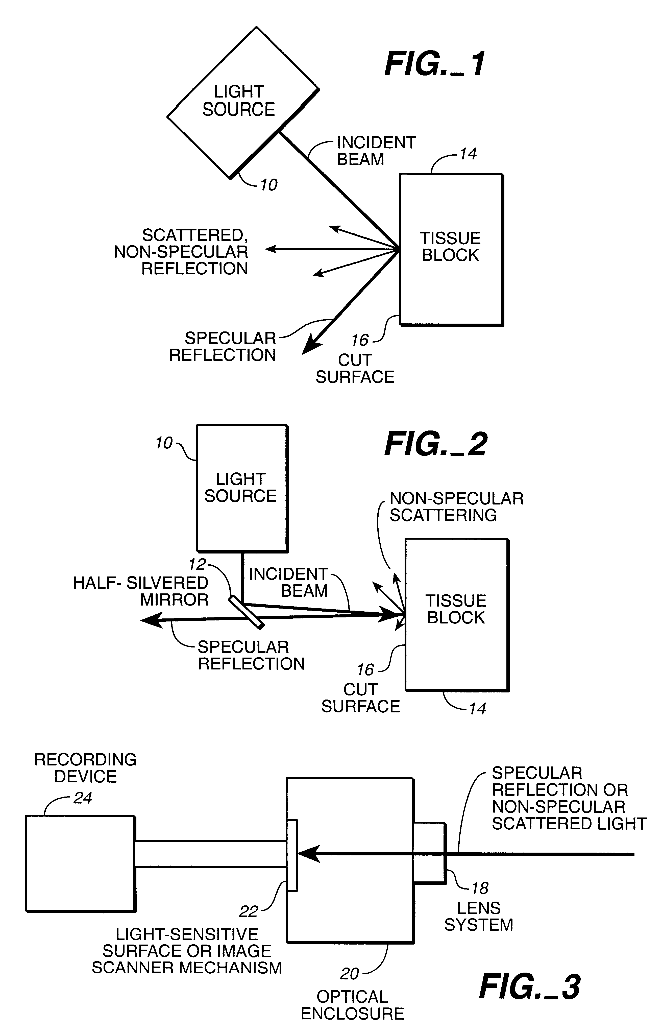
FIG. 1 is a schematic view of that part of the present invention for producing an image from the cut surface of a tissue sample histology block, illustrating a light source 10 and a tissue block 14, each as well known in the art. Light produced at the light source is directed onto the cut surface 16 of the tissue block, and thereafter from the cut face of the tissue block in a specular and / or non-specular manner.

FIG. 2 illustrates the preferred configuration depicting that part of the present invention for producing an image from the cut surface of a tissue sample histology block as more generally illustrated in FIG. 1. FIG. 2 depicts the specular image capture mode, illustrating a light source 10, partially reflective mirror 12, and tissue block 14, all as well known in the art. Light produced at the light source is reflected from the partially reflective mirror 12 onto the cut surface 16 of the tissue block, and thereafter from the cut face of the tissue block in a specular and / or...


PUM

PropertyMeasurementUnit
lengthaaaaaaaaaa
flatnessaaaaaaaaaa
widthaaaaaaaaaa

Abstract

A method and apparatus for monitoring and evaluating the performance and condition of histology laboratory microtomes and microtome accessories including knives, motor drives, and illumination devices. Irregularities in an image of the surface of a block mounted on the microtome are detected and characterized, the block having been subjected to mechanical sectioning by the microtome to produce a cut block face. The image of light reflected from the surface of the block, either specular or non-specular, is recorded and subjected to graphical analysis to extract, quantify, and interpret patterned features, including those indicating anomalies in the function of the microtome, its accessories, or the tissue block itself.

Description

1. Field of the InventionThe present invention relates generally to sectioning histology tissue samples using a microtome, and more particularly to a method and apparatus for measuring and evaluating the performance of histology laboratory microtomes and microtome accessories.2. Description of the Prior ArtDefinitions: As used herein, the term "microtome" refers to a device in which a block of sample or tissue is precisely cut such that a very thin layer of material is removed, or "sectioned" from the surface of the block. The term "microtomy" applies to the functioning of the microtome. While microtomes have been developed in a wide variety of configurations, most modern devices are arranged such that the block is fixed to one end of a vertically moveable arm and is engaged with a stationary blade; therefore, the term "vertical markings" applies to linear features aligned parallel to the motion of the block, and the term "horizontal markings" applies to linear features aligned in a...

Claims


Application Information

Patent Timeline
Patent Type & Authority Patents(United States)
IPC IPC(8): A61B1/00B23Q15/00B26D7/08G01N1/04G01N1/06G02B21/34G06K9/00A61B
CPCG01N1/06G06T7/0012G06T2207/10056G06T2207/30168
Inventor KERSCHMANN, RUSSELL L.BOLLES, MICHAEL E.HENDRICKSON, ANDREW D.
Owner MICROSCI GROUP
Why Eureka
Try Eureka