Devices for investing within ligaments for retracting and reinforcing the same

a technology of ligaments and devices, applied in the field of suturing tissue, can solve the problems of bowel herniation, possibility of omental trapping, and often time-consuming procedure for closure, and achieve the effect of accurate and consistent restraint of position and angle, rapid and safe, and accurate visualization

Inactive Publication Date: 2002-05-07
COOPERSURGICAL INC
View PDF38 Cites 236 Cited by
  • Summary
  • Abstract
  • Description
  • Claims
  • Application Information

AI Technical Summary

Benefits of technology

A further object of the invention is to provide a laparoscopic instrument that allows for the rapid control of bleeding from inferior epigastric lacerations or other lacerations of vessels in the outer (or abdominal) wall that may occur with placement of the laparoscopy trocars.
Another object of the invention is to provide a laparoscopic instrument that easily disassembles at the handle and at the interface between the tube member and handle for providing easy access to all the instrument components for cleaning and sterilization prior to surgery.
Yet another object of the present invention is to provide a surgical instrument that works in a manner similar to a needle driver without the requirement for the needle itself in passing suture easily through the fascial and peritoneal surfaces and for retrieving the suture for completing the suture procedure in a rapid, safe, and visualized manner.
It is another object of the invention to provide a guide to accurately and consistently restrain the position and angle of insertion of a laparoscopic instrument to provide for proper placement and retrieval of suture material at a predetermined location within the body.
Accordingly, it is an objective of the present invention to provide a method associated with an improved surgical instrument of the standard laparoscopic-type grasper that better suits the needs of a surgeon when suturing closed a surgical incision. In addition, it is the objective of the present invention to allow the passage of suture through tissue in order to suture or ligate vessels, approximate tissues, and perform all suturing that would require a separate needle driver in laparoscopic surgery.
Still another object of the invention is to provide a method of laparoscopically suturing the round ligament that supports a woman's uterus to shorten and strengthen the ligament to reposition and stabilize a misaligned uterus.

Problems solved by technology

This procedure for closure is frequently a time-consuming procedure requiring the identification of the fascia and closure of each fascial site with suture from an external point.
The necessity for closing these port sites in laparoscopic surgery is critical since suturing the incisions improperly can lead to bowel herniation through the port sites as well as the possibility of omental trapping if the fascial sites are not properly closed.
Intra-abdominal suturing is a time-consuming process for surgeons in part because a lot of manipulation and "fiddling" is associated with the needle attached to the suture material.
Among the drawbacks to Gilliams procedure is that it does nothing to improve the strength of thinned ligaments and they may again stretch so that the correction would be short-lived.
Additionally, Gilliams procedure can change the geometry of the lower pelvis when ligaments are reattached to the anterior abdominal wall, creating a pouch that may entrap the bowel which is a serious complication.
Changing the attachment point does nothing to improve the strength of these thinned ligaments, and they may be prone to restretch.
The procedures do not reinforce thinned ligaments which may stretch and loosen from the fixation.
This may lead to a slipping of a length of intestine into an adjacent portion, which may produce an obstruction.
As evident from the above description, the prior art devices are not suitable for use to retract and reinforce ligaments to better support organs as envisioned by Applicant.

Method used


Image

  • Devices for investing within ligaments for retracting and reinforcing the same
  • Devices for investing within ligaments for retracting and reinforcing the same
  • Devices for investing within ligaments for retracting and reinforcing the same

Examples


Embodiment Construction

)

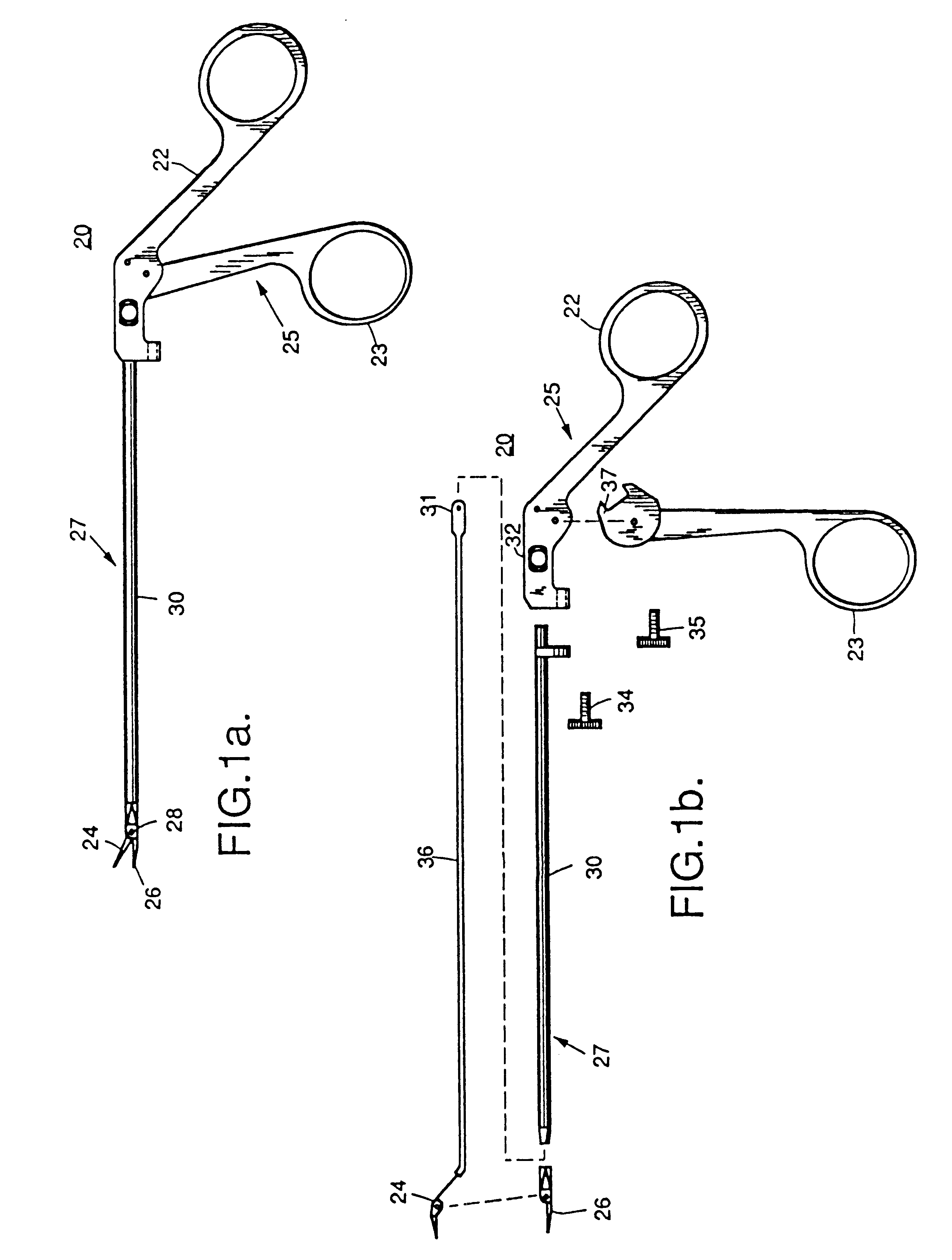
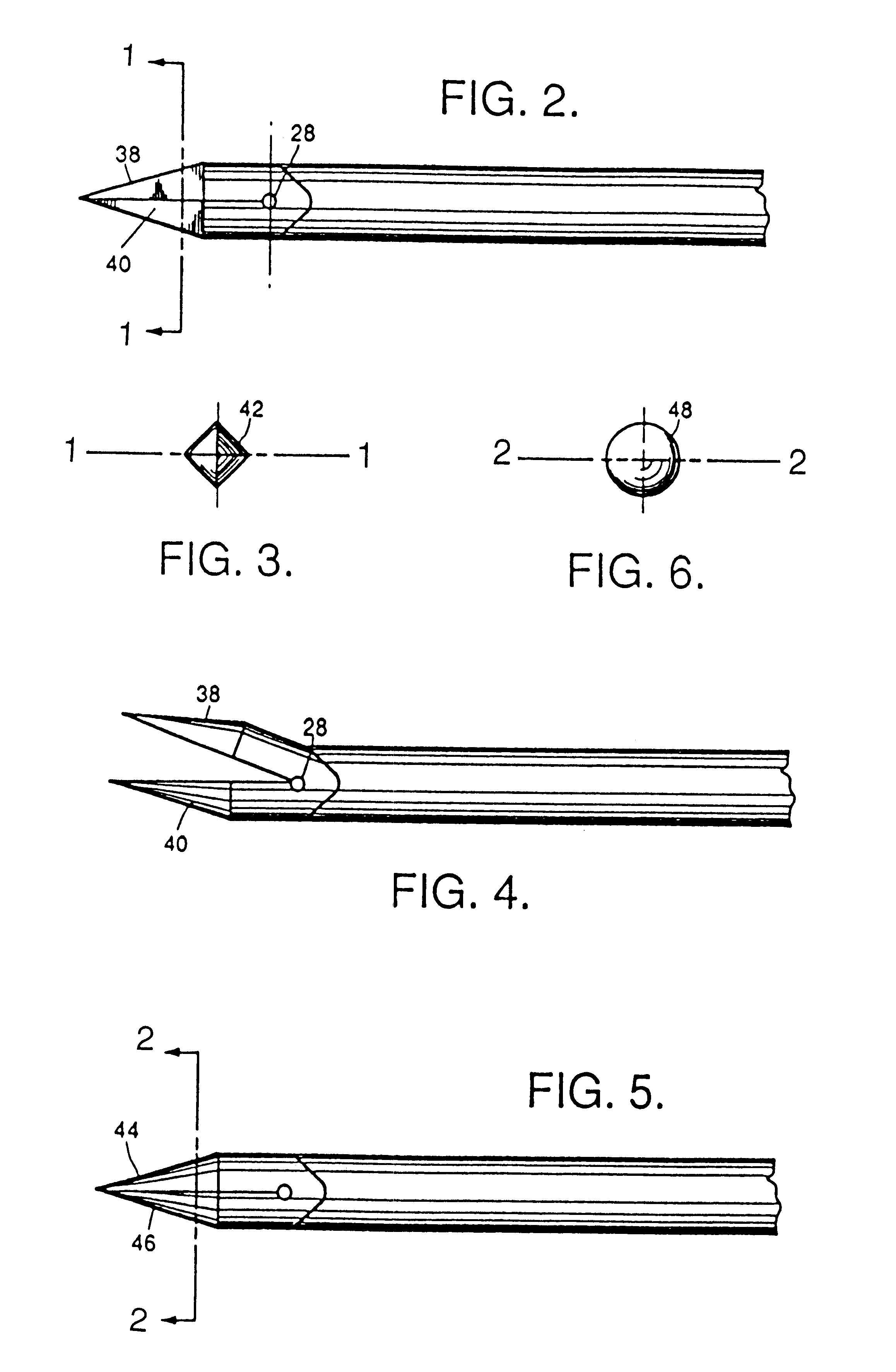
Referring now to the drawings wherein like reference numerals refer to like and corresponding parts throughout, the laparoscopic instrument is generally indicated by numeral 20. Referring now to FIGS. 1a and 1b, forceps jaws 24 and 26 are pivoted back and forth in double-action movement about an axis defined by pivot pin 28 when actuating rod 36 is reciprocated by a surgeon manipulating the scissor handles 22 and 23 providing a driving means 25 for driving forceps jaws 24 and 26 in a closed position through a patient's skin. Detachable means 27 comprise an elongated tube 30 concentrically sharing an axis with the actuating rod 36 having forceps jaws 24 and 26 engaged at a distal end.

As shown in FIG. 1b, the laparoscopic instrument 20 may be easily disassembled for sterilization prior to surgery by separating driving means 25 from detachable means 27 by loosening the knurled screw 34 on fixed handle housing 22, rotating the elongated tube 30 and forceps jaws 24 and 26 slightly, and ...


PUM


Abstract

The suture is released by opening and withdrawing the tip from the guide. The suture is recovered by using the guide to redirect the tip and puncturing the tissue opposite the first point of insertion. The tip grasps the suture and pulls the suture through the guide. The suture is pulled outside the wound, providing for rapid closure of the surgical incision. The guide is insertable within the wound to be closed and guides the surgical instrument at a predetermined angle from the longitudinal axis of the guide for optimum wound closure. The surgical instrument and method may be used to advantageously shorten and strengthen ligaments. For example, by gathering and reinforcing the round ligament that supports a woman's uterus with suture materials, surgeons can reposition and stabilize a retroverted uterus. Other devices may be also used in connection with retracting and reinforcing connective tissue. For example, rather than relying entirely on suture material to hold connective tissue in a retracted state, a device having one or more anchors may be inserted into connective tissue.

Description

1. Field of the InventionThe present invention relates to improvements in the procedure for suturing tissue during endoscopic / laparoscopic surgery, and to a method of suturing which utilizes a modified laparoscopic grasper and a guide. More particularly, the improved method relates to suturing of ligaments using a needle-point suture passer to retract and reinforce the ligaments, applications including uterine suspension and positioning. Devices suitable for insertion into ligaments or tendons for retracting and reinforcing the same in accordance with the improved method are also disclosed.2. Description of the Related ArtAn endoscopic / laparoscopy procedure involves making small surgical incisions in a patient's body for the insertion of trocar tubes thereby creating access ports into the patient's body. Thereafter, various types of endoscopic / laparoscopic instruments are passed through these access ports and the appropriate surgical procedures are carried out.After the surgical pro...

Claims


Application Information

Patent Timeline
Patent Type & Authority Patents(United States)
IPC IPC(8): A61B17/04A61B17/06A61B17/28A61B17/34A61B19/00A61B17/00A61B17/42
CPCA61B17/0057A61B17/0469A61B17/0482A61B17/0483A61B17/06061A61B17/06066A61B2019/4868A61B17/282A61B17/3417A61B17/42A61B2017/00349A61B2017/0046A61B2017/00637A61B2017/00663A61B2017/047A61B2017/2825A61B2017/4216A61B17/04A61B2090/0813
Inventor CARTER, JAMES E.COOK, JOHN D.GABLER, ROBERT A.JONES, LEE
Owner COOPERSURGICAL INC
Why Eureka
Try Eureka