Excisional biopsy needle and method for use with image-directed technology

a biopsy needle and image-directed technology, applied in the field of surgical instruments, can solve the problems of unsatisfactory scarring, small tissue recovery amount, stereotactic needle approach still does not recover a large tissue sample,

Inactive Publication Date: 2002-05-14
KIETURAKIS MACIEJ J
View PDF26 Cites 83 Cited by
  • Summary
  • Abstract
  • Description
  • Claims
  • Application Information

AI Technical Summary

Problems solved by technology

Such an open excisional biopsy causes trauma and may leave undesirable scarring.
Such needle-biopsy approaches cause little trauma but suffer from the disadvantage of recovering a only a small amount of tissue for biopsy purposes.
The stereotactic needle approach still does not recover a large tissue sample.

Method used


Image

  • Excisional biopsy needle and method for use with image-directed technology
  • Excisional biopsy needle and method for use with image-directed technology
  • Excisional biopsy needle and method for use with image-directed technology

Examples


Embodiment Construction

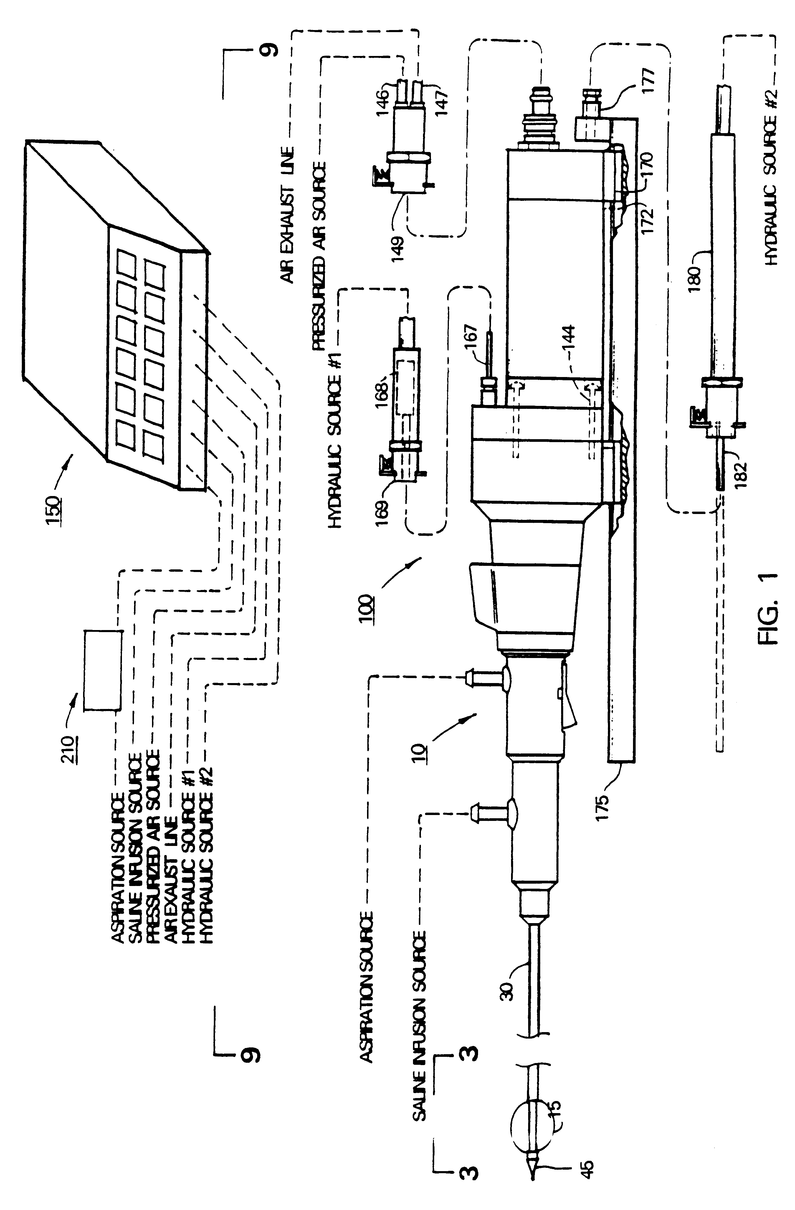
By way of example, FIG. 1 illustrates a excisional needle biopsy device or instrument 5 in accordance with the present invention. Instrument 5 is adapted for use with a conventional stereotactic needle apparatus, for example, a Model DSM unit (Digital Spot Mammography) made by Lorad, Inc. of Danbury, Mass.

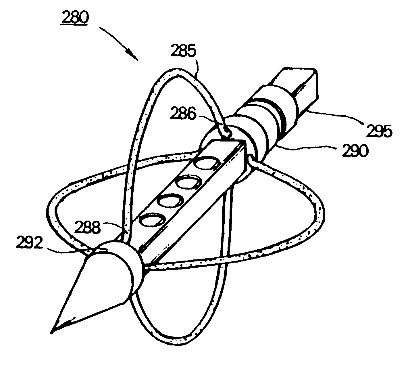
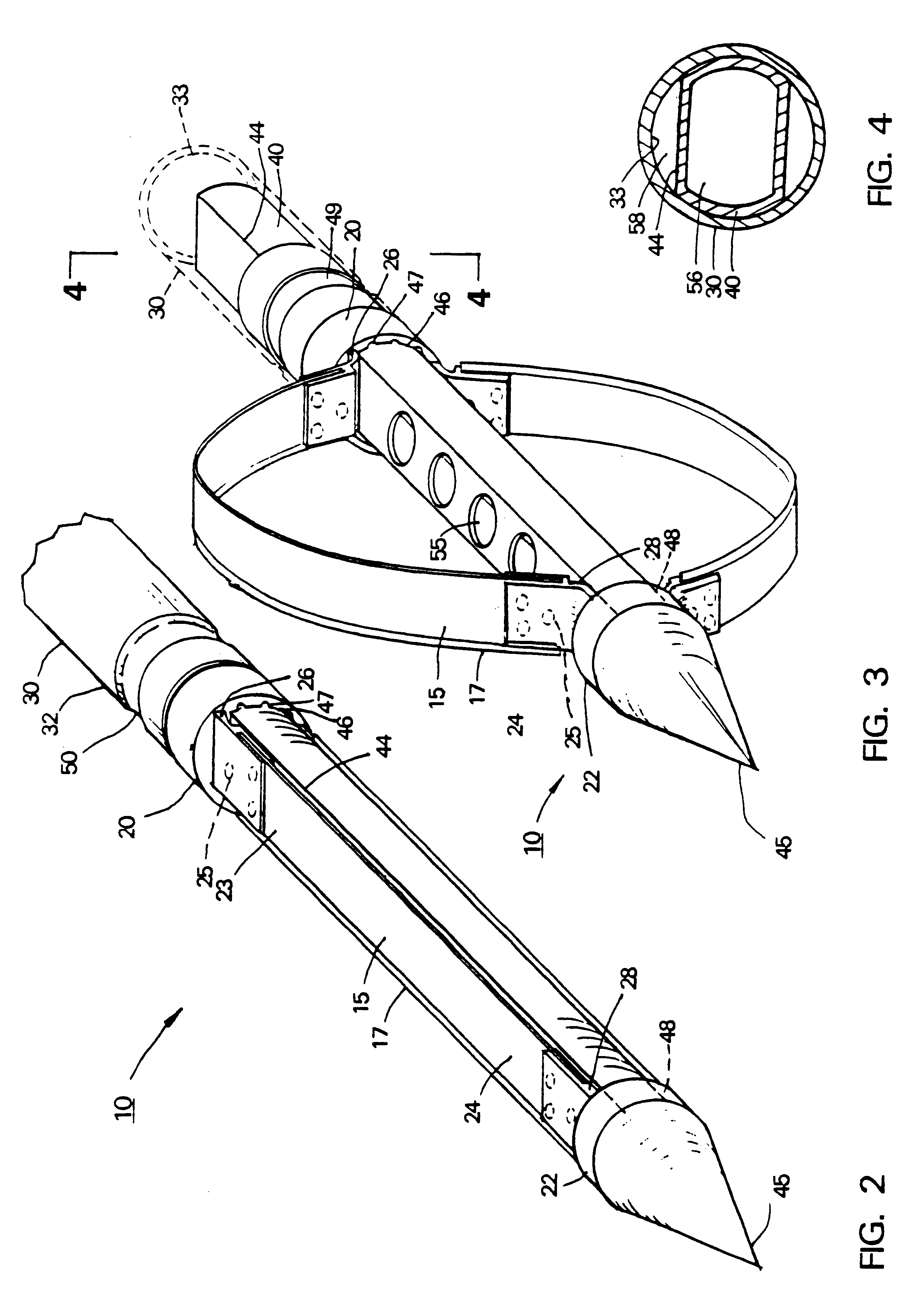
Instrument 5 includes a disposable flex-blade cutter subassembly 10 (see FIG. 1). FIGS. 2-3 are axionometric views of the distal tip of flex-blade cutter 10 in alternative positions. In FIG. 2. flex-blade cutter 10 is illustrated with flexible blades or flexors 15 in a contracted or first position as when cutter 10 is configured for axial piercing into a patient's body. In FIG. 3, cutter 10 is depicted with flexors 15 in an expanded or second position as when the cutter is rotating at high speed to excise and extract tissue from a toroidal-shaped region. Flexors 15 are fabricated from any suitable material such as stainless steel ribbon having a thickness ranging e.g. from 0.002" t...


PUM


Abstract

An excisional biopsy needle assembly for use in combination with a stereotactic platform for excising tissue from the interior of the body. The biopsy needle includes a rotatable flexible blade that is capable of transforming between a contracted configuration for piercing into the interior of the body and an expanded configuration in which the flexible blade is expanded relative to the shaft of the needle for excising tissue. The needle assembly may be mounted in a stereotactic needle for a breast biopsy procedure that digitally directs the needle tip into a lesion with the flexible blades in a contracted configuration, then expands the flexible blades to the expanded configuration and rotates the blades thus excising tissue in a region having a cross sectional dimension that is large in relation to the cross section of the needle shaft. The excised tissue is extracted from the needle tip through an aspiration channel.

Description

1. Field of the InventionThis invention relates to surgical instrumentation and more particularly to an instrument for use with an image-directed apparatus to perform an excisional biopsy, for example, of a lesion in a patient's breast. The invention and associated method relate to a manner in which biopsy tissue may be excised and extracted through a minimally invasive incision.2. Description of Prior ArtIt often is necessary to biopsy tissue from the interior of the body for diagnostic purposes. For example, when a physician discovers a suspicious breast lesion, a biopsy typically is performed on a small tissue sample to determine whether to remove the lesion or to perform another more invasive surgery. It remains common practice to use an open surgical approach to recover a tissue sample for biopsy purposes. Such an open excisional biopsy causes trauma and may leave undesirable scarring.In many cases, a needle biopsy approach is employed. A "fine needle aspiration" approach utili...

Claims


Application Information

Patent Timeline
Patent Type & Authority Patents(United States)
IPC IPC(8): A61B19/00A61B10/00A61B17/22A61B10/02
CPCA61B10/0266A61B19/20A61B10/0275A61B17/320725A61B10/0283A61B90/10
Inventor KIETURAKIS, MACIEJ J.
Owner KIETURAKIS MACIEJ J
Why Eureka
Try Eureka