Access and cannulation device and method for rapidly placing same and for rapidly closing same in minimally invasive surgery

a cannulation device and minimally invasive surgery technology, applied in the field of access and cannulation devices and methods for rapid placement and rapid closing of same in minimally invasive surgery, can solve the problems of obviating the need, requiring a considerable incision or port to conduct the operation, and difficult manipulation, so as to reduce or eliminate the problem of requiring a large incision or port, the type of access and cannulation can be easily selected, and the effect of reducing or eliminating

Inactive Publication Date: 2002-12-03
WESTSHAW CO LTD +3
View PDF74 Cites 288 Cited by
  • Summary
  • Abstract
  • Description
  • Claims
  • Application Information

AI Technical Summary

Benefits of technology

The cannulation device of the present invention can be placed and configured on the vessel or organ during minimally invasive surgery, and can be closed after use from outside the patient. By selecting from a plurality of different sleeves, the type of access and cannulation can be easily selected. For example, single or multiple cannulas can be accommodated by the device of the present invention.
Because the device is easily set and closed small sites can be accommodated, and virtually no blood will seep into the surgical field. Furthermore, since no hand suturing is required to close the site all of the above-discussed problems associated with suturing and hand suturing in particular, are eliminated. The mounting tines of the mounting ring anchor the ring in a manner that will not endanger tissue, especially fragile tissue, and thereby will reduce or eliminate the problems associated with damaging tissue due to placing and sealing an access port. The sleeve is flexible and isolates the cannula from the malleable ring and thus relieves stress that otherwise might be transferred from the cannula to the tissue.

Problems solved by technology

Many operations still require a considerable incision or port to conduct the operation, and many of these operations require hand suturing to perform the procedures.
This necessitates technologies which obviate the need for suturing and other fine motor tasks which require direct visualization and hand suturing since the suturing will be difficult with the limited access associated with minimally invasive surgery.
Still further, general access restrictions make manipulations difficult and blood in the surgical field is also a cause for concern.
Furthermore, blood leakage may cause a problem.
One problem with such devices is caused when the cannula is removed and the only way to re-establish patency of the vessels is to hand suture them closed.
This method is not well suited to the small incisions used in minimally invasive surgery since hand suturing requires more room to manipulate tools and also to enable the surgeon to visualize what he or she is doing.
In addition, older patients may have frail tissue which result in trouble pulling purse-string sutures tight.
In some cases, the sutures will cut the tissue causing further leaks which must be patched and sutured again.
As is especially the case with frail tissue, such anchoring may tend to tear the tissue.
Closure of purse-string sutures causes the tissue to be gathered and bunched resulting in a loss of cross-sectional area and / or a decreased function.
Still further, in manually sutured procedures a substantial amount of tissue is required to make a port closure.
For example, when the atrium of the heart or the aorta is cannulated with a purse-string suture the tissue inside the purse string is lost when the suture is tied.
This causes stretching and deformity of the heart or aorta when the purse-string is tied, sometimes leading to tears in the tissue and / or leaks.
However, the felt is secured with sutures and more holes and tearing can occur at the patch site.
However, these same drugs make it difficult to get large incisions in the vessel walls and heart muscle or other internal organs to seal.
In such situations sealing the tissue to the cannulation incision is very difficult.

Method used


Image

  • Access and cannulation device and method for rapidly placing same and for rapidly closing same in minimally invasive surgery
  • Access and cannulation device and method for rapidly placing same and for rapidly closing same in minimally invasive surgery
  • Access and cannulation device and method for rapidly placing same and for rapidly closing same in minimally invasive surgery

Examples


Embodiment Construction

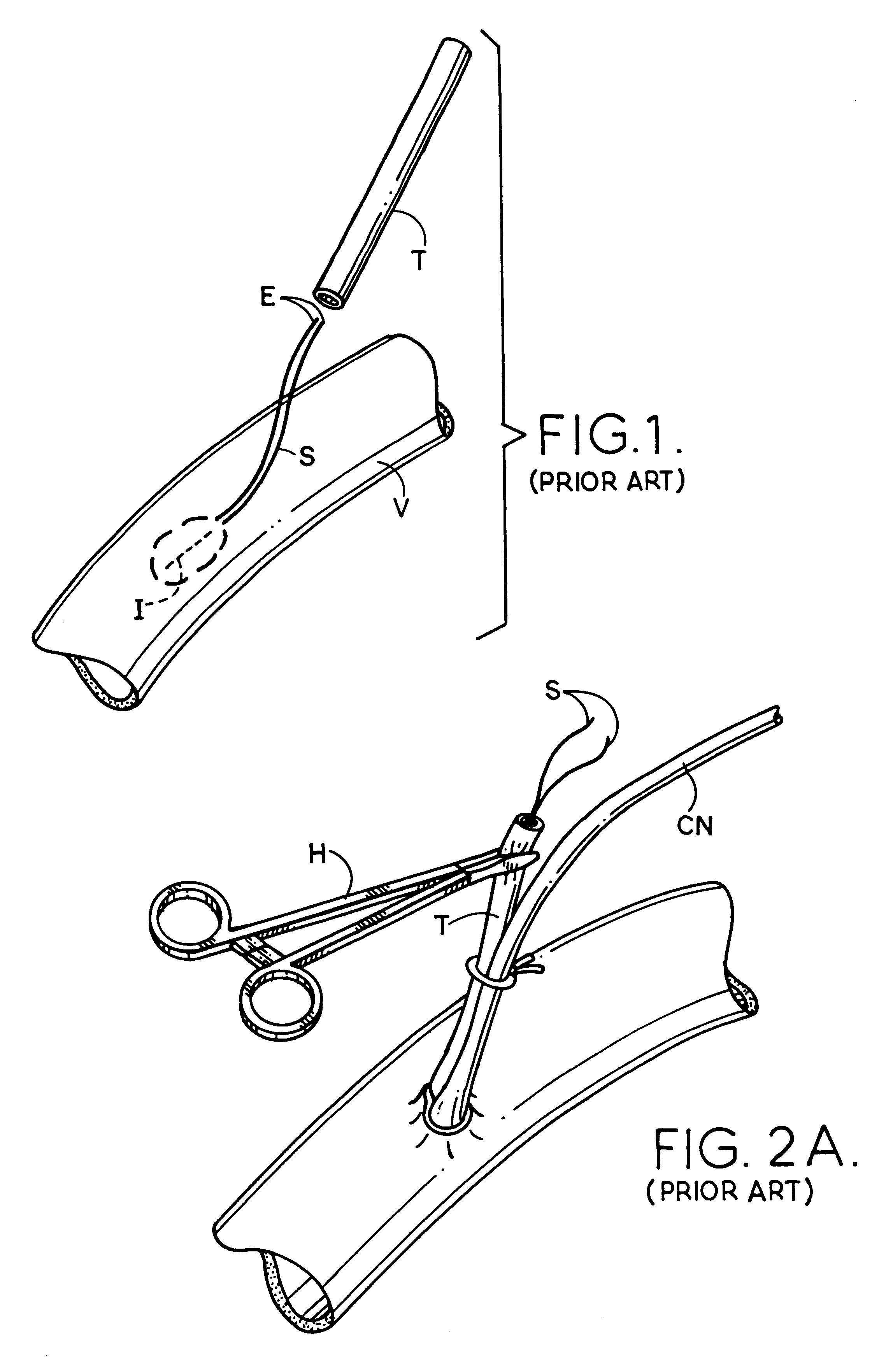
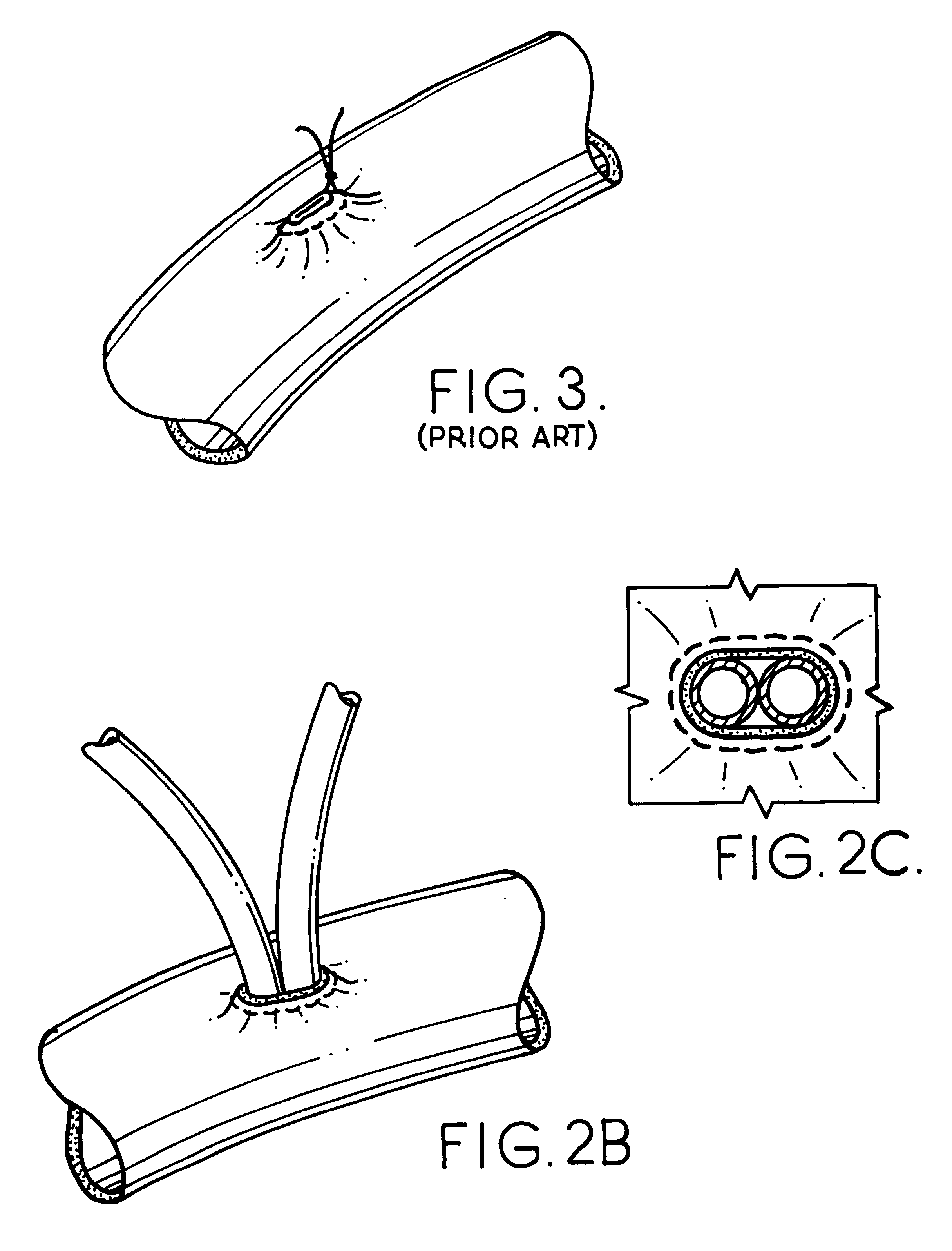
By way of background, the prior art method of cannulating a vessel will be described so the present invention will be appreciated. Referring to FIGS. 1-5, it can be understood that current surgical procedures employ a purse-string suture to tighten the tissue around a cannula tip. The procedure will be described below.

An incision I will be made in the wall of a vessel V. Before incision I is made, a running monofilament or braided suture S is made in a circle around an area that is approximately one to three cm in diameter. Ends E of sutures S are then fed through a piece of tubing T which is used as a garrote. Incision I is then made in the center of the circle of the stitches and the tip of cannula CN is inserted through the incision into the interior or vessel V.

The purse-string sutures are then pulled up tight while pushing down on tube T so the tissue is gathered and tightened down on cannula CN adjacent to the tip thereof. A hemostat H is then clamped onto the tube T which in ...


PUM


Abstract

An access and cannulation device includes a mounting element such as the anastomosis mounting element disclosed in U.S. application Ser. No. 08/714,615 (now U.S. Pat. No. 5,868,763) and U.S. application Ser. No. 09/200,796. The device is used to provide access therethrough and via an incision to the interior of a hollow anatomical structure such as a vessel, an organ or the like during surgery, especially minimally invasive surgery. It includes a flexible sleeve with a suture therein for closing the sleeve and locating edges of the structure adjacent to the incision in position for proper healing after completion of a surgical procedure. A tool is also disclosed for manipulating the device to configure it for the surgery and to close it after completion of the surgery whereby the edges of the wall of the structure adjacent to an incision are approximated to promote proper healing.

Description

The present invention relates to the general art of surgery, and to the particular field of minimally invasive surgery.As discussed in the incorporated documents, there is current interest in surgical techniques that are less invasive than previous techniques. This current interest has engendered interest in many areas that were previously abandoned including coronary fastening and valve placement among other areas that will occur to those skilled in the surgical art.Furthermore, many of these procedures include use of cardiopulmonary bypass for their execution. Cardiopulmonary bypass removes the venous blood from the heart and returns it to the circulation system of the patient through the patient's aorta or through one of its branches after it has been oxygenated. This bypass procedure makes it possible to remove the heart from the circulation system in order to perform corrections on the heart and also makes it possible to arrest the heart so that there is a non-moving field that...

Claims


Application Information

Patent Timeline
Patent Type & Authority Patents(United States)
IPC IPC(8): A61B17/115A61B17/11A61B17/03
CPCA61B17/11A61B17/1152A61B2017/1135A61B2017/1139A61B2017/3466
Inventor SPENCE, PAUL A.WILLIAMSON, IV, WARREN P.CHRISTAKIS, GEORGE
Owner WESTSHAW CO LTD
Why Eureka
Try Eureka