Percutaneous tissue dissection

a percutaneous tissue and tissue technology, applied in the field of surgical methods, can solve the problems of disc herniation or protrusion, traumatic dissection process, and various problems, and achieve the effect of less trauma, long incision and traumatic dissection process

Inactive Publication Date: 2005-01-04
SUDDABY LOUBERT
View PDF3 Cites 20 Cited by
  • Summary
  • Abstract
  • Description
  • Claims
  • Application Information

AI Technical Summary

Benefits of technology

An object of this invention is to provide a less traumatic method of fusing spinal elements whereby a lengthy incision and traumatic dissection process is not required. Utilizing the process described, both instrumented (pedicle screw) and non-instrumented fusions can be accomplished percutaneously with the assistance of an endoscope to provide light and visualization.

Problems solved by technology

As a result, the stresses acting upon the human backbone (or “vertebral column”), are unique in many senses, and result in a variety of problems or disease states that are peculiar to the human species.
Unfortunately, the repetitive forces acting on these intervertebral discs during repetitive day-to-day activities of bending, lifting and twisting cause them to break down or degenerate over time.
Disruption of a disc's internal architecture leads to bulging, herniation or protrusion of pieces of the disc and eventual disc space collapse.
In addition, loss of disc space height relaxes tension on the longitudinal spinal ligaments, thereby contributing to varying degrees of spinal instability such as spinal curvature or lithesis.
The process utilized is very traumatic to the muscle tissues and frequently results in considerable blood loss.
A long unsightly midline scar is the long-term consequence of such a spinal operation.

Method used


Image

  • Percutaneous tissue dissection
  • Percutaneous tissue dissection
  • Percutaneous tissue dissection

Examples


Embodiment Construction

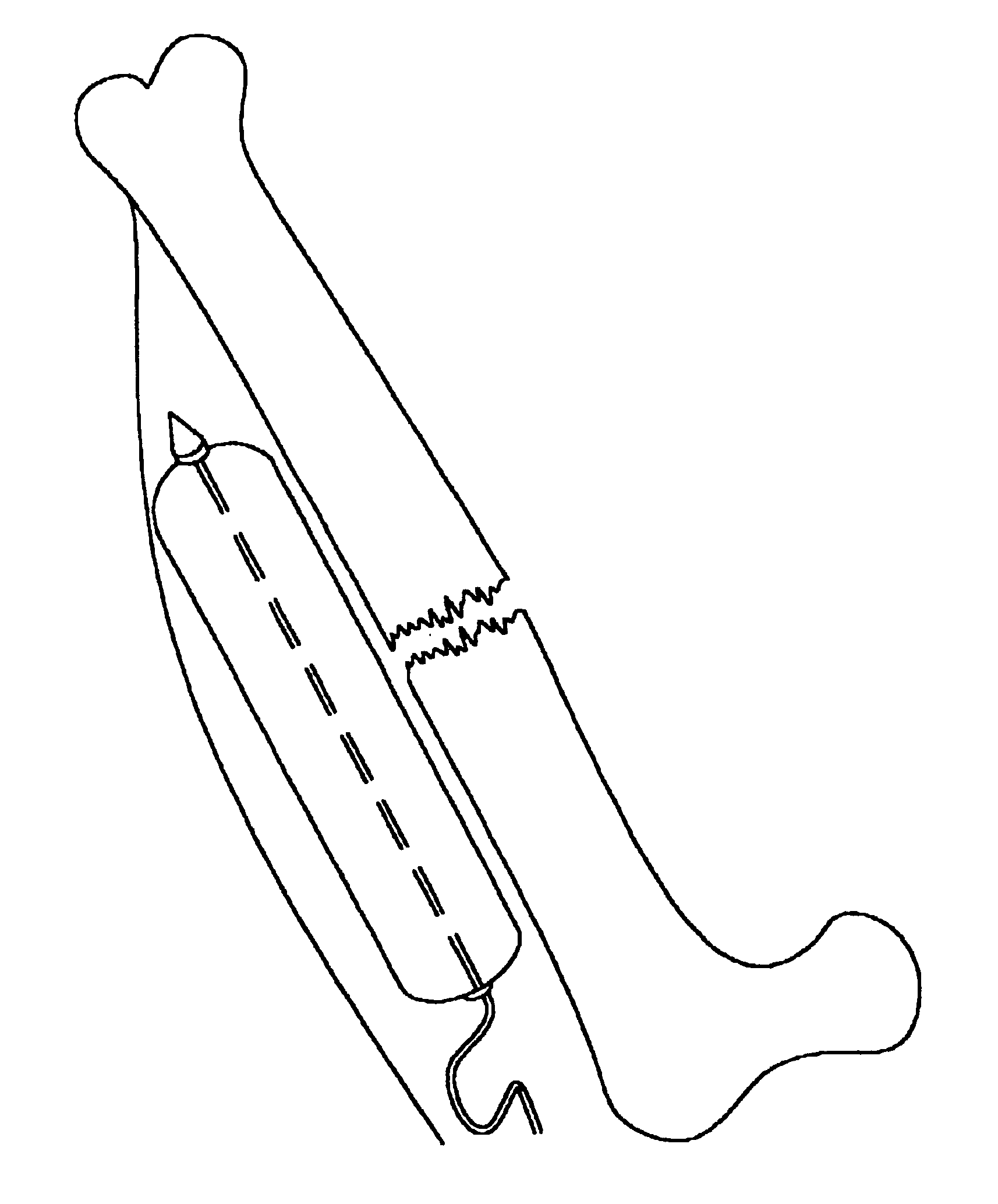
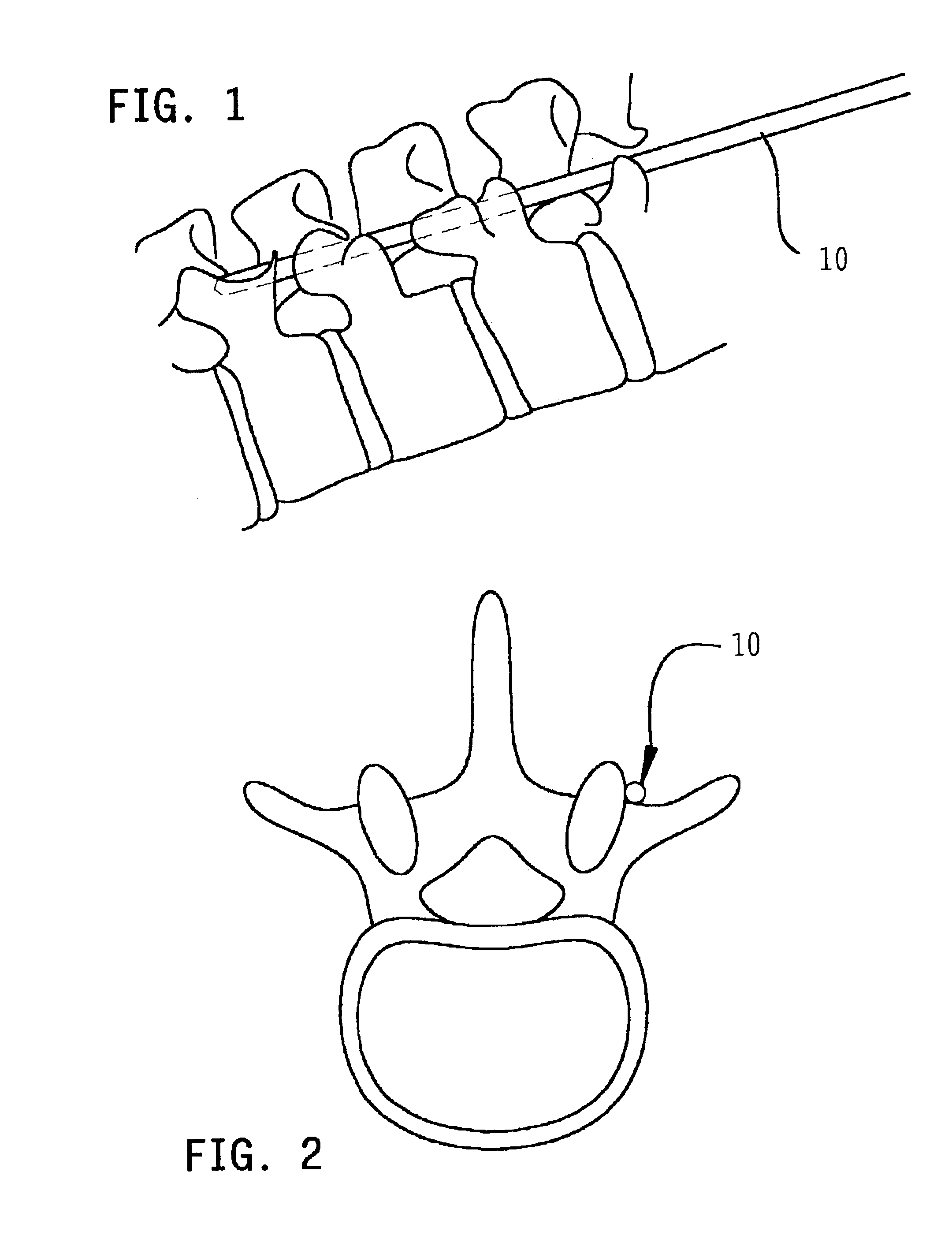
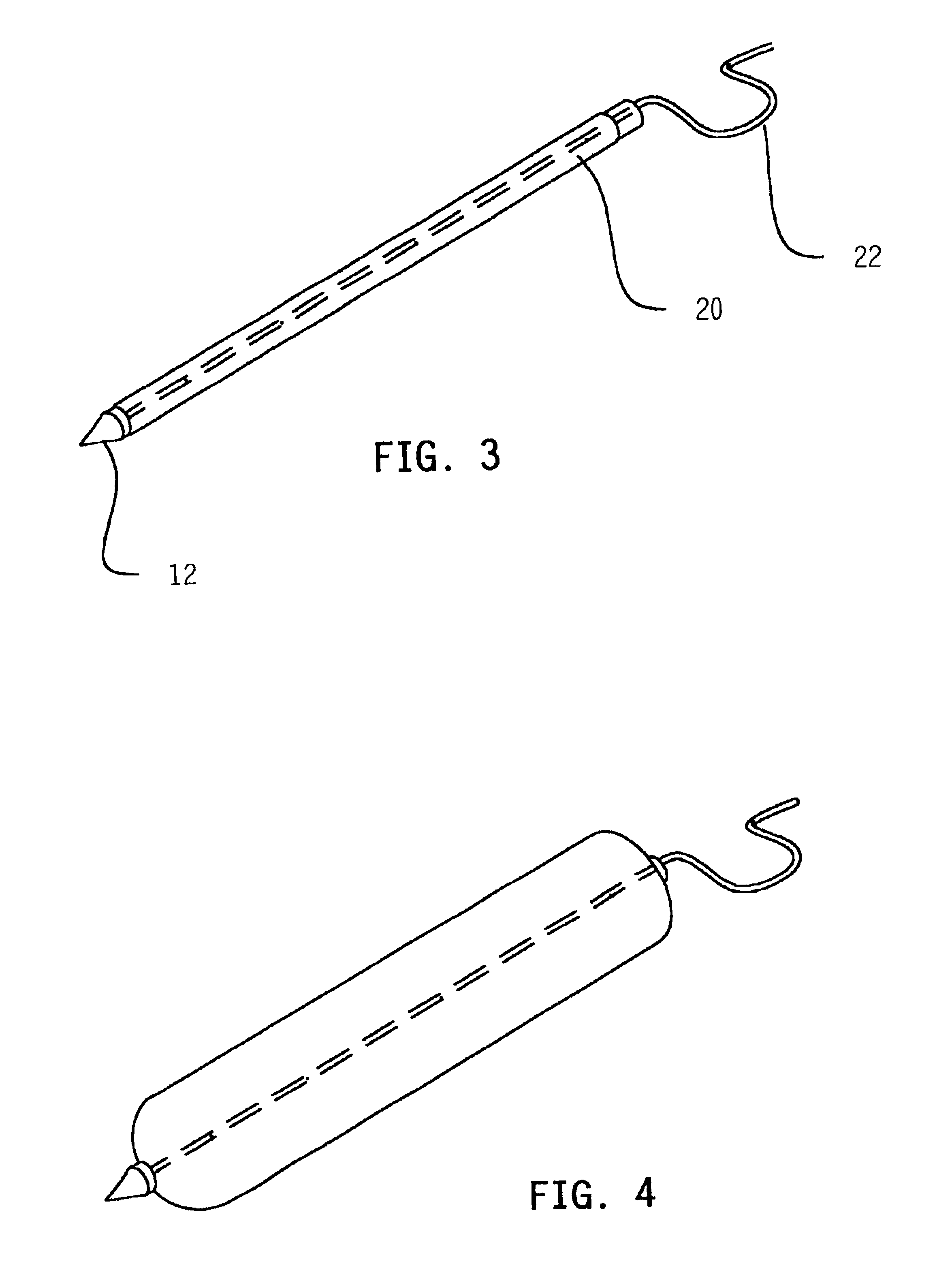
A malleable metal cannula 10 having an internal stylet 12 is first inserted into the body percutaneously (FIG. 1) and is guided fluoroscopically until the cannula lies along either the lamina of the transverse processes or the spinal elements to be fused (FIG. 2). The stylet is then removed, leaving the cannula in place. A generally cylindrical inflatable balloon 20 (FIG. 3), having an inflatable length approximating the distance between the vertebrae to be fused, is then inserted through the cannula.

The cannula 10 is then partially withdrawn, leaving the balloon in place along the spinal elements (FIG. 5). Next, the cylindrical balloon 20 is inflated (FIGS. 4, 6) by passing air or gas under pressure to the balloon via conduit 22. The expansion of the balloon elevates and dissects the muscle from either the lamina or the transverse processes, depending on the desired site for fusion. Once the muscle has been sufficiently separated from the bone, the balloon 20 is deflated and slid o...


PUM


Abstract

Tissue is retracted from a bone during spinal and other procedures by inserting an elongate balloon into a cannula, and guiding the cannula into position along between the tissue and the bone surface. The cannula is then partially withdrawn, and the balloon is inflated to retract the tissues. The method can be performed percutaneously, with minimal tissue damage.

Description

BACKGROUND OF THE INVENTIONThis invention relates to a surgical method, specifically to a balloon-assisted method of achieving a percutaneous posterior or posterolateral spinal fusion, or percutaneous surgical repair of bone fractures.Of all animals having a backbone, human beings are the only creatures who remain upright for significant periods of time. From an evolutionary standpoint, this erect posture has conferred a number of strategic benefits, not the least of which is freeing the upper limbs for purposes other than locomotion. From an anthropologic standpoint, it is also evident that this unique evolutionary adaptation is a relatively recent change and as such has not benefitted from natural selection as much as have backbones held in the horizontal attitude. As a result, the stresses acting upon the human backbone (or “vertebral column”), are unique in many senses, and result in a variety of problems or disease states that are peculiar to the human species.The human vertebr...

Claims


Application Information

Patent Timeline
Patent Type & Authority Patents(United States)
IPC IPC(8): A61B17/02A61B17/32
CPCA61B17/0218A61B2017/320048
Inventor SUDDABY, LOUBERT
Owner SUDDABY LOUBERT
Why Eureka
Try Eureka