
Apparatus and method for automatically sensing threshold histogram with differentiation of sinus from ectopic beats

a technology of automatic sensing and threshold histogram, which is applied in the field of programmable cardiac stimulation devices, can solve the problems of inability to accurately program these parameters, time-consuming, and difficult to achieve, and achieves the goal of detecting the signal amplitude of egm monitors in clinical settings. not well correlated

Inactive Publication Date: 2005-03-08
PACESETTER INC
View PDF19 Cites 38 Cited by
  • Summary
  • Abstract
  • Description
  • Claims
  • Application Information

AI Technical Summary

Benefits of technology

Still another feature of the present invention is to provide a historical record of event sensing thresholds and timing relationships to be available to a medical practitioner during patient follow-up visits. As used herein, an event sensing threshold is the highest numerical sensitivity setting at which an event is repeatedly sensed. Further, it would be advantageous to store a history of detected events, both sinus and non-sinus distinguished by sensing threshold and timing relationships, to be available for diagnostic procedures such that optimal pacing therapy can be delivered.
The present invention thus clearly accomplishes several objectives, among which are the following: 1) automatic monitoring of sensing threshold and adjustment of the sensitivity settings that can be accomplished, in one embodiment, without interrupting the function of a primary sense amplifier integral to the timing operations of the stimulation device; and 2) it provides a histogram record containing sensing thresholds of P-waves, PACs, and the coupling interval for conducted PACs.

Problems solved by technology

The programming of these parameters can be inaccurate and time consuming, and requires highly-skilled medical expertise to accomplish.
An added complication to the process of manually programming atrial or ventricular sensitivity is that signal amplitudes observed on EGM monitors in a clinical setting do not correlate well with the signal amplitudes sensed by the implanted pacemaker or ICD after the signal has been processed by internal circuitry which includes various filters and amplifiers.
Attempts to automate programming of sensitivity have not been completely successful, in part because of the natural fluctuation in the amplitudes of myopotentials and the incidence of noise associated with skeletal muscle depolarizations in unipolar systems and fluctuations in the amplitude of the intrinsic beats with marked differences between the sinus P wave and conducted ORS complex with respect to ectopic beats which cannot be assessed at the time of implantation or follow-up because they are not present at those times in both the unipolar and bipolar sensing configurations.
Selecting appropriate atrial sensitivity can be particularly challenging due to the low amplitude of atrial events, the small differences between sinus atrial event amplitudes and ectopic atrial event amplitudes, and the desire to sense and accurately detect low level signals associated with atrial fibrillation, flutter or ectopic foci.
The importance of correctly programming atrial sensitivity, however, cannot be over emphasized.
An incorrect high atrial sensitivity would predispose to over-sensing and inappropriate mode switching.
An overly low atrial sensitivity would prevent the correct detection of atrial fibrillation and would result in inappropriate ventricular pacing during atrial fibrillation associated with sensing and triggering on the intermittent atrial signals of sufficient amplitude, with serious hemodynamic consequences.
In addition, under-sensing of premature atrial contractions (PACs) and premature ventricular contractions (PVCs) can result in arrhythmia induction induced by competition.
The problem of automatically and accurately sensing P-waves and R-waves is even more pronounced when using an “A-V cross-chamber” electrode configuration, that is, an electrode configuration in which the stimulation device senses cardiac signals between an atrial tip electrode and a ventricular tip electrode, and stimulates each chamber in a unipolar fashion from the respective electrode to the housing (i.e., typically referred to as the case electrode).
While unipolar sensing configurations are more susceptible to extraneous noise, bipolar sensing configurations are also susceptible to problems of oversensing depending on electrode position and spacing.
The implantable device, if it is to perform its intended function, must correctly detect a sinus atrial depolarization (P-wave), a sinus-induced ventricular depolarization (R-wave), and it must not incorrectly detect a PVC or a PAC as a sinus P-wave or R-wave, or vice versa, and thus inappropriately adjust pacing parameters.
While it is well known that various blanking schemes may be used to block or blank out unwanted inappropriate physiologic signals such as far-field signals or retrograde P-waves by using different blanking intervals (i.e., PVARP, automatic PVARP extension, PVAB, etc.), and thereby prevent these far-field signals or retrograde P-waves from being falsely sensed as P-waves, such blanking schemes (based solely on timing considerations) have proven less than satisfactory because legitimate (anterograde) P-waves and PACs that need to be sensed, may and do occur during these blanking intervals.
An important limitation of this approach of adding a second physiological sensor is the added hardware required to implant in the patient and the additional circuitry required to interpret more than one physiological signal and relate them.
A major limitation of these methods is that in order to make automatic adjustments to sensitivity, a stable rhythm is required.
Thus, frequent premature beats or irregular rates might inhibit automatic sensing threshold tests and sensitivity adjustment and lead to prolonged periods of inappropriate sensing and even delivery of electrotherapies with adverse effects based on erroneous detections.
A further limitation of conventional devices is that the primary sensing circuitry is automatically adjusted such that only a target event of interest is detected, for example P-waves but not PACs.
Therefore, there is still an unsatisfied need to automatically monitor sensing thresholds of various cardiac events, such as P-waves and PACs, R-waves and PVCs, and to accurately detect and discriminate these events without interruption of the normal operation of the pacemaker.

Method used


Image

  • Apparatus and method for automatically sensing threshold histogram with differentiation of sinus from ectopic beats
  • Apparatus and method for automatically sensing threshold histogram with differentiation of sinus from ectopic beats
  • Apparatus and method for automatically sensing threshold histogram with differentiation of sinus from ectopic beats

Examples


Embodiment Construction

The following description is of a best mode presently contemplated for practicing the invention. This description is not to be taken in a limiting sense but is made merely for the purpose of describing the general principles of the invention. The scope of the invention should be ascertained with reference to the issued claims. In the description of the invention that follows, like numerals or reference designators will be used to refer to like parts or elements throughout.

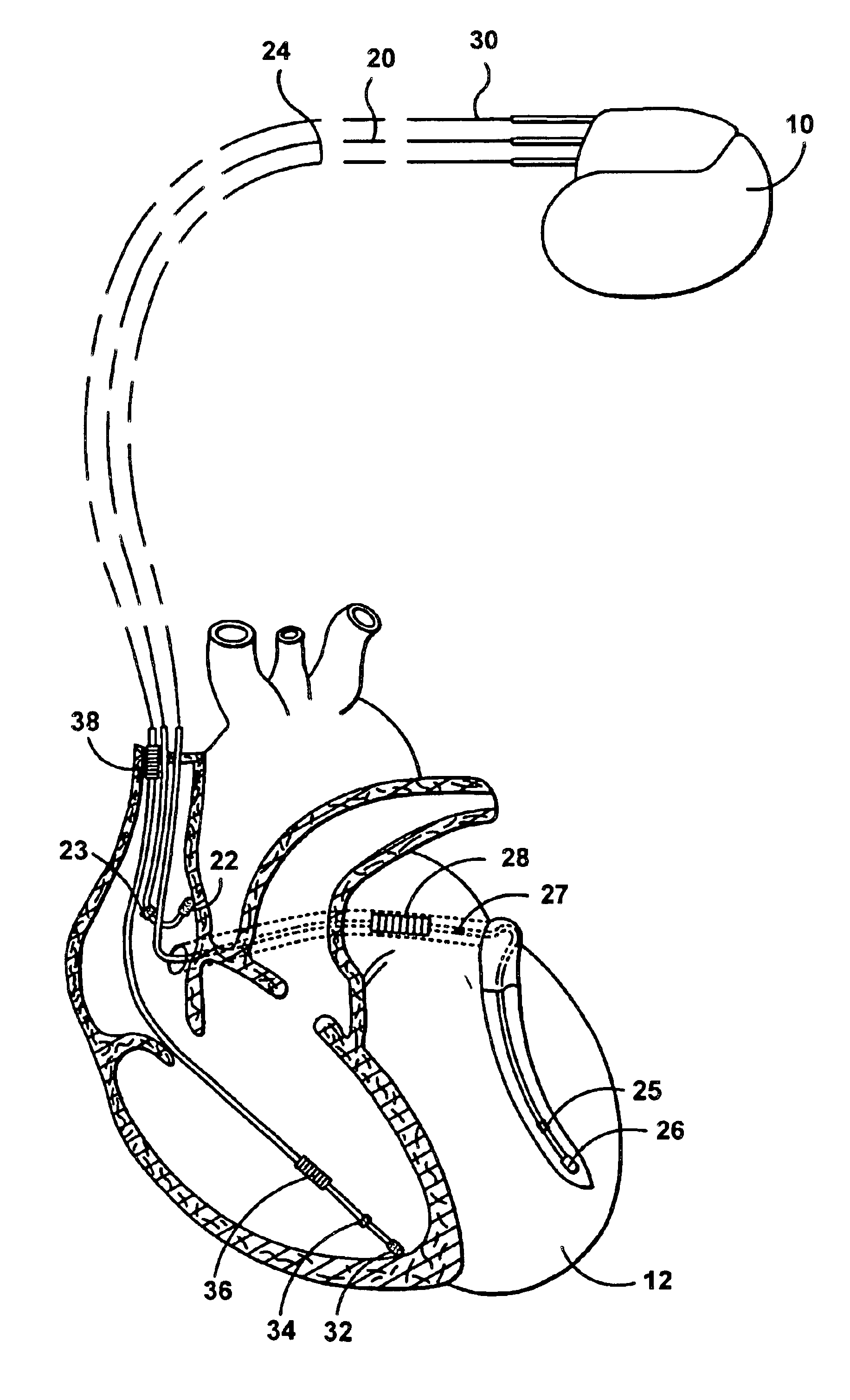
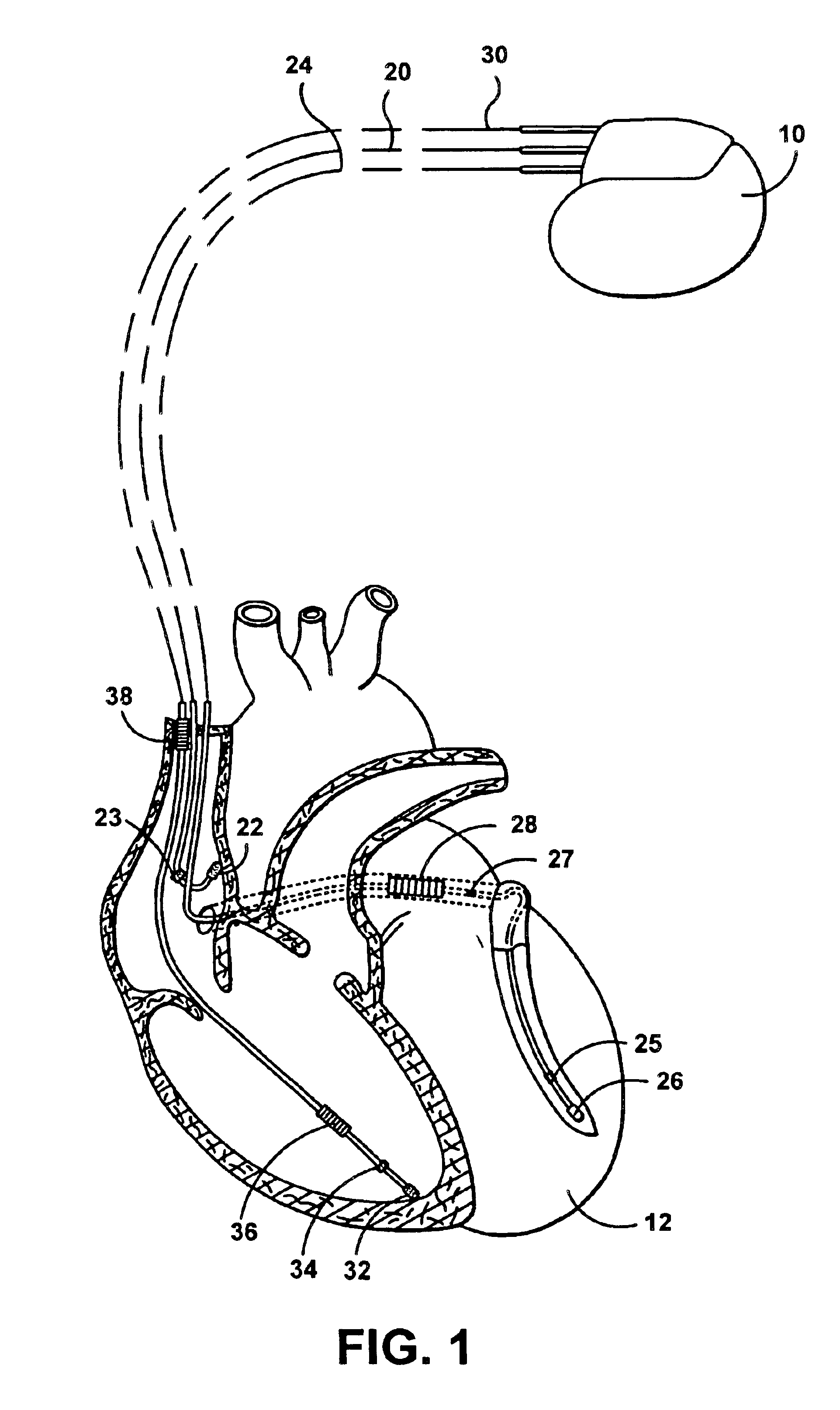
FIG. 1 illustrates a stimulation device 10 in electrical communication with a patient's heart 12 by way of three leads 20, 24 and 30 suitable for delivering multi-chamber stimulation and shock therapy. To sense atrial cardiac signals and to provide right atrial chamber stimulation therapy, the stimulation device 10 is coupled to an implantable right atrial lead 20 having at least an atrial tip electrode 22, which typically is implanted in the patient's right atrial appendage. The right atrial lead 20 may also have ...


PUM


Abstract

An implantable programmable cardiac stimulation device and associated method for differentiating between normal sinus events and ectopic beats. The stimulation device monitors the sensing thresholds of sinus and non-sinus cardiac events, and stores a history of these sensing thresholds along with temporal data for accurate event detection. The stimulation device further provides accurate and appropriate detection of sensed events including P-waves, non-conducted PACs, and conducted PACs and thus verifies correct detection of PVCs and R-waves. Furthermore, the present invention provides a history record of ectopic events, distinguished by sensing thresholds and timing intervals, giving a valuable diagnostic tool to the physician in optimizing rhythm management therapy. In addition, the stimulation device allows the sensitivity threshold to be set based on a single cardiac cycle and past history.

Description

FIELD OF THE INVENTIONThis invention relates generally to a programmable cardiac stimulation device for the purpose of differentiating between normal sinus events and ectopic beats. More specifically, the present invention is directed to an implantable stimulation device and associated method for automatically monitoring sensing thresholds of sinus and non-sinus cardiac events, and storing a history of these sensing thresholds along with temporal data for accurate event detection.BACKGROUND OF THE INVENTIONConventional pacemakers and ICDs require manual programming of numerous programmable parameters, including but not limited to: atrial sensitivity, ventricular sensitivity, post-ventricular atrial refractory period (PVARP), post-ventricular atrial blanking period (PVAB), and other parameters such as ventricular refractory period, ventricular output, atrial output, choice of pacing mode, upper rate limit, base rate, sleep rate, sensor slope, sensor threshold, and so forth. The progr...

Claims


Application Information

Patent Timeline
IPC IPC(8): A61B5/0452A61N1/37
CPCA61N1/3704
Inventor LEVINE, PAUL A.
Owner PACESETTER INC
Learn More