Methods of precisely forming bone tunnels in cruciate ligament reconstruction of the knee

a technology of cruciate ligament and bone tunnel, which is applied in the field of surgical instruments, can solve the problems of increasing patient discomfort, increasing invasiveness and trauma, and prolonging hospitalization and rehabilitation tim

Inactive Publication Date: 2005-04-12
MCGUIRE DAVID A +1
View PDF6 Cites 70 Cited by
  • Summary
  • Abstract
  • Description
  • Claims
  • Application Information

AI Technical Summary

Benefits of technology

Another object of the present invention is to provide a guide for precisely positioning a guide wire on a bone surface in relation to a reference edge on the bone surface engaged by the guide.
A further object of the present invention is to provide a femoral guide for precisely positioning a guide wire in the femur such that a bone tunnel formed along the guide wire has a longitudinal axis substantially aligned with the anatomic center of attachment of the cruciate ligament.
A still further object of the present invention is to provide a femoral guide for precisely positioning a guide wire on the femur such that a bone tunnel formed along the guide wire has a longitudinal axis approximately 6-7 millimeters anterior to a posterior edge or “over-the-top ridge” of a notch on the femoral condyle.
Furthermore, it is an object of the present invention to provide a method of precisely forming bone tunnels in a joint of the body endoscopically with a guide inserted at the joint from a portal of minimal size to position a guide wire on a surface of a bone of the joint such that the guide wire is a predetermined distance from a reference edge on the bone surface engaged by the guide allowing the guide wire to be driven into the bone for forming a bone tunnel along the guide wire.
Some of the advantages of the present invention are that accurate, isometric positioning of tibial and femoral bone tunnels in arthroscopic cruciate ligament reconstruction is enhanced, the time required to perform arthroscopic anterior cruciate ligament reconstruction is reduced, error in forming bone tunnels in arthroscopic cruciate ligament reconstruction is minimized, femoral bone tunnels can readily be located high in a notch formed in the femur, the use of blind, or closed-end, femoral bone tunnels as well as open-end femoral bone tunnels in arthroscopic anterior cruciate ligament reconstruction is permitted, the creation of a trough and not a tunnel when forming femoral bone tunnels is avoided, a cortical margin that is neither too wide nor too narrow is insured, tunnel placement can be verified prior to tunnel formation, the femoral isometric point can be accurately identified, the need for surgeons to estimate the position of drills and reamers when forming femoral bone tunnels along guide wires is eliminated, anteromedial and anterolateral portals can be used for inserting guide wires in arthroscopic anterior cruciate ligament reconstruction, the size of portals used in arthroscopic cruciate ligament reconstruction can be minimized, the femoral guide according to the present invention is streamlined in structure, right and left knee compatible and can be sterilized for repeated use, the tip of a guide wire positioned by the femoral guide on a bone surface can be viewed endoscopically, a footprint can be created on a bone surface as guided by a guide wire positioned by the femoral guide and examined prior to forming a bone tunnel allowing adjustments to be made if necessary, the femoral guide is designed to be accommodated in portals of very small size and the configuration of the femoral guide facilitates manipulation and maneuverability at the knee joint as well as arthroscopic visualization of the knee.

Problems solved by technology

However, open surgery possesses numerous disadvantages over closed, or least invasive surgery, including increased invasiveness and trauma, prolonged hospitalization and rehabilitation times, increased patient discomfort, possible violation of capsular mechanoreceptors, dessication of articular cartilage and delayed post-surgical mobility.
Where cruciate ligament reconstruction is performed as a closed, or endoscopic, surgical procedure, the small size of the portals limits access to and maneuverability at the knee joint making it relatively more difficult to precisely place the tibial and femoral bone tunnels at sites anatomically equivalent to the cruciate ligament.
Although the guide wires are effective in guiding instruments, such as drills and reamers, utilized to form the bone tunnels, problems can arise in arthroscopic cruciate ligament reconstruction in precisely positioning or locating the guide wires.
If the guide wires are not located and inserted at sites anatomically equivalent to attachment of the cruciate ligament, the tibial and femoral bone tunnels, as guided by the guide wires, will not be properly located, and ligament reconstruction will be impaired.
In arthroscopic anterior cruciate ligament reconstruction, it is difficult to position a guide wire on the femur at a position corresponding to the anatomic center of attachment of the anterior cruciate ligament.
However, it is extremely difficult in arthroscopic anterior cruciate ligament reconstruction to position and hold the guide wire from externally of the knee such that the tip of the guide wire is optimally, isometrically positioned on the femur; and, even when properly positioned, the guide wire can slip or shift prior to being driven into the femur resulting in an improperly positioned femoral bone tunnel and impaired placement of the graft or prosthetic ligament.
Where instruments are used to help guide or hold the guide wire, the instruments themselves can slip or shift causing displacement of the guide wire; and, frequently, the use of instruments does not eliminate the need for a surgeon to estimate where to place instruments such as drills or reamers when forming the femoral bone tunnel along the guide wire.

Method used


Image

  • Methods of precisely forming bone tunnels in cruciate ligament reconstruction of the knee
  • Methods of precisely forming bone tunnels in cruciate ligament reconstruction of the knee
  • Methods of precisely forming bone tunnels in cruciate ligament reconstruction of the knee

Examples


Embodiment Construction

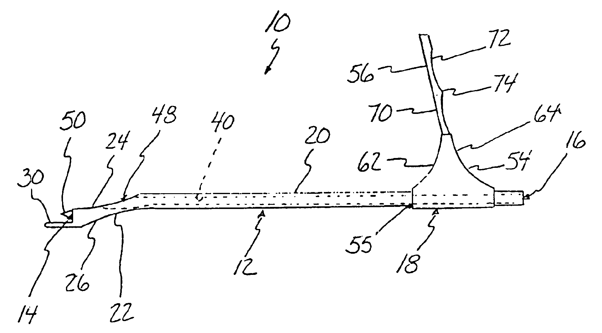
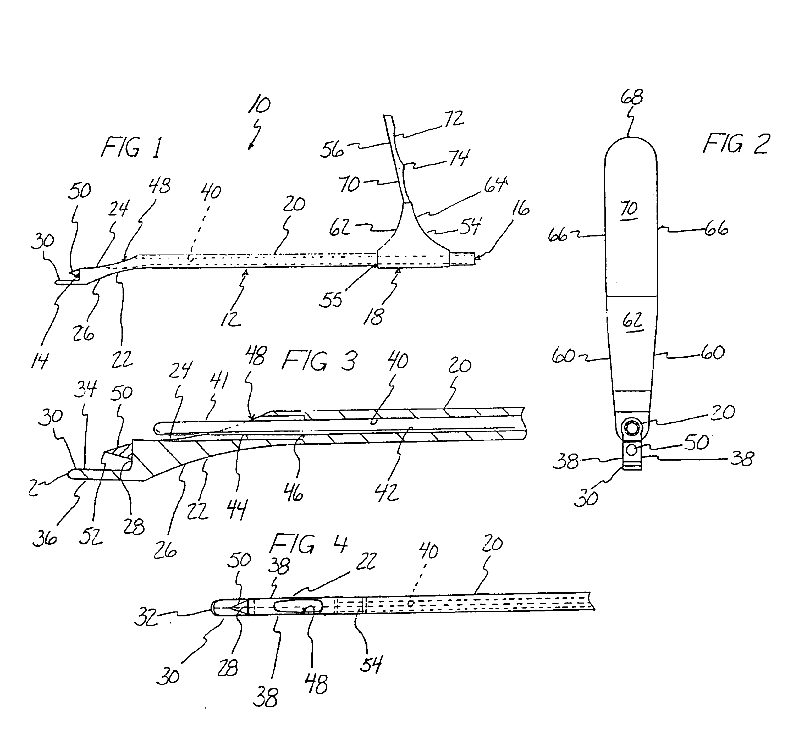
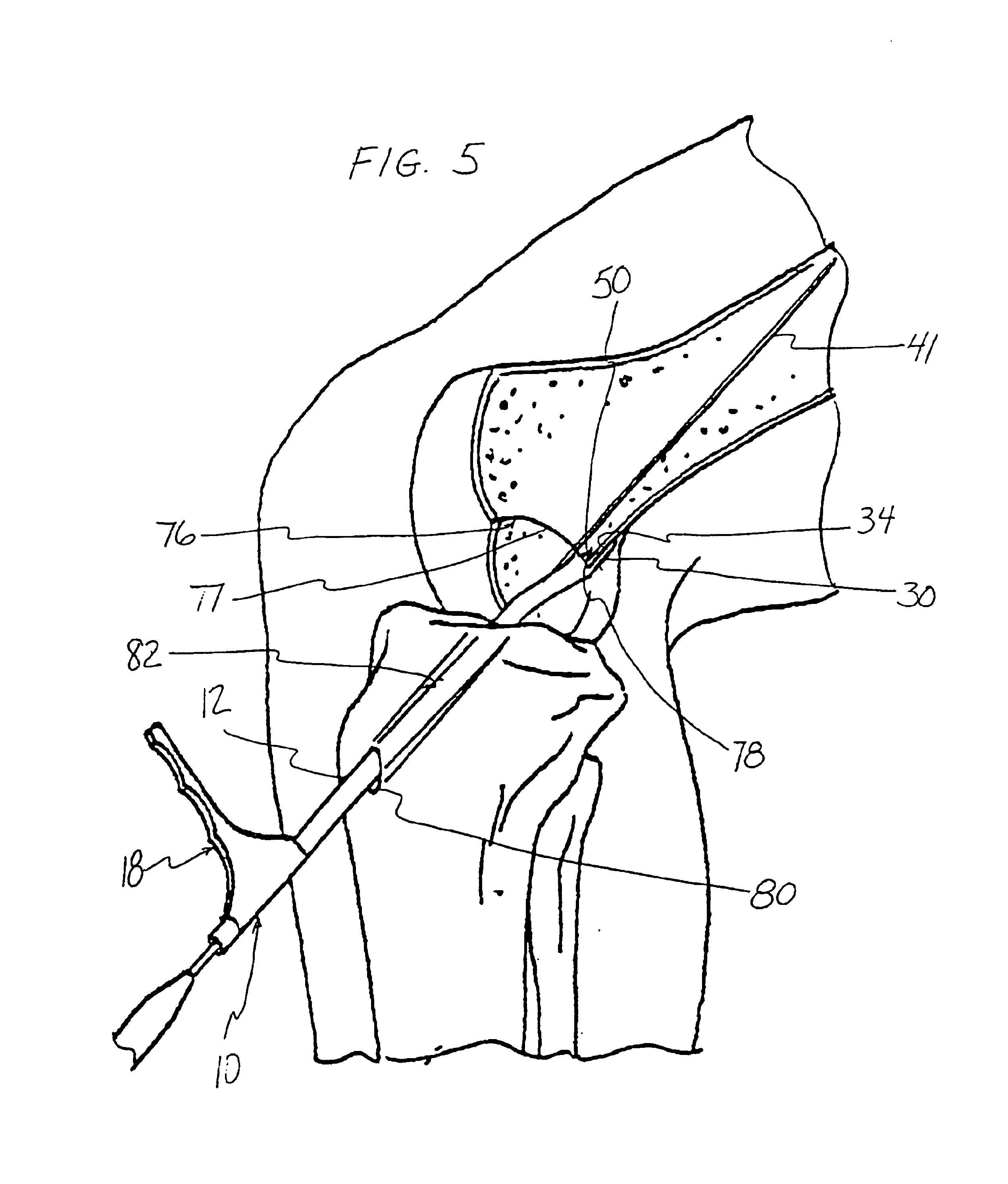
A femoral guide according to the present invention is shown at 10 in FIG. 1 and includes a body or probe 12 having a distal end 14, a proximal end 16 and a longitudinal axis and a handle 18 mounting the proximal end 16 of the body 12. The body 12 is preferably made of stainless steel and includes an elongate, cylindrical member or tube 20 terminating proximally at proximal end 16 and a tip 22 distally joined to the cylindrical member 20, the cylindrical member 20 having a longitudinal axis coaxially aligned with the longitudinal axis of the body 12. The tip 22 extends angularly, distally from the cylindrical member 20 and includes an arcuate surface 24 extending distally from a wall of the cylindrical member 20 with an inward curvature and an opposed arcuate surface 26 extending distally with an inward curvature from a wall of the cylindrical member 20. The arcuate surface 24 terminates distally at an end wall 28 at the distal end 14, the end wall 28 having a planar surface disposed...


PUM


Abstract

A femoral guide for precisely positioning a guide wire on a bone surface of the femur includes a body having a lumen for receiving a guide wire and a tongue protruding from the body for engaging an edge or reference point on the bone surface with the tongue being spaced a predetermined distance from a longitudinal axis of the lumen. The lumen includes an opening allowing a guide wire extending through the lumen to contact the bone surface at a location spaced from the edge substantially the predetermined distance with the tongue engaging the edge. A stylus on the body can be driven into the bone to secure and stabilize the femoral guide prior to driving the guide wire into the bone through the lumen. With the guide wire driven into the bone, a bone tunnel can be formed substantially concentrically or coaxially along the guide wire such that a longitudinal axis of the bone tunnel will be disposed from the edge substantially the predetermined distance. Methods of precisely forming bone tunnels include the steps of engaging an edge of a bone surface with a tongue of the femoral guide, inserting a guide wire through a lumen of the femoral guide, driving the guide wire into the bone through the lumen and forming a bone tunnel in the bone along the guide wire such that a longitudinal axis of the bone tunnel will be disposed from the edge engaged by the tongue a distance substantially equal to the distance that the tongue is disposed from a longitudinal axis of the lumen.

Description

BACKGROUND OF THE INVENTION1. Field of the InventionThe present invention pertains to surgical instruments for precisely positioning guide wires in bone allowing tunnels to be formed in the bone along the guide wires and, more particularly, to femoral guides for precisely positioning guide wires in the femur in cruciate ligament reconstruction of the knee allowing bone tunnels to be formed in the femur along the guide wires at sites anatomically equivalent to the cruciate ligament and to methods of precisely forming bone tunnels.2. Description of the Prior ArtVarious surgical procedures utilize graft or prosthetic ligaments to reconstruct natural ligaments that have been damaged by injury or disease. Where the ligaments to be reconstructed are found in joints or articulations of the body, i.e., the connections of the various surfaces of the bones in the body, graft or prosthetic ligaments are typically implanted and fixated in bones of the joint at sites anatomically equivalent to t...

Claims


Application Information

Patent Timeline
Patent Type & Authority Patents(United States)
IPC IPC(8): A61B17/17A61B17/16A61B17/88A61F2/08B25B13/00B25B13/48B25B23/00B25G1/04B25G1/00B25G3/26B25G3/00
CPCA61B17/1714A61F2/0805B25B23/0021B25G1/043B25G3/26B25B13/481A61B17/1796A61B17/1764
Inventor MCGUIRE, DAVID A.PATERSON, ROGER
Owner MCGUIRE DAVID A
Why Eureka
Try Eureka