Method and system for registering ultrasound image in three-dimensional coordinate system

a three-dimensional coordinate system and ultrasound image technology, applied in the field of medical imaging systems and methods, can solve the problems of lost mapping data and ablation locations of previous registered locations

Inactive Publication Date: 2005-05-24
BOSTON SCI SCIMED INC
View PDF35 Cites 149 Cited by
  • Summary
  • Abstract
  • Description
  • Claims
  • Application Information

AI Technical Summary

Benefits of technology

[0011]The location of at least one ultrasound transducer is determined within this first coordinate system and a second coordinate system (e.g., using a registration subsystem having one or more processors). Although not necessary, the second coordinate system is preferably fixed relative to the internal anatomical structure (e.g., by establishing the second coordinate system within the internal anatomical structure itself), so that the second coordinate system need not be modified if the internal anatomical structure moves.

Problems solved by technology

The main limitation of this method is that, because it reports the location of the transducer within the coordinate system of the 3-D image, the coordinate system will change as the position of the imaging device changes, and thus, any previously registered mapping data and ablation locations will be lost.

Method used


Image

  • Method and system for registering ultrasound image in three-dimensional coordinate system
  • Method and system for registering ultrasound image in three-dimensional coordinate system
  • Method and system for registering ultrasound image in three-dimensional coordinate system

Examples


Embodiment Construction

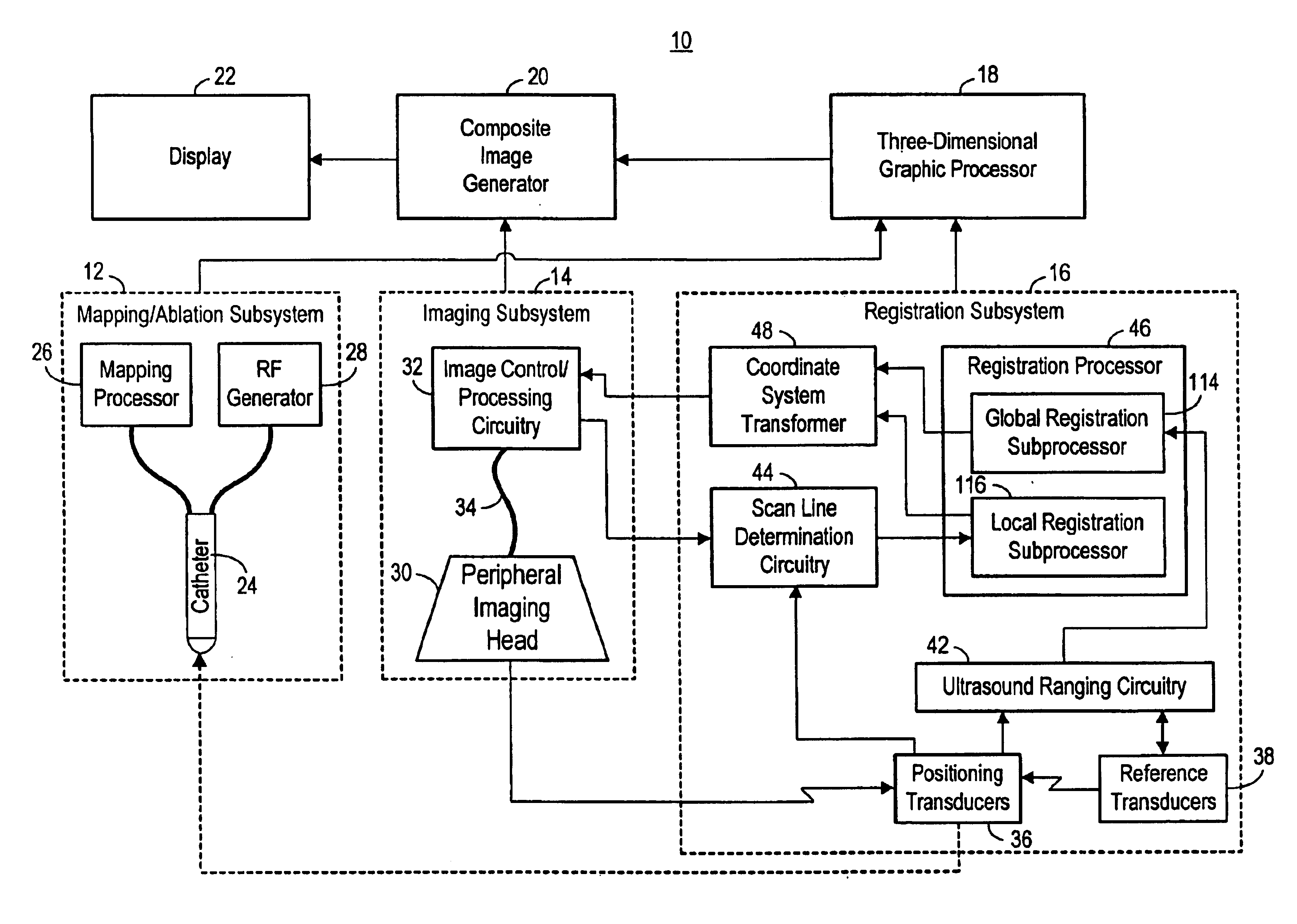
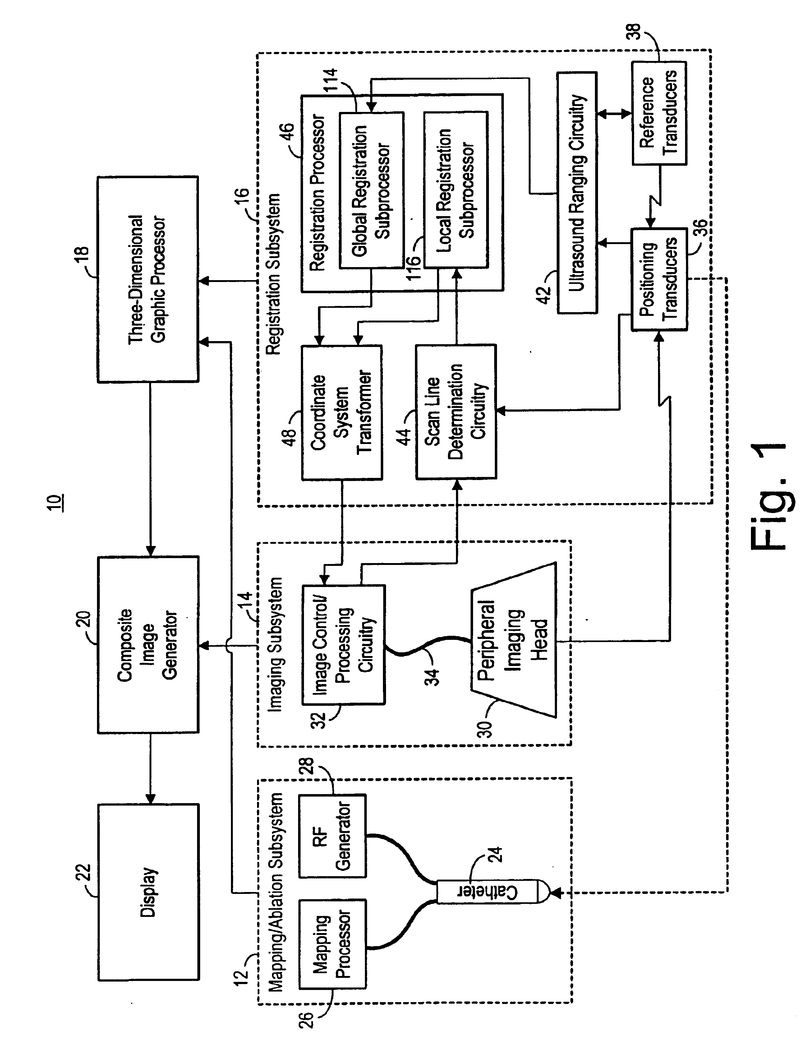
[0032]Referring to FIG. 1, an exemplary medical treatment system 10 constructed in accordance with the present inventions is shown. The treatment system 10 is particularly suited for imaging, mapping, and treating the heart. Nevertheless, it should be appreciated that it can be used for treating other internal anatomical structures, e.g., the prostrate, brain, gall bladder, uterus, esophagus and other regions in the body. The treatment system 10 generally comprises (1) a mapping / ablation subsystem 12 for mapping and ablating tissue within the heart; (2) an imaging subsystem 14 for generating image data of the heart; (3) a registration subsystem 16 for registering the image and mapping data within a 3-D graphical environment; (4) a 3-D graphical processor 18 for generating 3-D graphical data of the environment in which the imaged body tissue is contained; (5) a composite image generator 20 for generating a composite image from the registered image data and 3-D graphical data; and (6)...


PUM


Abstract

The present invention provides systems, methods, and devices for imaging an internal anatomical structure. Ultrasound image data of the anatomical structure is acquired within a first coordinate system, and graphical data (e.g., sites of interest) is acquired in a second coordinate system. The location of an ultrasound transducer within the first coordinate system and a second coordinate system is determined, and a transformation between the first and second coordinate systems is then performed based on the location of the ultrasound transducer within the first and second coordinate systems. Using this transformation, the ultrasound image data, which has previously been acquired in the first coordinate system, can be registered and displayed within the second coordinate system, along with graphical data. Or the graphical data, which has previously been acquired in the second coordinate system, can be registered and displayed within the first coordinate system, along with the image data. In this manner, the image and graphical data can be easily displayed together independent of any movement by the imaging device that acquires the image data.

Description

FIELD OF THE INVENTION[0001]The present inventions generally relate to medical imaging systems and methods, and more particularly to systems and methods for registering ultrasound images in a coordinate system.BACKGROUND OF THE INVENTION[0002]For purposes of diagnosis and treatment planning, imaging techniques are commonly used in medical procedures to view the internal anatomy of a patient's body. Although the technology for rendering real-time 3-D ultrasound images of most internal organs have been around for several years, real-time 3-D ultrasound in cardiology requires much higher frame rates for real-time acquisition and display to keep up with the beating heart or other cardiac motions. Until recently, 3-D rendered images of the heart have been generating on a non-real-time basis by sequentially acquiring two-dimensional and then using a workstation to input these images for volume rendering.[0003]Recent advancements in transducer and processing technology have enabled commerc...

Claims


Application Information

Patent Timeline
Patent Type & Authority Patents(United States)
IPC IPC(8): A61B8/08
CPCA61B8/0841A61B8/0833
Inventor WILLIS, PARKER
Owner BOSTON SCI SCIMED INC
Why Eureka
Try Eureka