Micro-invasive method for painless detection of analytes in extracellular space

a micro-invasive and painless technology, applied in the field of micro-invasive methods for painless detection of analytes in extracellular space, can solve the problems of reducing affecting the detection efficiency of analytes, and preventing the destruction of skin cells, so as to reduce or eliminate the delay time, reduce or eliminate the discomfort of the subj

Inactive Publication Date: 2005-06-07
BECTON DICKINSON & CO
View PDF19 Cites 293 Cited by
  • Summary
  • Abstract
  • Description
  • Claims
  • Application Information

AI Technical Summary

Benefits of technology

[0014]Another object is the provision of such a method and apparatus which substantially reduces or eliminates delay time before detection can be accomplished which in the prior art results from the necessary delay for sample collection.
[0015]A third object is the provision of such a method and apparatus which prevents the destruction of skin cells at the stratum corneum level.
[0016]A fourth object is the provision of such a method and apparatus which reduces or eliminates discomfort of the subject.
[0017]A fifth object is the provision of such a method and apparatus which reduces or eliminates the production of quantities of blood during the testing.

Problems solved by technology

However methods using various parts of the infrared spectrum for detecting some analytes are relatively unproven to date due to their inability to differentiate multiple occurrences of the analytes at different concentration levels in different body compartments using the available spectral data.
One drawback of this method is the initial time delay for retrieval of sufficient fluid for sampling.
Although this method has also produced data which correlates well with constituents found in blood, it suffers from a progressive closure of the aperture and a correlation between sufficient aperture size and the amount of discomfort caused by the initial ablation of the skin.
This method also requires several minutes more than present blood testing methods.
The Integ method may be prone to rupture of small capillaries that are found in the upper layers of the dermis and to contact with nerve endings that abut the dermal / epidermal junction.
There has also been controversial data produced as to whether glucose levels in particular track well between levels produced in the intradermal interstitial fluid and that found in capillaries or veins.

Method used


Image

  • Micro-invasive method for painless detection of analytes in extracellular space
  • Micro-invasive method for painless detection of analytes in extracellular space
  • Micro-invasive method for painless detection of analytes in extracellular space

Examples


Embodiment Construction

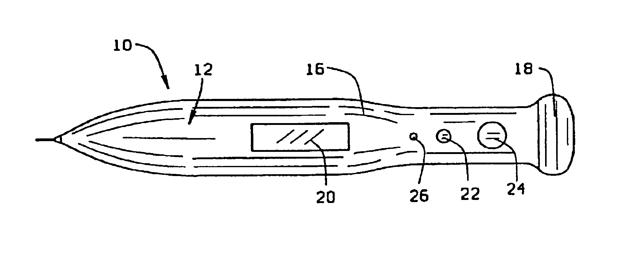
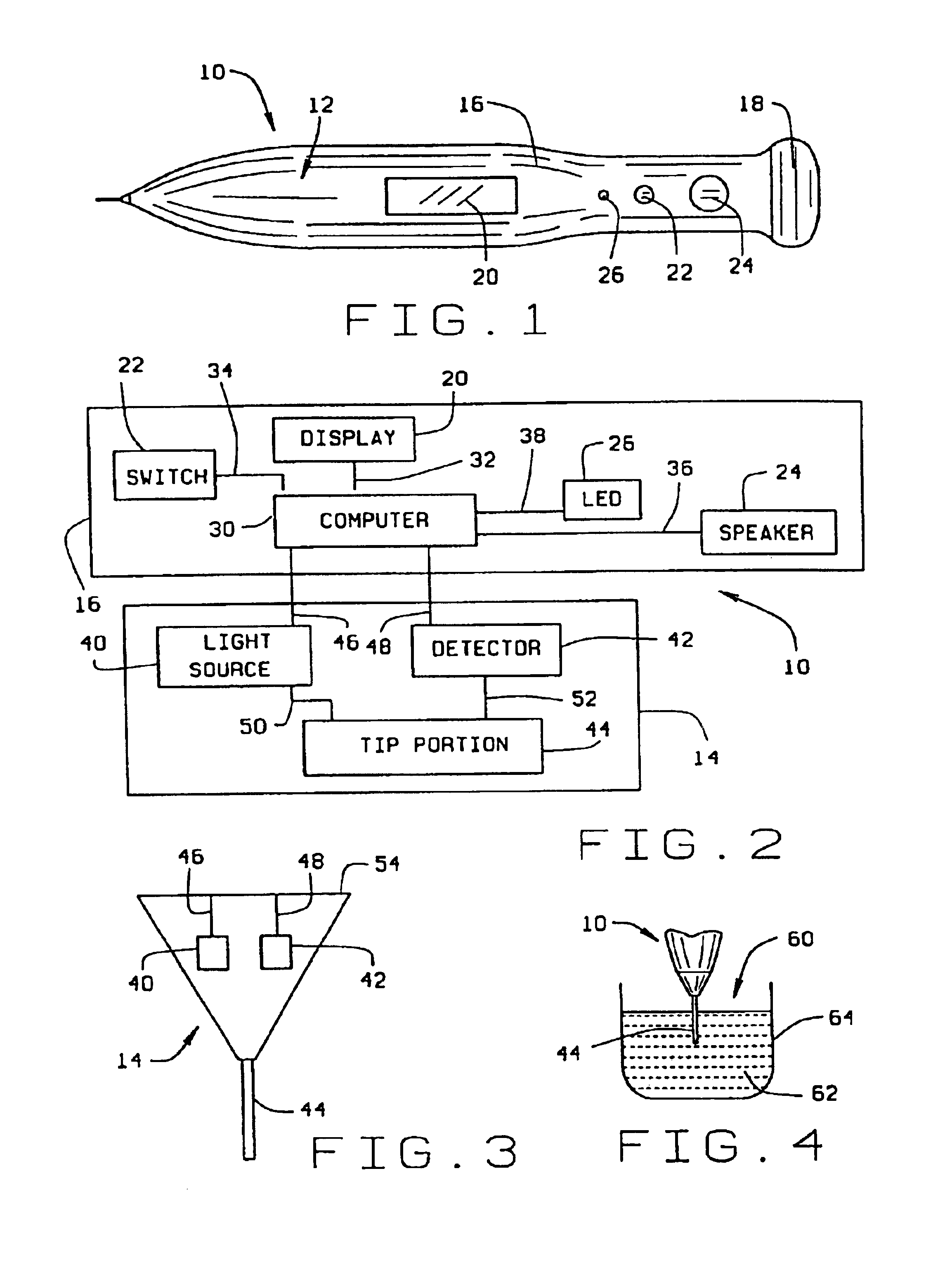
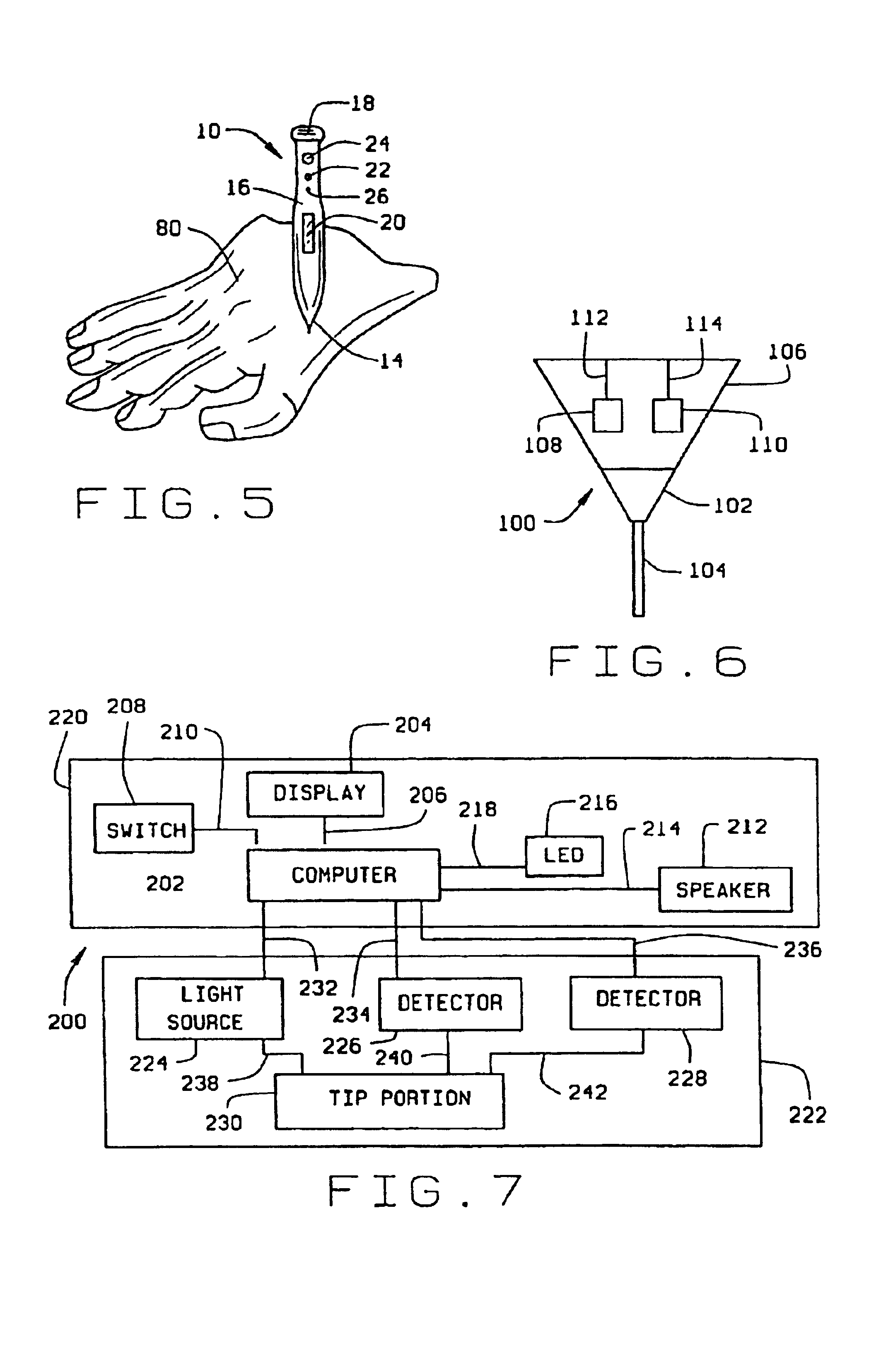
[0029]Referring now to the drawings, wherein like numbers refer to like items, number 10 identifies a preferred embodiment of a micro sensor device constructed according to the present invention. As illustrated in FIG. 1, the device 10 comprises a pencil or pen shaped body 12 which includes an integrated sensor head 14, a central body portion 16, and an end cap 18. The central body portion 16 further includes a display device 20, such as an LED (light emitting diode) type display or an LCD (liquid crystal display) type display, for displaying information. The end cap 18, which may be removable from the central body portion 16, is used to allow access into the interior of the central body portion 16. Batteries (not shown) can be inserted into the central body portion 16 to supply power to the device 10, as will be explained. The central body portion 16 may also include an ON / OFF switch 22 which may be used to operate the device 10, a speaker 24 which may be to audibly indicate certai...


PUM

PropertyMeasurementUnit
diameteraaaaaaaaaa
sizeaaaaaaaaaa
sizeaaaaaaaaaa

Abstract

A method of detecting at least one analyte in extra-cellular spaces includes the step of inserting a microprobe through the stratum corneum toward the stratum basale of the skin of a subject into extra-cellular spaces containing interstitial fluid having at least one analyte to be detected, said microprobe having a diameter at its tip no larger than approximately 10-50 microns. The method further includes optically testing for a predetermined analyte in the extra-cellular space adjacent the distal end of the microprobe without drawing a sample of the interstitial fluid. Preferably the microprobe body includes a sensor layer covering the distal optical tip of the microprobe body, the sensor layer being adapted to interact with a predetermined analyte to be detected in the interstitial fluid, and an optical detector responsive to interaction of the sensor layer with the predetermined analyte to signal detection of said predetermined analyte.

Description

CROSS-REFERENCE TO RELATED APPLICATION[0001]This is a continuation of U.S. patent application Ser. No. 09 / 668,703 filed Sep. 22, 2000, now U.S. Pat. No. 6,535,753, which is a continuation-in-part of U.S. patent application Ser. No. 09 / 143,258, filed Aug. 20, 1998, now U.S. Pat. No. 6,197,257.STATEMENT REGARDING FEDERALLY SPONSORED RESEARCH OR DEVELOPMENT[0002]Not Applicable.BACKGROUND OF THE INVENTION[0003]1. Field of the Invention[0004]This invention relates generally to a method and apparatus for detection of analytes, and more particularly to such method and apparatus for the painless detection of such analytes in extracellular space.[0005]2. Description of the Prior Art[0006]The past decade has seen an increased effort for new and less invasive means of detecting relevant compounds in biological systems. In the past such detection has involved clinical assays using in vitro diagnostics requiring invasive sampling methods. Newer philosophies in medicine and related disciplines ha...

Claims


Application Information

Patent Timeline
Patent Type & Authority Patents(United States)
IPC IPC(8): A61B5/00A61B10/00G01N21/85G01N33/487G01N33/48A61B5/145A61B5/1459A61B6/00G01N21/27G01N33/483
CPCA61B5/14514A61B5/14532A61B5/14546A61B5/1455G01N21/8507A61B5/6825A61B10/0045A61B5/1459A61B2010/008A61B2562/12B33Y80/00
Inventor RASKAS, ERIC JULES
Owner BECTON DICKINSON & CO
Why Eureka
Try Eureka