Autoclavable coupler for endoscopic camera system

a camera system and autoclave technology, applied in the field of medical devices, can solve the problems of inability condensation of lenses and other optical components, and inability of the coupler to transfer images from the scope to the camera,

Inactive Publication Date: 2005-08-23
STRYKER CORP
View PDF16 Cites 28 Cited by
  • Summary
  • Abstract
  • Description
  • Claims
  • Application Information

AI Technical Summary

Benefits of technology

[0009]A hermetically sealed optical coupler for an endoscopic camera system includes a coupler body that has a camera attached at one of its ends and partially contains a hermetically sealed lens rider.

Problems solved by technology

Instruments that will not survive the autoclave process are normally sterilized by less efficient techniques, such as immersion in sterilization liquid or gas.
Problems that occur with prior art couplers are that moisture from autoclaving is able to penetrate the interior of the optical chamber, causing fogging (condensation) of the lenses and other optica

Method used


Image

  • Autoclavable coupler for endoscopic camera system
  • Autoclavable coupler for endoscopic camera system
  • Autoclavable coupler for endoscopic camera system

Examples


Embodiment Construction

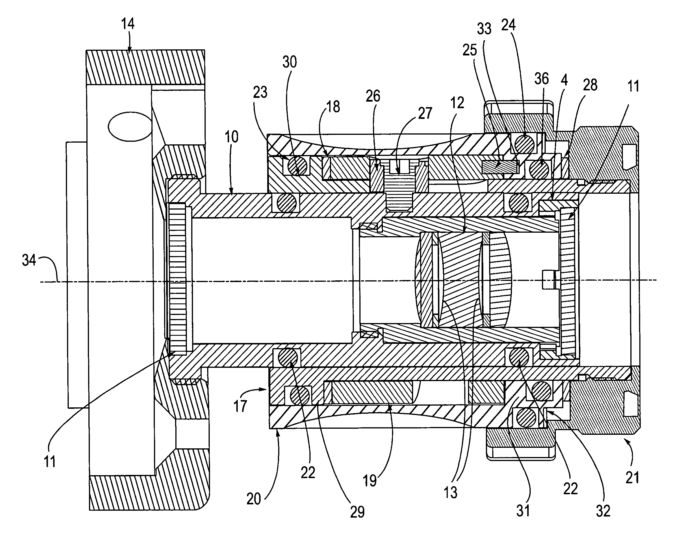
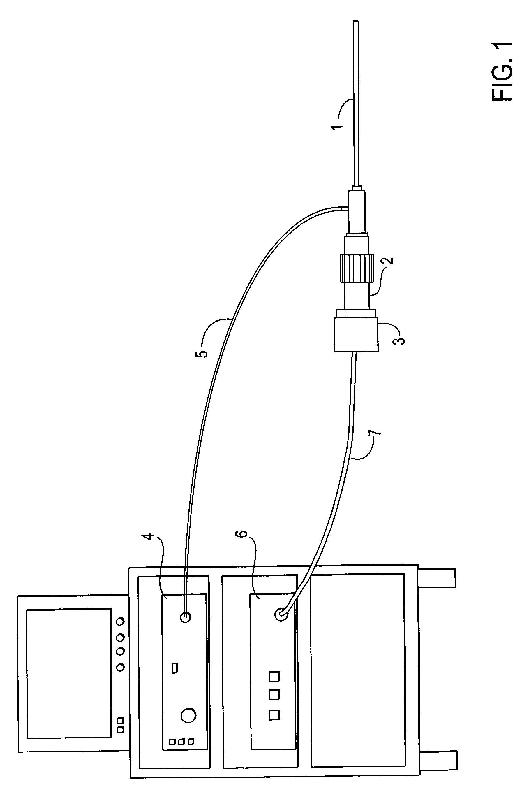
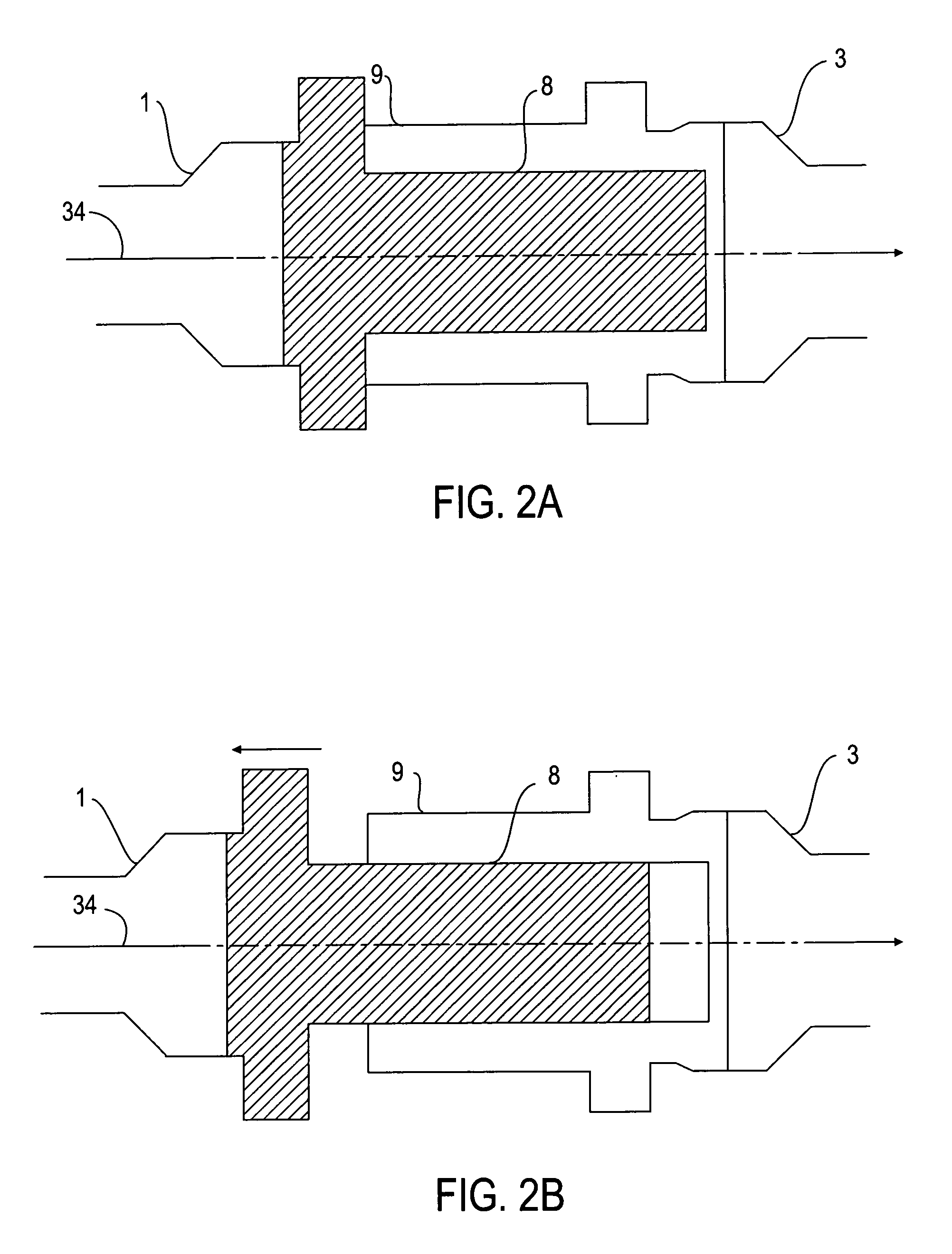
[0024]An autoclavable coupler for endoscopic camera systems is described. As will be described in greater detail below, the coupler includes a body containing one or more focusing lenses, which are hermetically sealed from the outside environment. The body is capable of coupling an endoscope at one side, and a camera head at an opposite side, so that the endoscope and the camera are situated in a path of optical communication with each other and with the lenses located inside the coupler body. The body is constructed so that the distance between the focusing lens or lenses inside the body and the CCD of the camera head can be varied by a simple manipulation of a focus ring. It is through this manipulation that an image being sent from the endoscope is focused onto the CCD, allowing a focused image to be sent from the camera to, and displayed on, a video monitor. The coupler is capable of withstanding repeated autoclave sterilization treatments without moisture penetrating the hermet...


PUM


Abstract

An autoclavable endoscopic optical coupler includes the capability to optically couple an endoscope and a camera, so that a focused image will be communicated to the camera head. Furthermore, the coupler has the capability of withstanding multiple autoclave sterilization treatments without allowing moisture to penetrate the interior. The coupler includes a lens holder assembly that has an endobody on one portion and a second portion that is inserted into and coupled to a coupler body. The coupler body has a camera attached at one of its ends. A focus ring is physically coupled to the coupler body, which is coupled to the lens holder assembly. Turning the focus ring causes a translation motion of the lens holder assembly and endoscope with respect to the camera head.

Description

FIELD OF THE INVENTION[0001]The present invention pertains to the field of medical devices. More particularly, the present invention relates to an optical coupler for endoscopic camera systems.BACKGROUND OF THE INVENTION[0002]Endoscopy is a medical field which allows the acquisition of high-quality video images of internal features of a human body, without the need for invasive surgery. A basic tool of endoscopy is an endoscopic camera system, which includes a scope that is inserted into the body of a patient. Some endoscopic procedures involve the use of a flexible scope, as in the field of gastroenterology, for example. Other procedures, such as arthroscopy or laproscopy, involve the use of a rigid scope. The scope is normally coupled to a camera head that includes electronics for acquiring video image data through the scope.[0003]The coupled scope and camera head may be held and manipulated during endoscopic surgery by a human surgical assistant or by a holding tool, such as a ro...

Claims


Application Information

Patent Timeline
IPC IPC(8): A61B1/00A61B1/04
CPCA61B1/04A61B1/00126A61B1/00128
Inventor PANG, CHIEN MIENNG, YANPENGCHANG, WILLIAM H. L.
Owner STRYKER CORP
Why Eureka
Try Eureka