Speculum having ultrasound probe

a technology of ultrasound probes and speculum, which is applied in the field of medical procedures, can solve the problems of inability to adequately visualize difficulty in locating embryos, and difficulty in adequately observing the cervical canal and uterine cavity, and achieve the effect of superior transvaginal ultrasound imaging

Inactive Publication Date: 2005-11-01
WONG IRWIN LANE +1
View PDF23 Cites 42 Cited by
  • Summary
  • Abstract
  • Description
  • Claims
  • Application Information

AI Technical Summary

Benefits of technology

[0012]The present invention solves the foregoing problems by providing a speculum equipped with an ultrasound probe, for permitting ultrasound visualization during gynecological procedures, such as IVF. The inventive system provides the best of both worlds; i.e. superior transvaginal ultrasonographic imaging without interference by the ultrasound probe with usual embryo transfer techniques, and without the need for an additional assistant. In other words, the inventive device provides a speculum which has the capability of providing simultaneous hands-free sonographic information by integrating the sonographic probe with the speculum. Thus, the inventive speculum, with integrated ultrasound probe permits physical access to a procedural site, and simultaneous real-time sonographic visualization of the site, as well as allowing the operator to have two hands free to interact with the site, e.g. to perform a transcervical transfer of embryos into the uterine cavity.
[0013]In its broadest aspect, the invention provides a physical self-retaining device for separating tissue, into which an ultrasound imaging capability has been incorporated. This unique combination provides an ability to physically and visually access a procedural site with simultaneous sonographic imaging and both of the practitioner's hands free to operate on the site.

Problems solved by technology

Clinical studies also indicate that, using current techniques, some embryos may be inadvertently placed within the cervical canal.
This transabdominal approach makes it very difficult to adequately visualize the cervical canal and uterine cavity.
The pubic bone can also obstruct visualization.
Thus, the sonographic image of the cervical canal and the uterine cavity obtained by the transabdominal probe is more uncomfortable and usually inferior in quality to that obtained by use of the transvaginal probe.
This approach provides an improved visualization of the cervical canal and uterine cavity relative to transabdominal ultrasound, but interferes with the most common embryo transfer techniques by virtue of the ultrasonic probe being physically present in the vagina.
1) Placing the speculum in the vagina first. Then, the catheter is threaded through the cervix and into the uterine cavity by feel. While holding the catheter in position, the speculum is removed. The transvaginal probe is then placed into the vagina while still holding the catheter in place, while hoping that the probe's placement will not disturb the catheter's placement unduly.
2) Placing the speculum in the vagina first. Then, the catheter is threaded through the cervix and into the uterine cavity by feel. While holding the catheter, and with the speculum still in place, an assistant places the transvaginal probe into the vagina. This awkward method thus requires two people, one to hold the transvaginal probe in position to obtain visualization of the cervical canal and uterine cavity, and the second to hold the catheter, and then, while the catheter tip is in the optimum position, to depress the syringe attached to the catheter and thus release the embryos.
3) Placing the speculum in the vagina first. Then, the transvaginal probe is placed through the speculum into the vagina. Finally, an attempt is made to thread the catheter through the external cervical os into the uterine cavity while the transvaginal probe is also within the speculum. This approach is very difficult because the probe within the speculum physically and visually blocks access to the external cervical os.
As is obvious from the above descriptions, each of these described suboptimal approaches creates difficulty and unnecessary complications, thereby reducing the opportunity for a successful outcome.
Yet a third visualization approach sometimes employed in certain procedures is transrectal ultrasound, but this technique for visualization is cumbersome, compromising, and uncomfortable.

Method used


Image

  • Speculum having ultrasound probe
  • Speculum having ultrasound probe
  • Speculum having ultrasound probe

Examples


Embodiment Construction

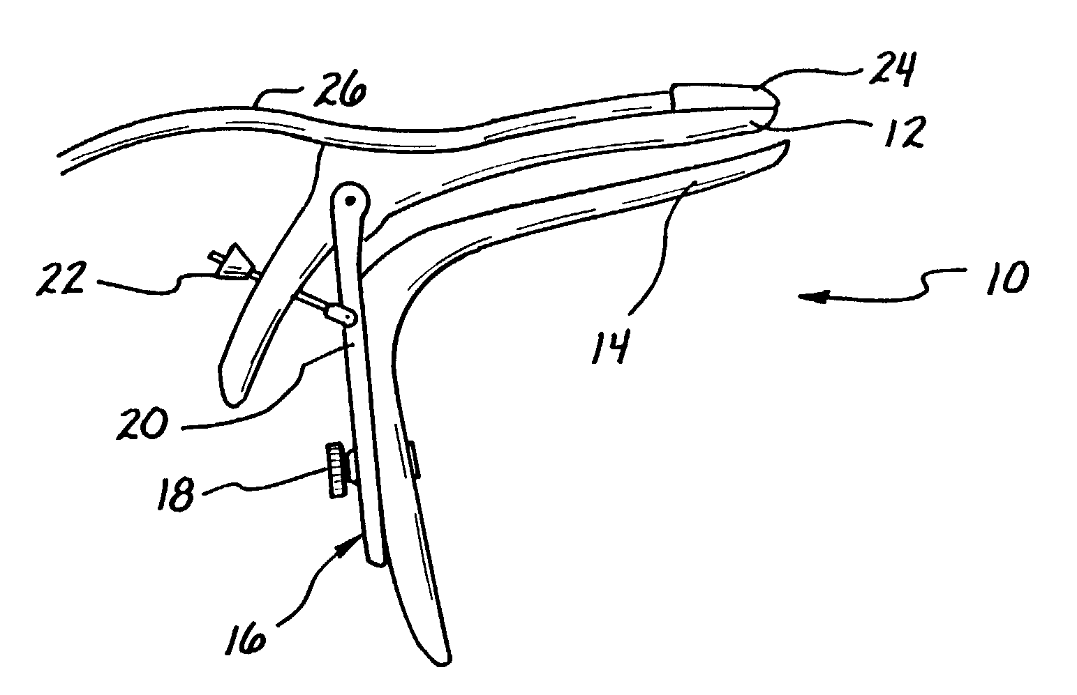
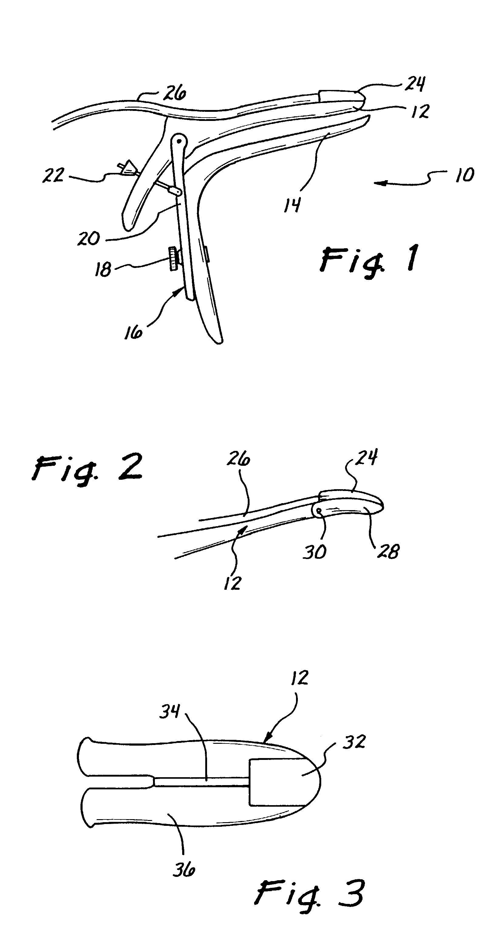
[0029]Now with reference more particularly to the drawing figures, there is shown in FIG. 1 a first embodiment of a medical instrument or speculum 10 constructed in accordance with the principles of the present invention. For purposes of defining terminology in the appended claims, the term “speculum” is broadly defined as a physical device for separating tissue to allow visualization of an internal bodily structure, such as the cervix. The speculum 10 in the illustrated preferred embodiment may be of a generally conventional design, except as described below, and comprises an anterior (upper) blade 12, as well as a posterior (lower) blade 14. As is well known in the art, the two opposed blades 12, 14 are movable between a closed position for insertion and withdrawal, and an open position for performing a pelvic examination or procedure. A downwardly depending handle portion 16 operates to effect blade movement, as desired, in a manner also well known in the art. A screw 18 on the h...


PUM


Abstract

A speculum is equipped with an ultrasound probe, for permitting ultrasound visualization during procedures, such as transcervical embryo transfer with In Vitro Fertilization, thus providing superior transvaginal ultrasonographic imaging without interference by the ultrasound probe with usual embryo transfer techniques, and without the need for an additional assistant. The probe may be disposed on either of the posterior blade and the anterior blade, depending upon physiological factors varying with each patient and procedure.

Description

BACKGROUND OF THE INVENTION[0001]The present invention relates to methods and devices for performing medical procedures, including, but not limited to, gynecologic, otologic, neurosurigical, and gastrointestinal procedures, and move particularly to medical devices such as a speculum, having a built-in capability for permitting ultrasound visualization of a procedural site.[0002]There are a number of gynecological procedures for which it would be advantageous to have the capability of direct transvaginal ultrasound visualization, for the purpose of permitting the medical practitioner to guide catheters and other instruments into the cervical canal and uterine cavity. For example, with the common infertility treatment known as In Vitro Fertilization (IVF), the crucial last step is the transfer of the fertilized eggs (embryos) through the cervix into the uterine cavity. This step is identified in the practice as transcervical embryo transfer. Embryos are usually transferred by first pl...

Claims


Application Information

Patent Timeline
Patent Type & Authority Patents(United States)
IPC IPC(8): A61B1/303A61B8/12
CPCA61B8/12
Inventor WONG, IRWIN LANEHATASAKA, HARRY
Owner WONG IRWIN LANE
Why Eureka
Try Eureka