Method of cancer screening primarily utilizing non-invasive cell collection, fluorescence detection techniques, and radio tracing detection techniques

a cancer and cell collection technology, applied in the field of cancer screening, can solve the problems of complex equipment, special medical expertise, and many references, and achieve the effects of reducing patient trauma along with the cost of the procedure, avoiding invasiveness, and avoiding invasiveness

Inactive Publication Date: 2006-01-10
ADAIR EDWIN L
View PDF51 Cites 21 Cited by
  • Summary
  • Abstract
  • Description
  • Claims
  • Application Information

AI Technical Summary

Benefits of technology

[0020]In addition to providing the above described photo sensitive compounds alone, the photo sensitive compounds may be complexed with a radioisotope, such as 64Cu or 67Cu. Accordingly, these complexes then provide the ability for not only conducting fluorescence detection and photo-therapy, but also provides the ability for observation of malignancies through positron emission tomography (for 64Cu) or single photon emission computed tomography (for 67Cu). Additionally, for 67Cu, it provides a source of beta radiation for selective destruction of malignancies. As well understood by those skilled in the art, positron emission tomography (PET) is a scanning technique which enables precise mapping of tissue which uptakes the radioactive marker. Single photon emission computed tomography (SPECT) is closely related to PET scanning and also provides an exact means to locate targeted tissue which uptakes an introduced radio tracer.
[0021]For those cells which are not dislodged through exfoliation and are therefore left within the body, those cells are doubly tagged not only for fluorescence, but for radioactivity as well. It may be, therefore, desirable under some circumstances to not only remove cells from the body for exuluo analysis, but also to investigate believed malignancies through fluorescence guided endoscopy, or to view the suspect cells through the appropriate tomography technique. One advantage of attaching a radioisotope to the photosensitive compounds is that through the use of recently available tomography devices one can, in effect, conduct a complete body scan of all potentially malignant areas within the body without having to conduct separate endoscopic procedures for each potentially malignant site found. The images taken from the scanning technique can then be provided to the particular specialist for follow-on treatment, which may include surgical intervention or radiation treatment.
[0022]By the method of this invention, a quick and reliable means is provided for cancer screening. Because non-invasive or minimally invasive techniques are used for cell collection, patient trauma is reduced along with the cost of the procedure. Because the method of this invention provides the option of introducing the compounds ex-vivo, the concern for any possible allergic reaction or phototoxic reaction by a patient's exposure to the sun is eliminated. Furthermore, because no tissue biopsies are taken, the method of this invention eliminates the inherent hazard in administering local and / or general anesthesia. Cell marking by use of the above-identified compounds is extremely reliable in terms of differentiating healthy cells from pre-malignant or malignant cells. The segregation, counting and analysis of the fluorescent cells may be achieved with commercially available flow cytometers and supporting equipment. The results of the cancer screening procedure may be forwarded to a pathologist who may wish to conduct additional tests to further determine the exact nature of the malignancy. The fluorescing cells may be photographed to provide documentation of malignancy.

Problems solved by technology

Furthermore, many of the references require complex equipment, and special medical expertise in order to conduct the procedures and to make the diagnoses.

Method used


Image

  • Method of cancer screening primarily utilizing non-invasive cell collection, fluorescence detection techniques, and radio tracing detection techniques
  • Method of cancer screening primarily utilizing non-invasive cell collection, fluorescence detection techniques, and radio tracing detection techniques
  • Method of cancer screening primarily utilizing non-invasive cell collection, fluorescence detection techniques, and radio tracing detection techniques

Examples


Embodiment Construction

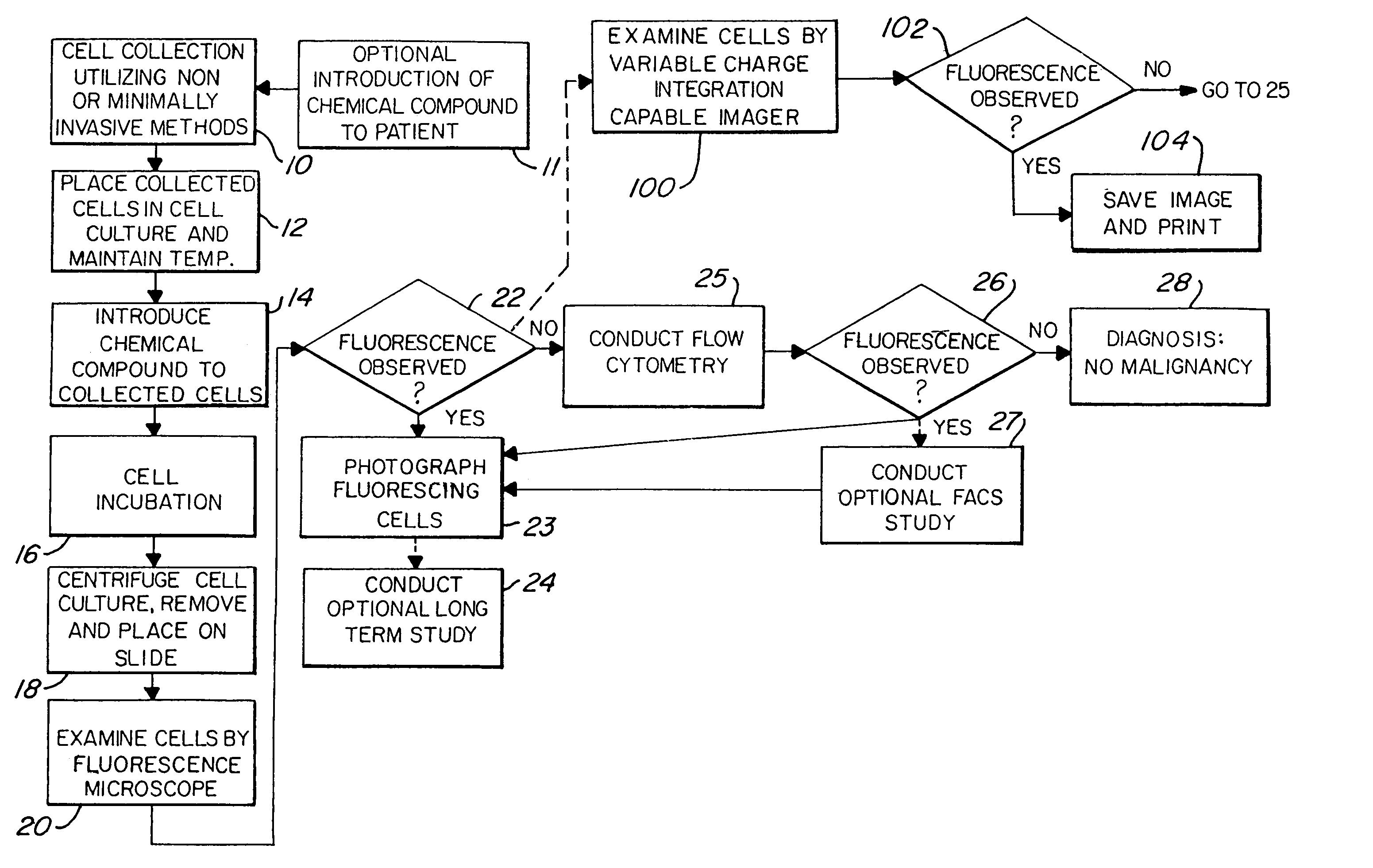
[0027]FIG. 1 illustrates the major steps in one aspect of the method of this invention. As shown, the process begins with non-invasive means or minimally invasive means of cell exfoliation / collection 10. Once collected, the cells are then placed in a culture media or cell transport media, shown at step 12. The goal is to keep the collected cells alive and viable after removal from the body. Thus, even at this intermediate step, the cells should be maintained at approximately 37° C. by use of a water bath or other means. Next, the collected cells are exposed to a chemical which induces fluorescence in pre-malignant and malignant cells, illustrated as step 14. As discussed above, the chemical compounds which are contemplated for use in this invention include, but are not limited to, 5-ALA, protoporphyrin IX, TCPP, hematoporphyrin derivative, photofrin, and photofrin II. Other possible compounds which may be used include uroporphyrin; coproporphyren; tetraphenylporphinesulfonate (TPPS)...


PUM

PropertyMeasurementUnit
wave lengthsaaaaaaaaaa
temperatureaaaaaaaaaa
incubation timeaaaaaaaaaa

Abstract

A cancer screening method is provided, wherein the method is characterized by administering a compound to a patient, the compound being a complex of a fluorescent marker and a radioactive marker, in a dose emitting between 5 Gy and 20 Gy radiation. Cells are then collected preferably through non-invasive or minimally invasive means. If fluorescence is observed in the exfoliated cells, tomographic scanning is conducted to further locate and/or confirm suspected malignant areas or metastatic areas. Further observation or treatment may be conducted either through fluorescence guided endoscopy, photo-dynamic therapy, and/or radiation treatment.

Description

CROSS REFERENCE TO RELATED APPLICATION[0001]This application is a continuation-in-part of U.S. patent application Ser. No. 10 / 176,558 filed Jun. 21, 2002 U.S. Pat. No. 6,750,037 which is a continuation-in-part of co-pending PCT application Ser. No. PCT / US00 / 34121 filed on Dec. 16, 2000, which is a continuation of U.S. patent application Ser. No. 09 / 472,945, filed on Dec. 27, 1999, now U.S. Pat. No. 6,190,877.TECHNICAL FIELD[0002]This invention relates to a method of cancer screening and, more particularly, to a method primarily utilizing non-invasive cell collection techniques, and a combination of fluorescence detection techniques and radio tracing techniques for positive identification of malignant cells.BACKGROUND ART[0003]There are a number of prior art methods and apparatuses which are used in the detection and treatment of cancer. Fluorescent markers have been used to help identify cancerous tissue within a patient. Radio tracers or markers have also been used in the detection...

Claims


Application Information

Patent Timeline
Patent Type & Authority Patents(United States)
IPC IPC(8): C12Q1/02C12Q1/00G01N33/53A61K49/00A61K51/04G01N33/50G01N33/574
CPCA61K49/0021A61K49/0036G01N33/5091G01N33/574A61K51/0485Y10S435/968
Inventor ADAIR, EDWIN L.
Owner ADAIR EDWIN L
Why Eureka
Try Eureka