
Dental shade mapping

a technology for tooth color and mapping, applied in the field of dental color measurement, can solve the problems of inability to accurately map tooth color, difficulty in initial matching procedure, and a number of problems, and achieve the effect of promoting intraoral camera adaption and accurate mapping of tooth color

Active Publication Date: 2012-01-19
CARESTREAM DENTAL TECH TOPCO LTD
View PDF6 Cites 40 Cited by
  • Summary
  • Abstract
  • Description
  • Claims
  • Application Information

AI Technical Summary

Benefits of technology

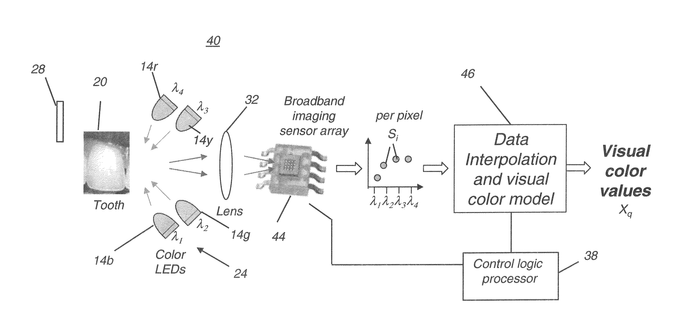
[0019]An advantage of the present invention is that it employs an imaging array for obtaining spectrophotometric measurements over the full image. This promotes adaption for intraoral camera use. In addition, the output spectrophotometric color data that is provided is not subject to metamerism, which affects solutions that use colorimetric and RGB color matching techniques. The approach employed in particular embodiments of the present invention obtains spectral reflectance data for each pixel of the tooth image, allowing accurate mapping of tooth color, including variation in color over different portions of the tooth.
[0020]These objects are given only by way of illustrative example, and such objects may be exemplary of one or more embodiments of the invention. Other desirable objectives and advantages inherently achieved by the disclosed invention may occur or become apparent to those skilled in the art. The invention is defined by the appended claims.
[0021]According to one aspect of the invention, there is provided a method for obtaining a color mapping for a dental object, the method comprising: directing illumination toward the dental object over at least a first, a second, and a third wavelength band, one wavelength band at a time; capturing, on an imaging array, an image of the dental object at each illumination wavelength band to form a set of images of the dental object; for each of a plurality of pixels in the captured set of images: (i) obtaining an image data value for the pixel corresponding to each of the at least first, second, and third wavelength bands; (ii) calculating a plurality of interpolated image data values proportional to the spectral reflectance of the dental object, according to the obtained image data values and according to image data values obtained from a reference object at the at least first, second, and third wavelength bands; obtaining spectral distribution data for a viewing illuminant; reconstructing the visual color of the dental object according to the calculated interpolated image data values and the obtained spectral distribution of the viewing illuminant; and storing at least the reconstructed visual color as data in a computer-accessible memory.

Problems solved by technology

The visual shade matching process can be highly subjective and subject to a number of problems.
The initial matching procedure is often difficult and tedious, and it is not unusual for the process to take twenty minutes or longer.
The problem of accurately modeling the color of a tooth is more complex than obtaining a close color match using shade tabs.
One result of this complexity is that color appearance and color measurement are greatly influenced by lighting geometry, surrounding colors, and other environmental factors.
As a further complication, color within a single tooth is generally not uniform.
Color non-uniformities can result from spatial variations in composition, structure, thickness, internal and external stains, surface texture, fissures, cracks, and degree of wetness.
As a result, measurements taken over relatively large areas produce averaged values that may not be representative of a tooth's dominant color.
In addition, natural color variations and non-uniformities make it unlikely that a given tooth can be matched exactly by any single shade tab.
Further, tooth color is seldom uniform from tooth to tooth.
Therefore, the ideal color of a restoration may not be in visual harmony with that of an adjacent tooth or of any other single tooth in a patient's mouth.
Understandably, they are quite intolerant of restorations that appear inappropriate in color.
It is often difficult to decide which tab matches most closely (or, conversely, which has the least mismatch) and to provide accurate information on color variation over the tooth surface.
Frequently, the practitioner determines that the patient's teeth are particularly difficult to match, requiring that the patient then go in person to the orthodontics laboratory that will be fabricating the restoration.
Considering the relative difficulty of the color matching task, and the further complexity of color mapping, a high rate of failure is not at all surprising.
Visual color evaluation of relatively small color differences is always difficult, and the conditions under which dental color evaluations must be made are likely to give rise to a number of complicating psychophysical effects such as local chromatic adaptation, local brightness adaptation, and lateral-brightness adaptation.
Moreover, shade tabs provide at best a metameric (that is, non-spectral) match to real teeth; thus, the matching is illuminant-sensitive and subject to variability due to normal variations in human color vision, such as observer metamerism, for example.
Maintaining accuracy tends to be difficult and measurements are compromised due to metamerism, in which the color measured is highly dependent upon the illuminant.
This is particularly troublesome since dental measurement and imaging are generally carried out under conditions that differ significantly from natural lighting conditions.
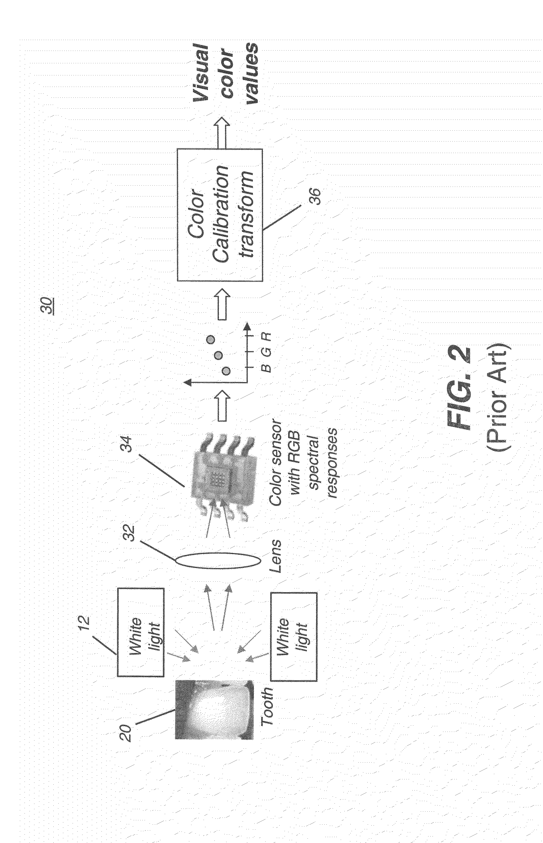
As with RGB-based devices described in (i), measurements from this type of device also suffer from metamerism.
It is significant to note, however, that the spectrophotometer is not an imaging device.
Although the data obtained using the spectrophotometric approach provides advantages for color matching over colorimetric and RGB approaches, including elimination of metamerism, this approach has been found difficult to implement in practice.
The use of a light scanning component for measuring different spectral components, generally employing a grating or filter wheel, tends to make the spectrophotometric system fairly bulky and complex.
This makes it difficult to measure teeth toward the back of the mouth, for example.
Attempts to alleviate this problem have not shown great success.
However, as is true of spectrophotometric devices in general (iii, above), this device measures only a small area of the tooth surface at a time.
The image capture process is time-consuming and does not provide consistent results.
Color mappings can be inaccurate using such an approach, since there can be considerable sensitivity to illumination and image capture angles and probe orientation during the imaging process.
In general, conventional methods that employ color filters, either at the illuminant end or at the sensor end, can be less desirable because they are subject to the limitations of the filter itself.

Method used


Image

  • Dental shade mapping
  • Dental shade mapping
  • Dental shade mapping

Examples


Embodiment Construction

[0034]The following is a detailed description of the preferred embodiments of the invention, reference being made to the drawings in which the same reference numerals identify the same elements of structure in each of the several figures.

[0035]In the context of the present application, the term “narrow band” is used to describe LED or other illumination sources that emit most of their output light over a narrow range of wavelengths, such as 20-50 nm wide. The term “broadband” is used to describe a light sensor that exhibits high sensitivity to incident light over a wide wavelength range extending at least from about 400 nm to about 700 nm. Because this type of sensor responds to light but does not distinguish color, it is often referred to as a “monochrome” sensor or, somewhat inaccurately, as a “black-and-white” sensor.

[0036]In the context of the present application, the term “pixel”, for “pixel element”, has its common meaning as the term is understood to those skilled in the imag...


PUM


Abstract

A method and apparatus for obtaining a color mapping of a dental object. Illumination is directed toward the object over at least first, second, and third wavelength band, one band at a time. An image of the dental object is captured at each wavelength band to form a set of images of the dental object. For pixels in the captured set of images, an image data value for the pixel corresponds to each of the wavelength bands and calculates interpolated image data values proportional to the spectral reflectance of the dental object, according to the obtained image data values and according to image data values obtained from a reference object at the wavelength bands. Spectral distribution data for a viewing illuminant is obtained and the visual color of the dental object reconstructed according to the calculated interpolated image data values and the obtained spectral distribution of the viewing illuminant.

Description

CROSS REFERENCE TO RELATED APPLICATIONS[0001]Reference is made to U.S. Ser. No. ______, entitled “DENTAL SHADE MAPPING”, filed on even date in the names of Wong et al., and which is commonly assigned.FIELD OF THE INVENTION[0002]This invention relates generally to methods and systems for dental color measurement and more particularly relates to a digital method and system for determining color shade information for natural teeth, reference shade samples, and fabricated dental prostheses.BACKGROUND OF THE INVENTION[0003]Modem restorative dental procedures often require accurate color matching, such as for filling materials and for the fabrication of restorations such as crowns, implants, fixed partial dentures, and veneers. The materials used for these procedures, such as ceramics and other materials, can be skillfully formed and treated to closely match the shape, texture, color and translucency of natural teeth.[0004]A widely used technique for determining and communicating tooth co...

Claims


Application Information

Patent Timeline
Patent Type & Authority Applications(United States)
IPC IPC(8): G06K9/00H04N7/18A61B1/24A61B6/51
CPCG01J3/0264G01J3/46G01J3/462G01J2003/466G01J3/50G01J3/501G01J3/508G01J3/463A61C19/04
Inventor WONG, VICTOR C.MILCH, JAMES R.
Owner CARESTREAM DENTAL TECH TOPCO LTD
Learn More