System and method of determining a user-defined region-of-interest of an imaging subject for x-ray flux management control

a technology of flux management and imaging subject, applied in the field of diagnostic imaging, can solve the problems of increasing the overall manufacturing cost of the imaging system, reducing the image quality and dose efficiency, and unable to accommodate the numerous patient sizes and shapes that may be encountered

Active Publication Date: 2006-01-24
GENERAL ELECTRIC CO
View PDF32 Cites 162 Cited by
  • Summary
  • Abstract
  • Description
  • Claims
  • Application Information

AI Technical Summary

Problems solved by technology

However, fashioning an imaging system with a sufficient number of attenuation filters to accommodate the numerous patient sizes and shapes that may be encountered can be impractical given the variances in a possible population.
Additionally, manufacturing an imaging system with a multitude of attenuation filters would increase the overall manufacturing cost of the imaging system.
However, improper patient centering and / or bowtie filter selection can significantly degrade image quality and dose efficiency because x-ray attenuation is misapplied to the particulars of the subject.
However, this is not always the case, e.g. when the subject is mis-centered in the scanner.
However, if the patient 16 is not centered with respect to the bowtie center and the corresponding isocenter, significant image degradation can occur.
Specifically, photon incidence or flux at the edges of the patent may increase image noise to a level that may be prohibitively high for diagnostically valuable images.
However, image degradation may occur if the bowtie opening created is too small for a large patient since useful x-ray needed for imaging is attenuated by the bowtie thereby causing high image noise.
A properly sized bowtie configuration, however, does not ensure acceptable image quality.
If the subject is mis-centered, image degradation may still persist.
First, if subject mis-centering is caused by mis-elevation of the subject with respect to the bowtie filter then the calculation of tube current will result in an underestimate of the subject size.
As a result of the lower tube current, excessive image noise will be present relative to the user's selection.
Secondly, patient mis-centering with respect to elevation may also position the thickest part of the patient such that x-rays for lateral projections pass through the thickest part of the bowtie which results in over-attenuation of the imaging beam.
Such mis-centering can result in an additional image noise increase by as much as 70%.
These errors can cause images of such high noise that the diagnostic value is compromised.
Furthermore, traditional edge detection methods rely on identifying the center of the patient indirectly by detecting the edges of the patient, which can be particularly susceptible to error.
However, while PC and ED CT systems have the potential to realize numerous advantages over traditional CT detectors, the systems may be impractical for some scan protocols.

Method used


Image

  • System and method of determining a user-defined region-of-interest of an imaging subject for x-ray flux management control
  • System and method of determining a user-defined region-of-interest of an imaging subject for x-ray flux management control
  • System and method of determining a user-defined region-of-interest of an imaging subject for x-ray flux management control

Examples


Embodiment Construction

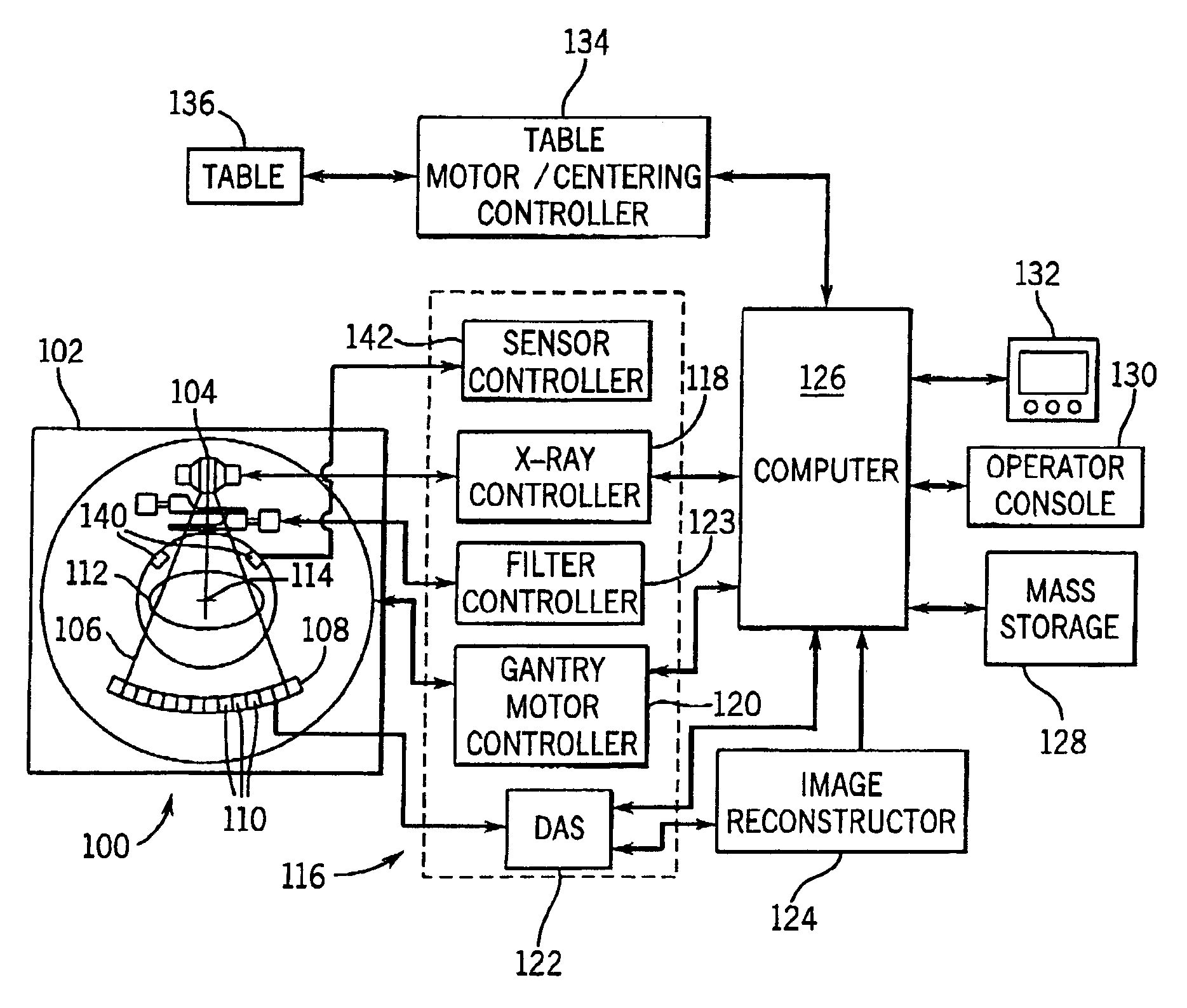
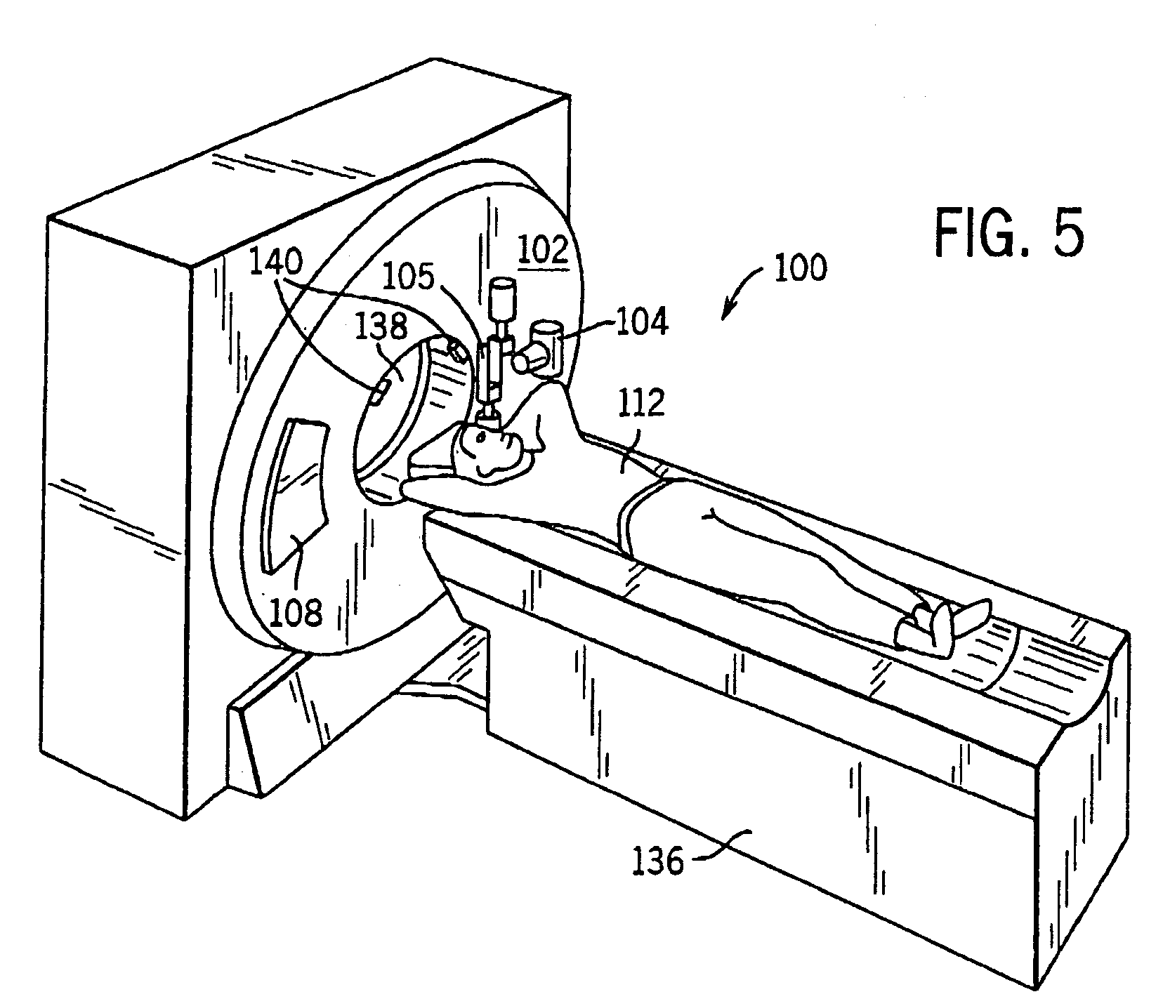
[0042]The present invention is directed to a method and system that automatically determines the patient's size, shape, and centering within an imaging volume and dynamically controls x-ray flux accordingly. Preferably, one or two scout scans together with a plurality of sensors integrally formed with the CT scanner provide patient particulars. The present invention uses the information to provide centering information to the user, allow user input, automatically re-center the patient elevation, correct projection area measurements for dynamic tube current control and select the correct bowtie filter for the optimum dose efficiency.

[0043]The operating environment of the present invention is described with respect to a four-slice computed tomography (CT) system. However, it will be appreciated by those skilled in the art that the present invention is equally applicable for use with single-slice or other multi-slice configurations. Moreover, the present invention will be described wit...


PUM


Abstract

A system and method of diagnostic imaging is provided that includes positioning a subject in an imaging device, performing at least one scout scan, and marking a user-defined region-of-interest. An attenuation characteristic of an attenuation filter is then automatically adjusted based on the user-defined region-of-interest. The present invention automatically selects a proper attenuation filter configuration, corrects patient centering, and corrects noise prediction errors, thereby increasing dose efficiency and tube output.

Description

CROSS-REFERENCE TO RELATED APPLICATION[0001]The present invention claims the benefit of Provisional Application No. 60 / 514,711 filed Oct. 27, 2003.BACKGROUND OF THE INVENTION[0002]The present invention relates generally to diagnostic imaging and, more particularly, to a method and apparatus to optimize dose efficiency by dynamically filtering radiation emitted toward the subject during radiographic imaging in a manner tailored to the position and / or shape of the subject to be imaged.[0003]Typically, in computed tomography (CT) imaging systems, an x-ray source emits a fan-shaped beam toward a subject or object, such as a patient or a piece of luggage. Hereinafter, the terms “subject” and “object” shall include anything capable of being imaged. The beam, after being attenuated by the subject, impinges upon an array of radiation detectors. The intensity of the attenuated beam radiation received at the detector array is typically dependent upon the attenuation of the x-ray beam by the s...

Claims


Application Information

Patent Timeline
Patent Type & Authority Patents(United States)
IPC IPC(8): G21K1/12A61B6/03
CPCA61B6/032A61B6/4035A61B6/488A61B6/544A61B6/469A61B6/4488A61B6/583A61B6/4241
Inventor TOTH, THOMAS L.HOFFMAN, DAVID M.
Owner GENERAL ELECTRIC CO
Why Eureka
Try Eureka