Upright CT scanner

a ct scanner and ct scanner technology, applied in the field of tomography equipment, can solve the problems of inability to eliminate the bed the design of conventional ct scanners is not safe, and the difficulty of detecting minute abnormalities, etc., to achieve the effect of reducing the cost of the bed, and reducing the risk of accidents

Inactive Publication Date: 2006-02-21
CHEN WILLIAM BARRY +1
View PDF8 Cites 32 Cited by
  • Summary
  • Abstract
  • Description
  • Claims
  • Application Information

AI Technical Summary

Benefits of technology

[0008]The object of the present invention is to provide a unique upright CT scanner that produces clinical quality images and is affordable enough for use in small hospitals, outpatient clinics, and doctors' offices. Instead of lying on a bed, a patient is either seated or standing during the examination. The X-ray source and detector are mounted on a platform instead of a gantry, translating while rotating. The distance between the X-ray source and detector, as well as the height at which they operate, is adjustable. The distance adjustment is particularly desirable because X-ray dosage can be reduced when the detector in close proximity to the patient.
[0009]Several factors make the system affordable. Clearly the cost of the bed can be eliminated. Also, employment of the 2D digital X-ray detector eliminates the need for rapid rotation and thus the mechanical components and associated control systems are simplified. In addition, the step-and-shot scan mode of the detector makes it possible to utilize a pulse X-ray tube with less power, which reduces the cost of the tube as well as cooling and power generation systems. In addition, the small footprint of the invention reduces the cost associated with the installation, shielding and maintenance.
[0010]More importantly, the present invention brings a variety of clinical benefits. The unique upright patient position provides some clinical advantages, such as detecting small anomalies in cardiac imaging. The gantry-less design allows for safe and easy access for both the patient and medical personnel. The high-resolution digital X-ray detector and stable mechanical structure produce quality high-resolution CT images that exceed the image quality of even the most expensive current multi-slice CT scanners. Coupled with an EKG device, the present invention is particularly suitable for cardiac imaging, as the field of view can be centered at the heart and one view of the image is large enough to cover the entire heart. Finally, the present invention can be used as a radiography machine. The ability to take high-resolution radiographs and CT scans in a single setting is highly desirable for many examinations, particularly in the field of urology.

Problems solved by technology

This is not the optimal position for to examine for cardiovascular diseases because when a patient is lying down, his organs take more relaxed positions and thus it becomes more difficult to detect minute abnormalities.
However, it is not possible to eliminate the bed for conventional CT scanners.
Practically, however, his design has safety concerns and is probably more expensive than conventional CT scanners.
Despite many advantages of C-arm CT scanner, structural instability of C-arm during the oscillating rotation causes the problem of image registration.
In addition, C-arm CT scanners may not be able to scan a large patient or part of body that is far from the axis of rotation center without a large, expensive detector.

Method used


Image

  • Upright CT scanner
  • Upright CT scanner
  • Upright CT scanner

Examples


Embodiment Construction

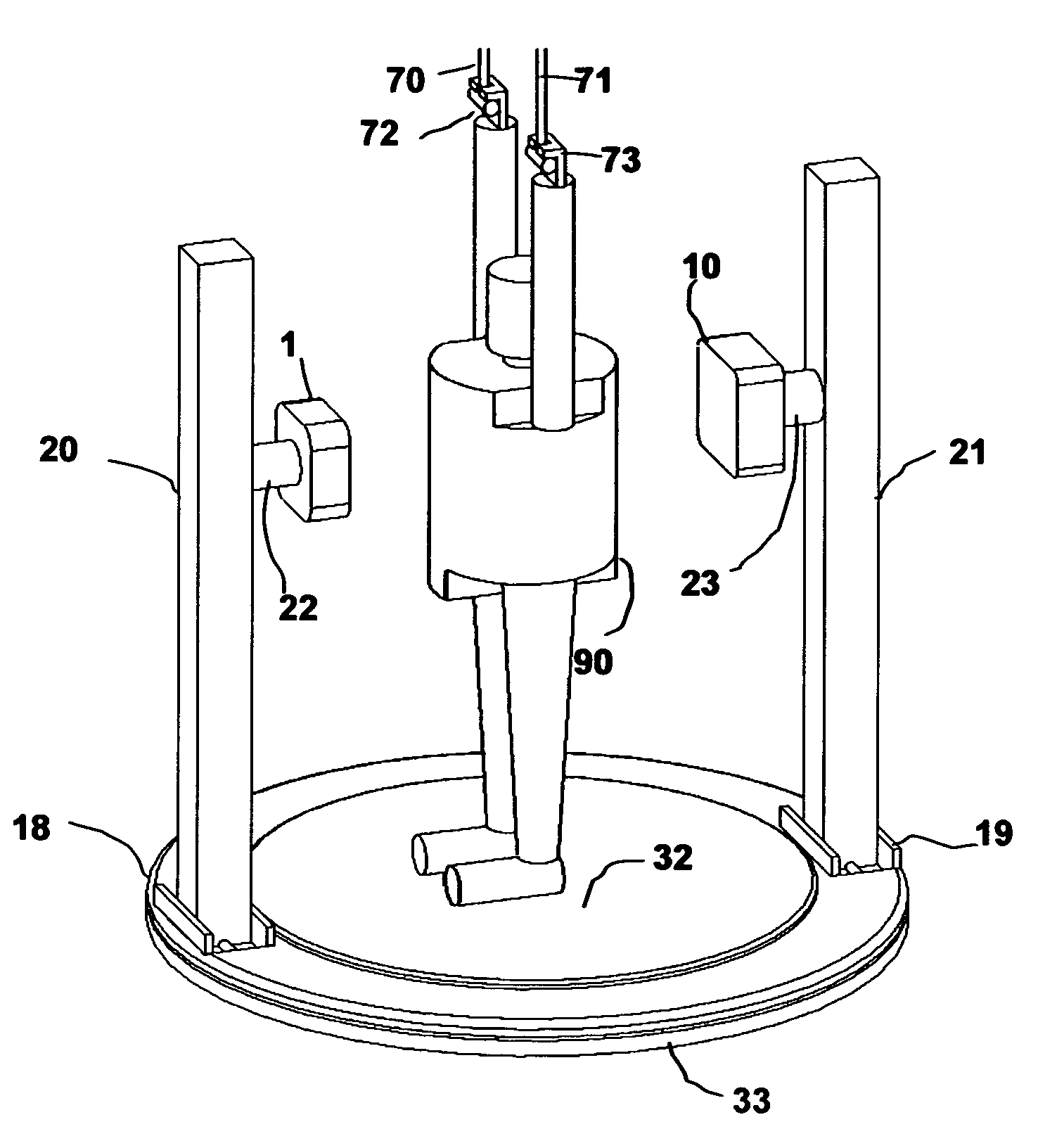
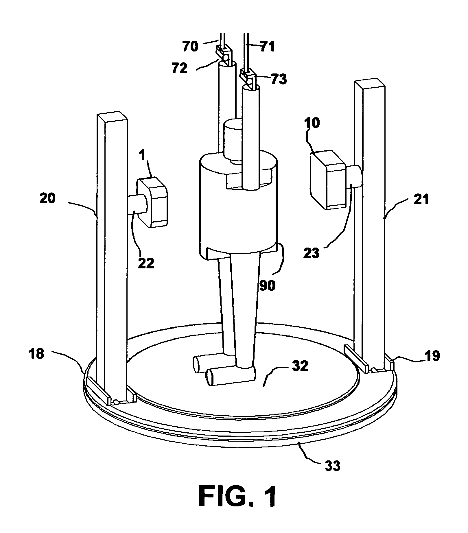
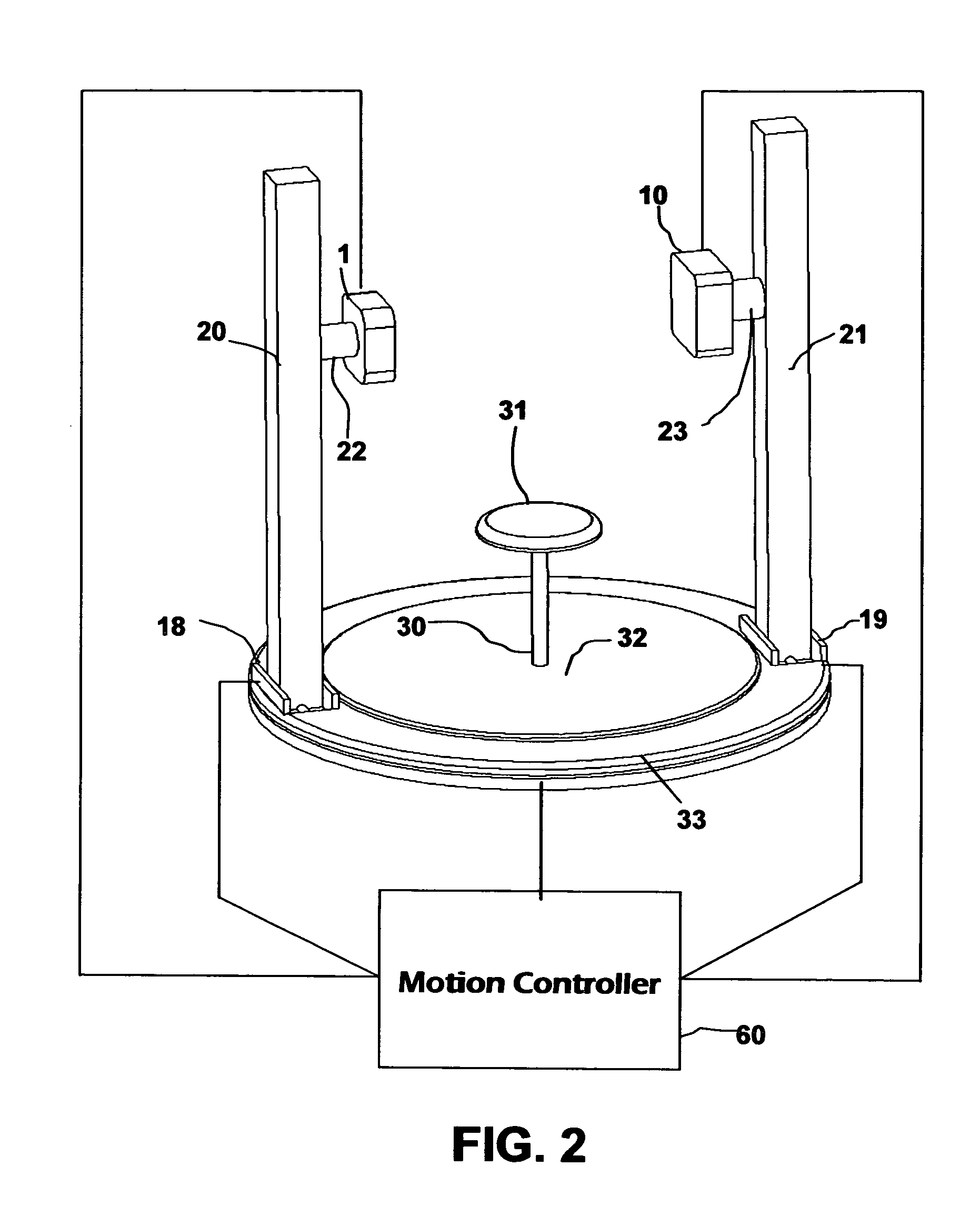
[0014]The principles of the invention will be discussed first in reference to FIG. 1. In this figure, patient 90 is standing on a stationary base platform 32 and holding handles 72 and 73, which are mounted to the ceiling by structures 70 and 71. The X-ray source is designated generally by the reference numeral 1. The two-dimensional digital X-ray detector is designated generally by the reference numeral 10. The X-ray source 1 is mounted to a horizontal linear motion mechanism 22, while the X-ray detector 10 is mounted to another horizontal linear motion mechanism 23. The combination of the linear motion from mechanisms 22 and 23 adjusts the distance between the X-ray source 1 and the detector 10. The mechanisms 22 and 23 can be controlled to move either individually or in tandem.

[0015]The linear motion mechanisms 22 and 23 are mounted to two vertical linear motion mechanisms, designated as columns 20 and 21, respectively. The joint motion provided by columns 20 and 21 adjusts the h...


PUM


Abstract

In an upright CT scanner, an X-ray source and a two-dimensional digital X-ray detector are mounted on a rotating platform with a vertical axis of rotation. During scanning, the X-ray source and the detector are on opposite sides of the rotational axis for orbiting about a body to thereby scan it with pyramid-shaped X-ray beam. During scanning, the source and detector can be jointly translated along a horizontal axis that is perpendicular to the rotation axis and parallel to the detector surface, to scan a specific part of a body that is not centered or a body that is too large. The mounting height and distance of source and the detector can also be adjusted.

Description

STATEMENT REGARDING FEDERALLY SPONSORED RESEARCH[0001]Not applicable.BACKGROUND OF THE INVENTION[0002]1. Field of Invention[0003]This invention relates examining the human body with X-ray by means of computed tomography, and more specifically, to a tomography apparatus that allows a patient to be scanned with his torso upright.[0004]2. Description of the Prior Art[0005]X-ray computed tomography is one of the most frequently utilized modalities in diagnostic imaging. Recent developments in multi-slice CT technology have proven the diagnostic value of CT in areas such as cardiology. Since cardiovascular disease is currently the leading cause of death in the United States, it is desirable that cardiac CT scanners become a routine examination tool just as X-ray examinations have become, capable of being utilized in small hospitals, outpatient clinics, and doctors' offices. This requires a new generation of CT scanners that are more affordable, convenient to use, and able to detect minut...

Claims


Application Information

Patent Timeline
Patent Type & Authority Patents(United States)
IPC IPC(8): A61B6/04A61B6/03A61B6/00
CPCA61B6/04A61B6/00A61B6/4435
Inventor CHEN, WILLIAM BARRYCHEN, JASON ANDREW
Owner CHEN WILLIAM BARRY
Why Eureka
Try Eureka