Determination of cell viability and phenotype
a cell viability and phenotype technology, applied in the field of cell viability and phenotype determination, can solve the problems of inability to determine the overall amount of color, the inability to separate the viability of the cell from the intracellular facs method, and the use of fixation kills all cells in a sample, etc., to achieve accurate and straightforward quantification of viable cells
- Summary
- Abstract
- Description
- Claims
- Application Information
AI Technical Summary
Benefits of technology
Problems solved by technology
Method used
Image
Examples
example 1
[0078]Materials and Methods
[0079]All materials may be obtained from any suitable source, including those available from multiple commercial entities. Many pre-dyes are available from Molecular Probes as well as other vendors.
[0080]As an alternative to the following general protocol, the IntraCyte™ Intracellular FACS Kit from Orion Biosolutions (Vista, Calif.) may be used to conduct steps beginning with the fixation step.
[0081]General Protocol
[0082]For cells that are not normally in suspension, prepare single-cell suspension by enzymatic digestion or dissociation, preferably sufficiently mild to minimize cell damage. As a non-limiting example, adherent cells may be incubated cells with trypsin / EDTA in a buffered salt solution at physiological pH, such as by use of trypsin / EDTA in Hank's Balanced Salt Solution (HBSS) without Ca++ / Mg++ or phenol red (Sigma Cat. # H-6648). Trypsin may be present from 0.2 to 0.01% (w / v) while EDTA may be present from 0.02 to 0.001% (w / v) as non-limiting ...
example 2
[0104]Intracellular FACS Analysis of Rat Neural Stem Cells
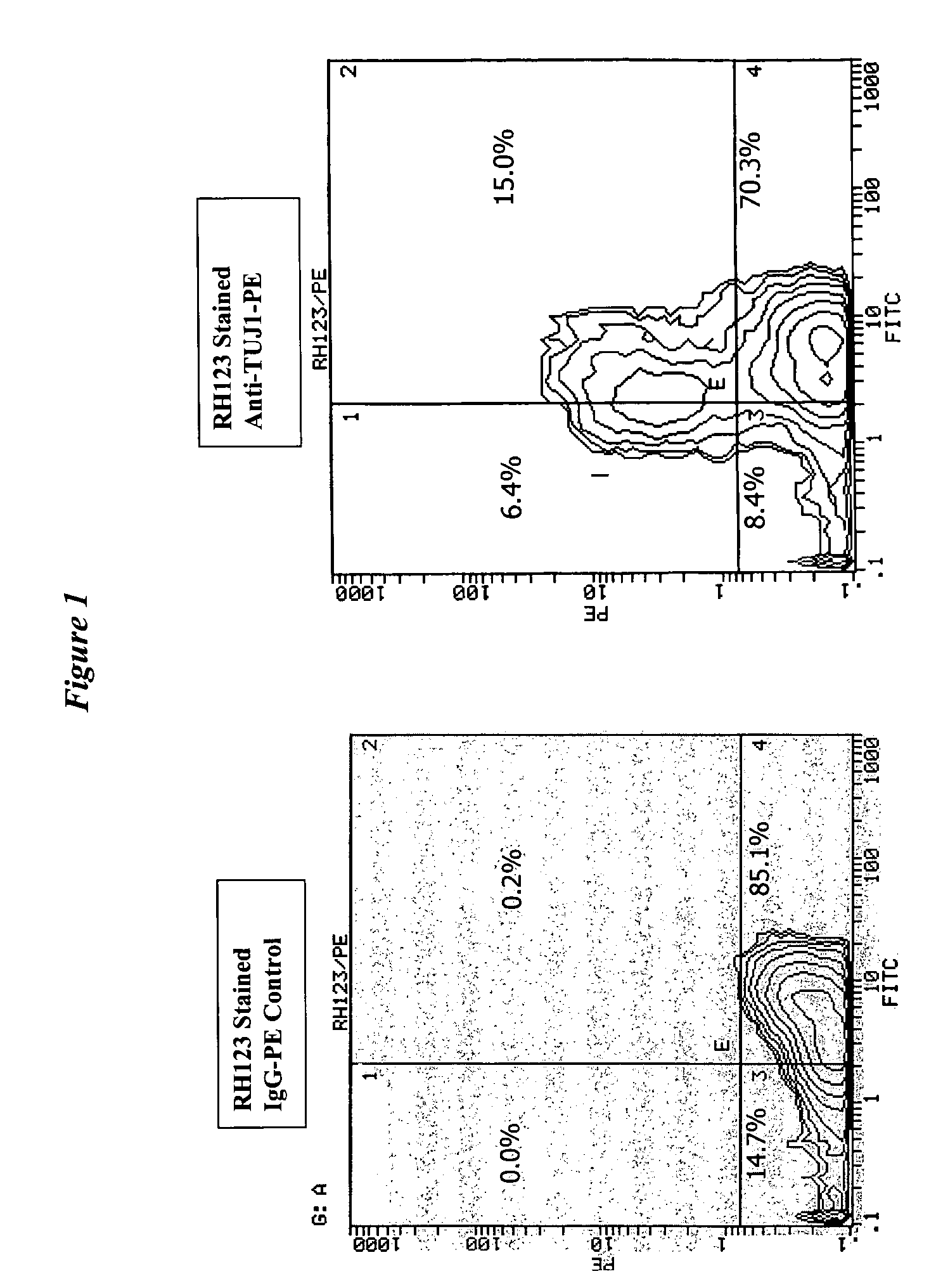
[0105]Rat neural stem cells were obtained and treated essentially as described in Example 1. The cells were exposed to retinoic acid and growth factor withdrawal for 5 days followed by contact with dihydrorhodamine. The cells were then fixed and permeabilized followed by contact with a PE conjugated antibody against beta-tubulin III (TuJ1), the expression of which is used to identify a phenotype of differentiating cells.
[0106]The results are shown as dual-parameter contour plots in FIG. 1, where the horizontal axes are rhodamine 123 fluoresence and the vertical axes are cell counts. The figure shows the portion of TuJ1-positive neuronal cells (upper quandrants in right panel) in comparison to the use of a PE conjugated IgG (left panel) as a negative control. 15% of total cells were TuJ1 positive as well as rhodamine 123 (RH123) positive and thus were viable. 6.4% of neuronal cells were TuJ1 positive but rhodamine 123 negative...
PUM
| Property | Measurement | Unit |
|---|---|---|
| temperature | aaaaa | aaaaa |
| length of time | aaaaa | aaaaa |
| temperature | aaaaa | aaaaa |
Abstract
Description
Claims
Application Information
 - R&D
- Intellectual Property
- Life Sciences
- Materials
- Tech Scout
- Unparalleled Data Quality
- Higher Quality Content
- 60% Fewer Hallucinations
Browse by: Latest US Patents, China's latest patents, Technical Efficacy Thesaurus, Application Domain, Technology Topic, Popular Technical Reports.
© 2025 PatSnap. All rights reserved.Legal|Privacy policy|Modern Slavery Act Transparency Statement|Sitemap|About US| Contact US: help@patsnap.com

