
Method of and apparatus for detecting atrial fibrillation

a technology of atrial fibrillation and detection method, which is applied in the field of detection method and apparatus for atrial fibrillation, can solve the problems of irregular heartbeat pattern, many rhythm abnormalities, and high risk of heart failure, and achieve the effect of improving the detection accuracy and accuracy of atrial fibrillation

Inactive Publication Date: 2006-03-28
WIESEL JOSEPH
View PDF30 Cites 50 Cited by
  • Summary
  • Abstract
  • Description
  • Claims
  • Application Information

AI Technical Summary

Benefits of technology

[0033]The present invention provides a method and apparatus that determines the presence of atrial fibrillation (i) by detecting the pulse beat intervals over a short time period to determine whether the intervals form a random, irregular pattern (ii) determines whether this pulse beat pattern indicates possible atrial fibrillation and then (iii) communicates this information to the user so that a medical practitioner may be consulted for further testing and / or treatment. The present invention also provides a method of and an apparatus for detecting irregular pulse rhythms during a time period and storing this information for comparison with the pulse rhythm at later time periods. The present invention may also detect patterns over multiple time periods and compare the patterns over various time periods.
[0036]The present invention allows for the elimination of at least one or more of the initial pulse beat intervals detected during deflation of a blood pressure cuff. This is necessary due to the respiratory variation in the systolic pressure of sinus beats even in normal individuals.

Problems solved by technology

Many rhythm abnormalities may be present.
The atrioventricular node acts as a filter and allows a reduced number of these impulses to reach the ventricles which results in a highly irregular heartbeat pattern.
These conditions may stretch or scar the atria, thereby causing irregularities in the heart system.
Atrial fibrillation may also accompany lung problems or thyroid gland disorders and is also associated with significant morbidity and possible mortality.
The most serious complication of atrial fibrillation is formation of a blood clot in the left atrium which may result in a stroke.
The literature, however, is generally limited to disclosing instructions for manually taking one's pulse accompanied with additional descriptive information.
A ventricular contraction that cannot be palpated as a pulse in the arterial system results in what is called a “pulse deficit.” This is very common in atrial fibrillation.
This pulse deficit means that an irregular pattern of heartbeats in the ventricle may result in a less irregular pulse beat pattern since the shortest intervals between heartbeats may not be detected in the pulse.
Therefore, any method used to determine the presence of atrial fibrillation by analyzing the time intervals between beats in the ventricles may not be valid when applied to the pulse beats.
At the highest sensitivity setting, this device would not be useful to detect atrial fibrillation in the home setting.
U.S. Pat. No. 6,095,984 did not describe methods and apparatus to look for atrial fibrillation and would not be useful for home monitoring of atrial fibrillation, because the algorithm can result in multiple sources of false readings and the apparatus was not specifically set for optimal detection of pulse beats in atrial fibrillation.
There are several devices available that measure both blood pressure and pulse rate, but none of these devices is capable of monitoring, detecting and / or communicating whether or not an irregular heartbeat pattern is present to indicate possible atrial fibrillation.
The commercially available devices measure the number of pulse beats over a preset time interval, usually ten (10) seconds, but these devices neither analyze nor determine the presence of irregular heartbeat rhythms.
However, a pulse beat can only be detected by these devices when the systolic pressure of the beat exceeds a minimum value.
Therefore, when the cuff of these automatic sphygmomanometers is inflated to a high pressure it will only detect pulses with higher systolic pressures.
Even though this variation in systolic pressures is usually less than 10 mm Hg, it may be enough to cause some normal sinus beats not to be detected when the blood pressure cuff is inflated to high pressures.

Method used


Image

  • Method of and apparatus for detecting atrial fibrillation
  • Method of and apparatus for detecting atrial fibrillation
  • Method of and apparatus for detecting atrial fibrillation

Examples


Embodiment Construction

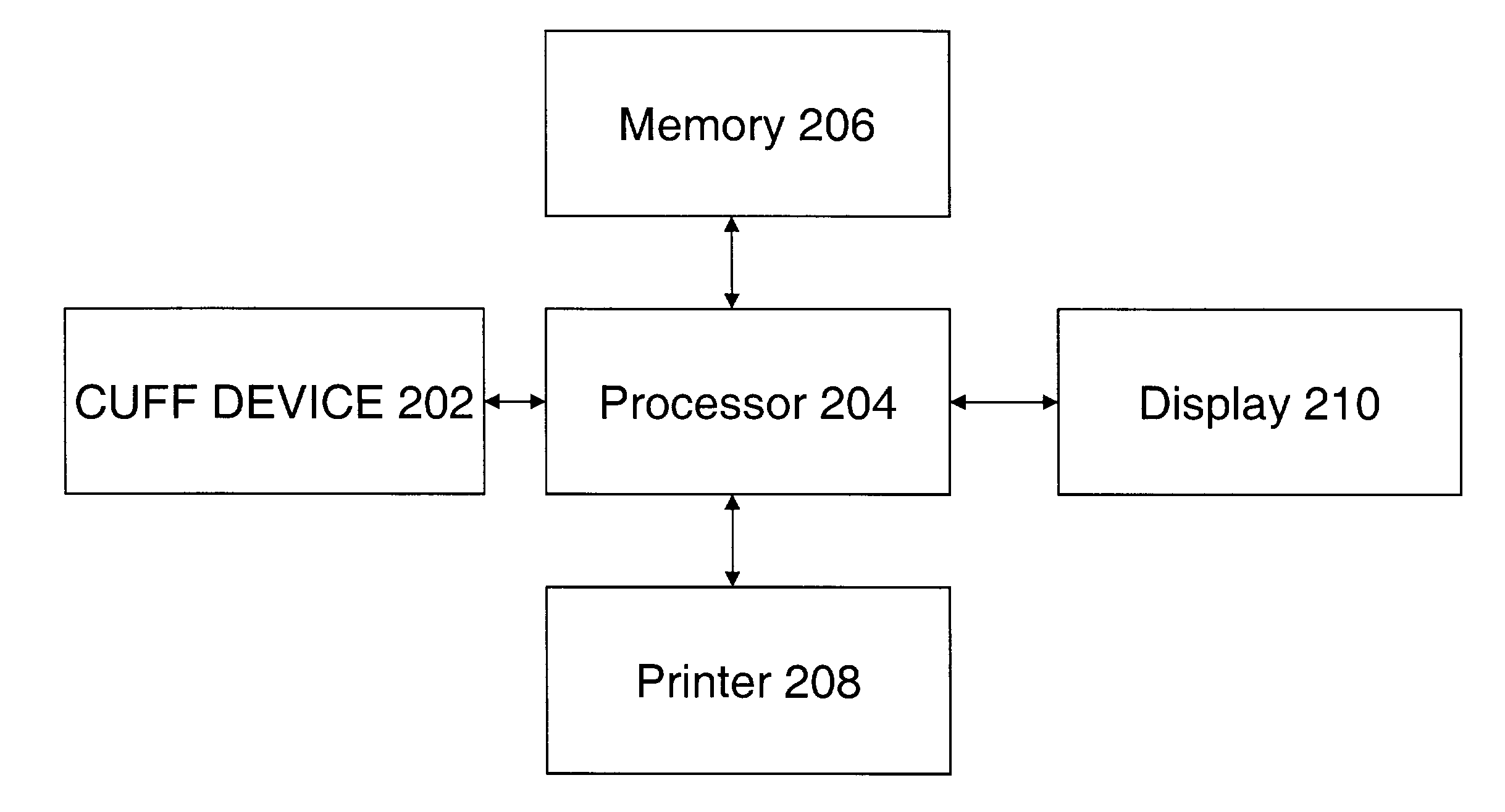
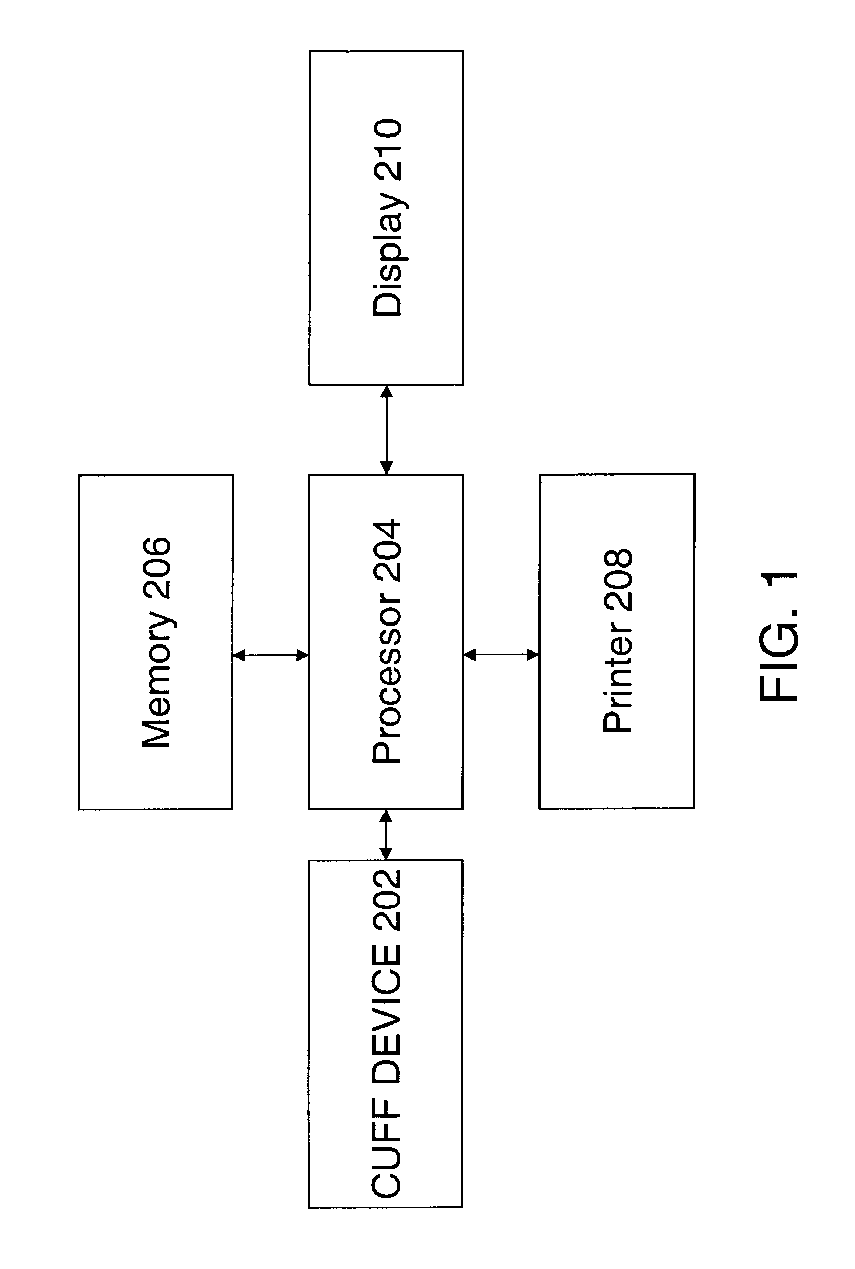
[0045]FIG. 1 shows an embodiment of the invention in which pulse beats are detected using an inflatable cuff device 202. The inflatable cuff device 202 may be a known apparatus used to measure blood pressure using oscillometric or auscultatory means.

[0046]The inflatable cuff device 202 is placed around an appendage such as an arm and inflated above systolic pressure. While the cuff is deflated, the pulse beats are detected. The cuff deflation may be stopped and the cuff may remain at a fixed pressure to allow for monitoring of the pulse beats during a constant cuff pressure. The time of each pulse beat is delivered to a processor 204, which includes instructions that carry out the method described above.

[0047]Further, the processor 204 stores the time of each pulse beat, the intervals between pulse beats and other information in a memory 206. The memory 206 may include RAM or other device memory or include a hard disc, a floppy disk or other memory devices. The processor 204 may com...


PUM


Abstract

A method and apparatus to determine possible atrial fibrillation that includes detecting irregular pulse rhythms from a succession of time intervals each corresponding to a respective interval of time between successive pulse beats of a sequence of the pulse beats; analyzing the detected irregular pulse rhythms to make a determination of possible atrial fibrillation; indicating the possible atrial fibrillation from the determination. If a sphygmomanometer is used, a plurality of pulse beats may be detected with it including the sequence of pulse beats; and respiratory variation in systolic pressure of the pulse beats may be compensated by designating the succession of time intervals to exclude at least an initial one of the plurality of time intervals.

Description

CROSS-REFERENCE TO COPENDING PATENT APPLICATIONS[0001]This is a continuation-in-part of Ser. No. 09 / 467,233, filed Dec. 20, 1999, now U.S. Pat. No. 6,519,490.BACKGROUND OF THE INVENTION[0002]1. Field of the Invention[0003]The present invention generally relates to a method of and an apparatus for detecting atrial fibrillation by monitoring and analyzing pulse beats.[0004]2. Discussion of the Related Art[0005]The heart is the major muscle that functions as the primary pump for blood flow throughout the body. The heart contains two upper chambers called atria and two lower chambers called ventricles. The right atrium receives oxygen-depleted blood while the left atrium receives blood enriched with oxygen from the lungs. When the atria are full, the outlet valves within the heart open and the atria squeeze blood into the ventricles. The right ventricle then pumps oxygen-depleted blood to the lungs while the left ventricle pumps oxygen-enriched blood to all parts of the body. In this fa...

Claims


Application Information

Patent Timeline
IPC IPC(8): A61B5/04A61B5/361
CPCA61B5/046A61B5/361
Inventor WIESEL, JOSEPH
Owner WIESEL JOSEPH
Learn More