Supplementary endo-capsular lens and method of implantation

a technology of endocapsular lens and supplementary lens, which is applied in the field of surgical techniques, can solve the problems of static refracting power, reduced contrast of the focused part of the image, and reduced the ability of the eye to obtain good resolution for all viewing distances, and achieve the effect of increasing the accommodative capacity of the ey

Inactive Publication Date: 2006-06-13
THE VISION CRC LTD +1
View PDF14 Cites 17 Cited by
  • Summary
  • Abstract
  • Description
  • Claims
  • Application Information

AI Technical Summary

Problems solved by technology

These procedures are prone to complications, such as retinal detachment, and, in the case of extracapsular cataract extraction, opacification of the posterior capsule.
Common disadvantages are that these refracting powers are static and they are restricted to specific gaze positions.
A common disadvantage of these devices is that the contrast of the focused part of the image is reduced by the poorly focussed portions of the image.
The defocussed images reduce the contrast of the focused image and therefore reduce the ability of the eye to obtain good resolution for all viewing distances.
Alternatively, crosslinking may be effected through the cornea, but such procedures may damage corneal tissues.
However, a reduced cross-linking density of these polymers results in an unacceptable gummy product having low resilience.
Injection of such partially polymerized materials through a cannula may cause shear stress, which results in rough areas of the polymerized material that impair the function of the synthetic lens.
Leakage of such materials into the anterior chamber of the eye may cause a number of ocular problems, and endanger delicate ocular structures.
Additionally, leaching of non-endogenous liquids or gels from the capsular bag may cause glaucoma, due to blockades of trabeculae and associated increases in intraocular pressure due to increased volumes of aqueous humor.
Undesirable conditions, such as interference with motion of the iris and impairment of the optics of the eye due to glare are also known to occur upon escape of viscous liquids and gels introduced to the capsular bag.
Such damage to the anterior rim leaves the rim vulnerable to tearing radially when the rim is stressed, particularly upon insertion of instruments for manipulating the capsular lens matrix.
Tearing of the lens capsule during capsulorhexis increases the likelihood of untoward leakage of materials injected into the evacuated capsular bag during lens refilling.
However, tears may arise despite taking such precautionary measures.
Although generally successful, certain drawbacks exist with this process, including expansion of the capsulorhexis opening during filling, causing intraoperative leakage.
Moreover, Nishi et al. reported difficulties achieving a reproducible, centrally positioned circular capsulorhexis of an appropriate size for securely holding the inserted synthetic lens in the capsular bag.
Furthermore, patients receiving such implantation developed capsular bag distention causing blurred vision.
The protrusion of this implant may mechanically interfere with motion of the iris, impairing pupillary opening and closing.
Contact of the inner surface of the iris causes drag, which may interfere with ocular accommodation.
In addition, the protruding tube may scratch the corneal endothelium upon rubbing of the patient's eye containing the implant.
Such implants are susceptible to biocompatibility problems, and may cause severe inflammatory reactions within the eye.
However, such methods have not been completely successful.

Method used


Image

  • Supplementary endo-capsular lens and method of implantation
  • Supplementary endo-capsular lens and method of implantation
  • Supplementary endo-capsular lens and method of implantation

Examples


example 1

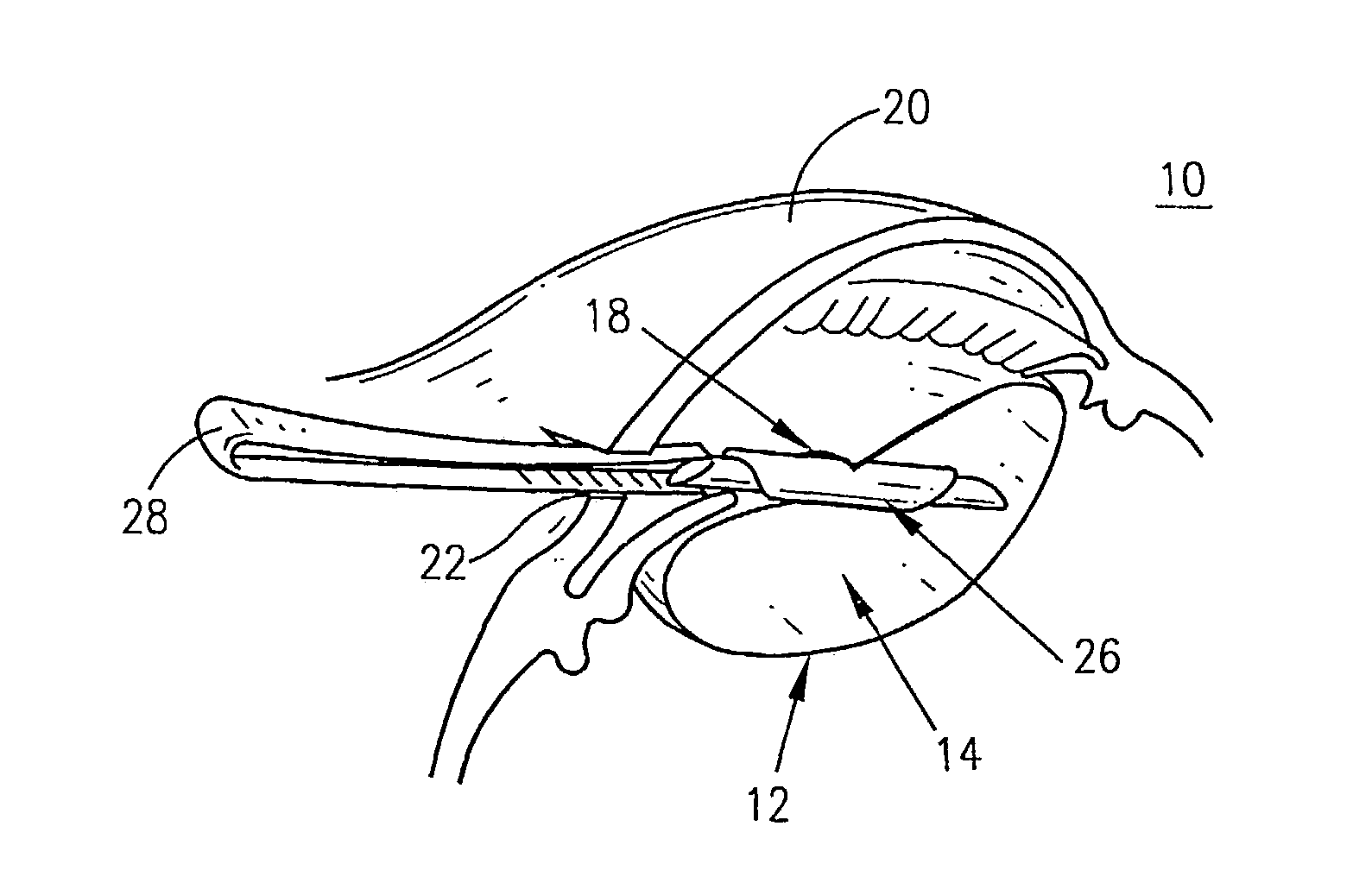
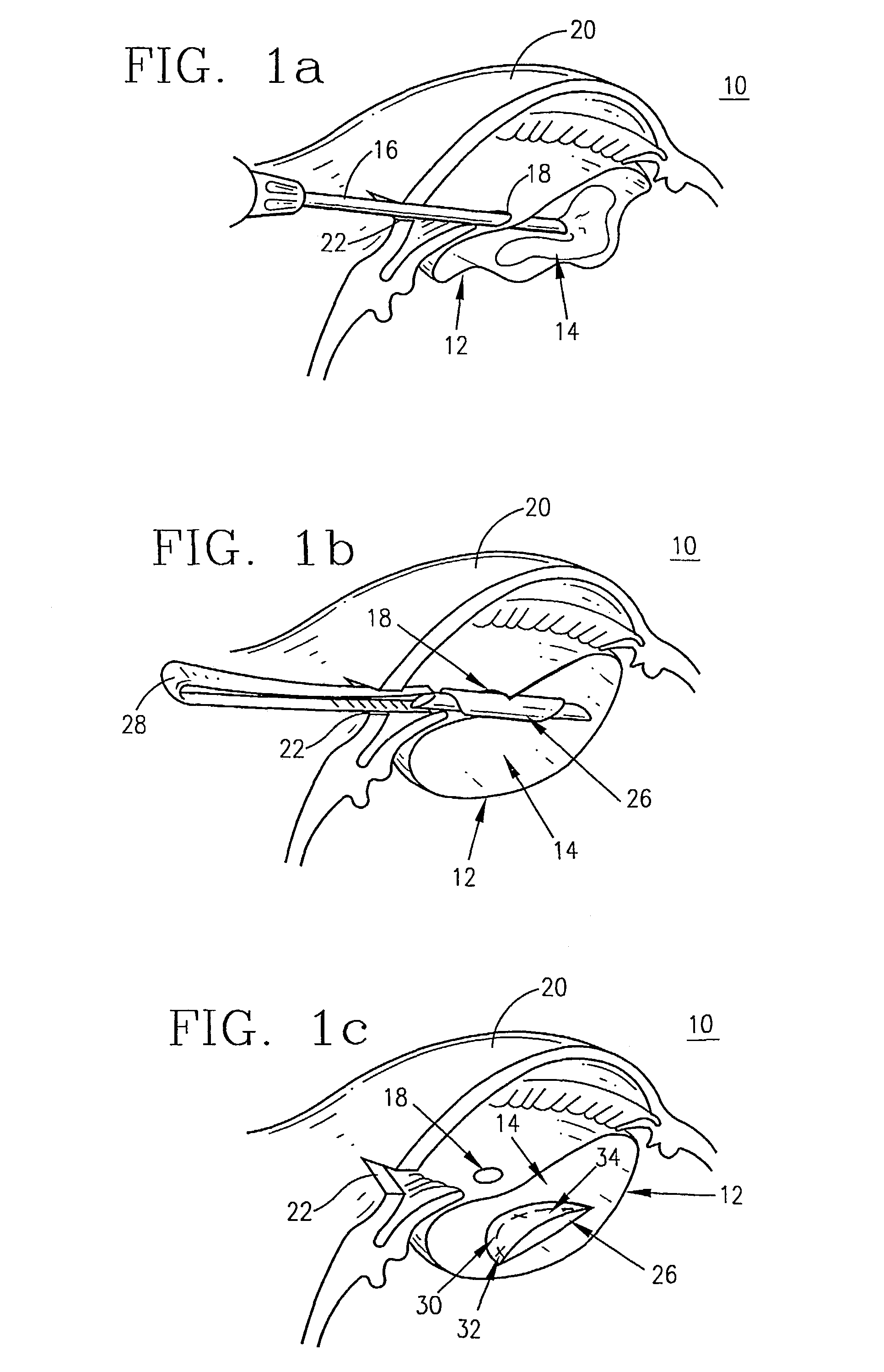
Method for Implantation of a SECL in the Lens Capsule and Lens Refilling Procedure (phaco-erzatz)

[0090]Dilation drops Mydriacyl 0.5% in the amount of 1 drop in each eye were added. Five (5) minutes later 1 drop in each was added of 1% atropine sulfate. The eye was then anesthetized with 35 mg / kg of ketamine and 5 mg / kg of xylazine with 0.75 mg / kg of acepromazine. After preparing the subject for surgery in aseptic conditions an eyelid speculum was inserted. A two step incision was made at the corneal periphery (1.5 mm in width). A mini-capsulorhexis was made (0.8–1.2 mm diameter). The lens material was aspirated. A cannula was then inserted (loaded with a pre-rolled SECL) through the mini-capsulorhexis into the lens capsule and disengaged from the cannula. The SECL then unfolded. A mini-capsulorhexis valve (MCV) was then placed across the capsule opening. A polymeric gel was then injected from a cannula through the capsulorhexis and into the lens capsule. When necessary, the gel was ...

example 2

In-Vitro Method for Verifying the Efficacy of a SECL in Correcting Ametropia Without Significantly Degrading the Accommodative Amplitude

[0091]The instructions outlined in this Example present one method by which the optical efficacy of a SECL may be demonstrated in situ. It is understood that a wide range of optometric measurement methods may be used for the same purposes. The method described here is chosen as it involves only simple componentries.

[0092]A working SECL would typically have a diameter sufficiently large to cover the entire pupil of the eye in most illumination conditions. Further, individual eyes would differ in their amount of ametropia. Given these two constraints, it is extremely difficult and certainly impractical to demonstrate the difference in optical performance (refraction and accommodation) of a post-ersatz lens against a post-ersatz lens which also has had a SECL implant.

[0093]Therefore the simplest demonstration of a SECL's optical performance is by using...


PUM

PropertyMeasurementUnit
diameteraaaaaaaaaa
specific gravityaaaaaaaaaa

Abstract

The present invention is directed to a supplemental endo capsular lens (SECL) and the method of inserting and embedding the SECL within either a gel or polymer, inside the capsule of the crystalline lens, during phaco-ersatz or similar surgical procedures in order to supplement the refractive power of the eye with a view to (1) correcting ametropia while (2) maintaining a useable amplitude of accommodation.

Description

[0001]This application is a 371 of PCT / US01 / 14728 filed May 8, 2001.FIELD OF THE INVENTION[0002]The present invention is directed to the field of surgical techniques and apparatuses designed to restore accommodation to eyes that have sustained lens removal.BACKGROUND OF THE INVENTION[0003]The human eye comprises a roughly spherical organ having essentially three distinct layers of tissue divided into three basic chambers. The tough outer sclerotic coat serves as a protective barrier for the eye, and forms the transparent cornea through which light passes into the eye. The sclerotic coat is composed of dense collagenous tissue. The middle choroid coat forms the iris, a diaphragm that controls the amount of light admitted into the interior of the eye through the pupil. The transparent crystalline lens is immediately posterior to the iris, and is held in place by zonular fibers attached to ciliary processes surrounding the crystalline lens. The zonular fibers collectively culminate in ...

Claims


Application Information

Patent Timeline
Patent Type & Authority Patents(United States)
IPC IPC(8): A61F2/14A61F2/16
CPCA61F2/1616A61F2/1664Y10S623/905
Inventor HO, ARTHURERICKSON, PAULMANNS, FABRICEPAREL, JEAN-MARIE
Owner THE VISION CRC LTD
Why Eureka
Try Eureka