System and method of characterizing vascular tissue

a vascular tissue and system technology, applied in the field of vascular tissue, can solve the problems of loss of image resolution, inaccurate evaluation of a vessel and its plaque content, and errors in analysis if the screen setting is changed, and achieve the effect of removing histological preparation artifacts

Active Publication Date: 2006-07-11
THE CLEVELAND CLINIC FOUND
View PDF60 Cites 219 Cited by
  • Summary
  • Abstract
  • Description
  • Claims
  • Application Information

AI Technical Summary

Problems solved by technology

This process, however, loses image resolution since the videotape typically has a lower resolution than the originally collected backscatter data.
Losing image resolution may result in an inaccurate evaluation of a vessel and its plaque content.
Since plaque (or tissue type) is identified by its appearance on the screen, errors may occur in the analysis if the screen settings have been modified.
Another drawback is that certain information (e.g., tissue composition, etc.) cannot readily be discerned from an IVUS image (at least not to any degree of certainty).

Method used


Image

  • System and method of characterizing vascular tissue
  • System and method of characterizing vascular tissue
  • System and method of characterizing vascular tissue

Examples


first embodiment

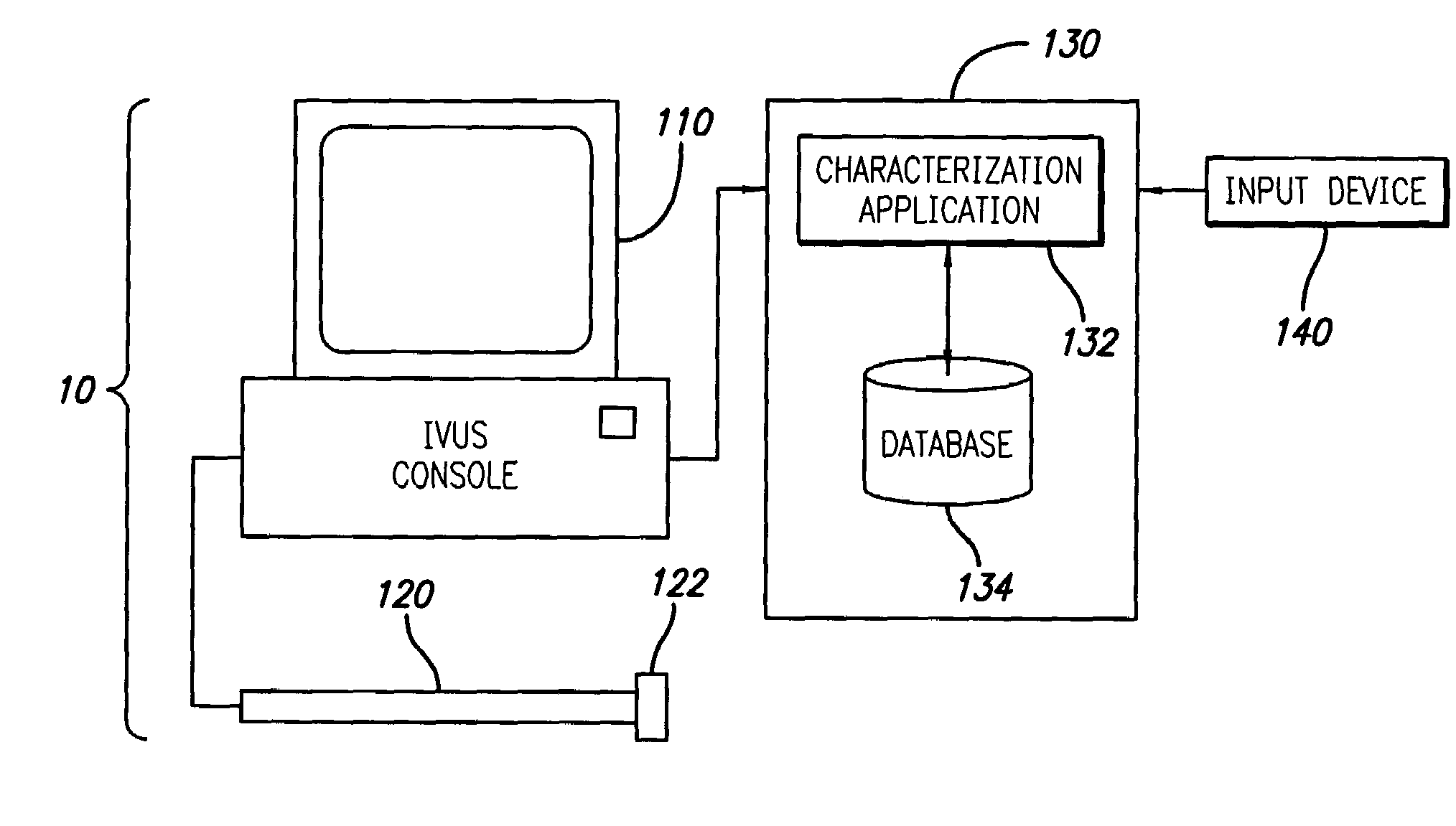
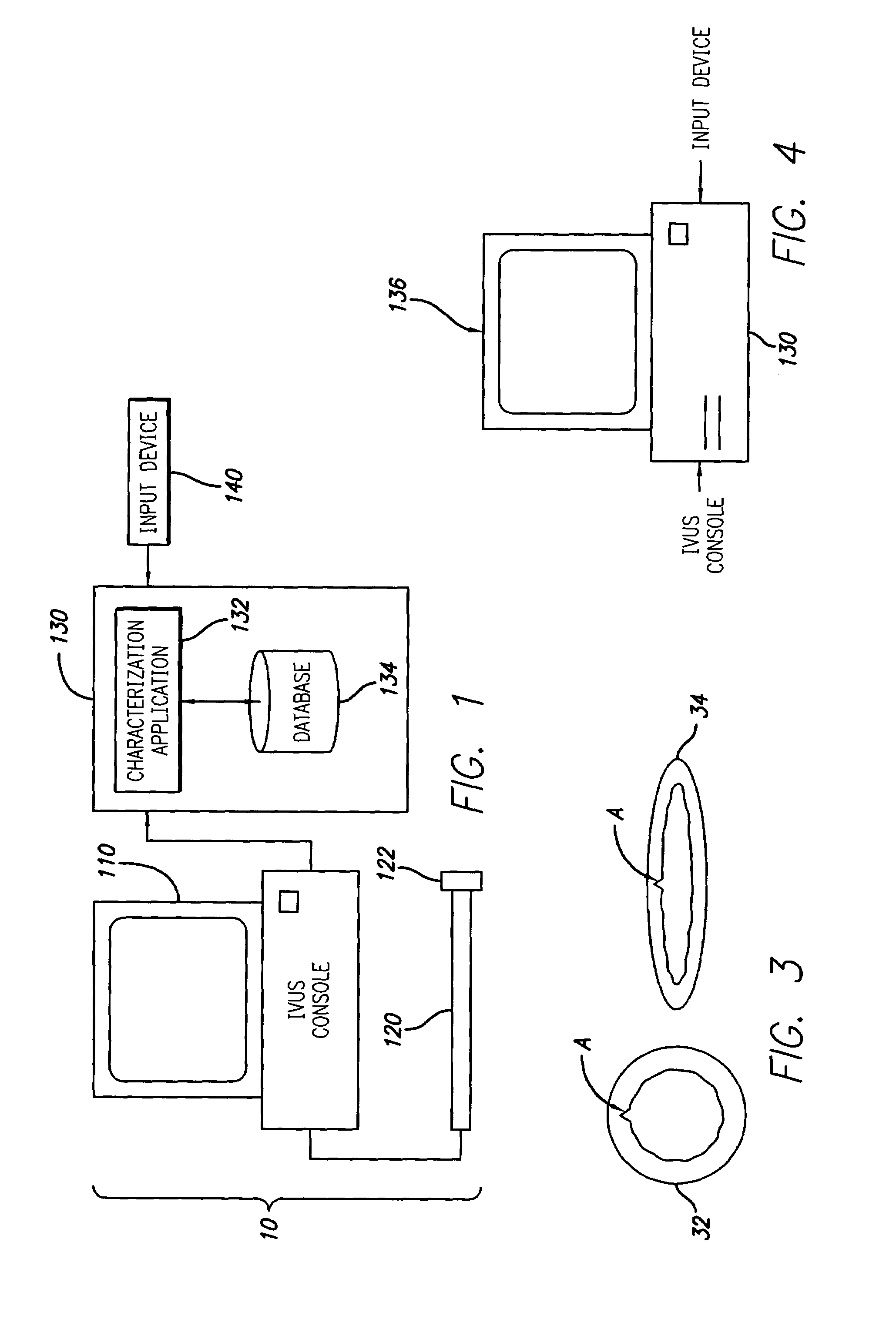
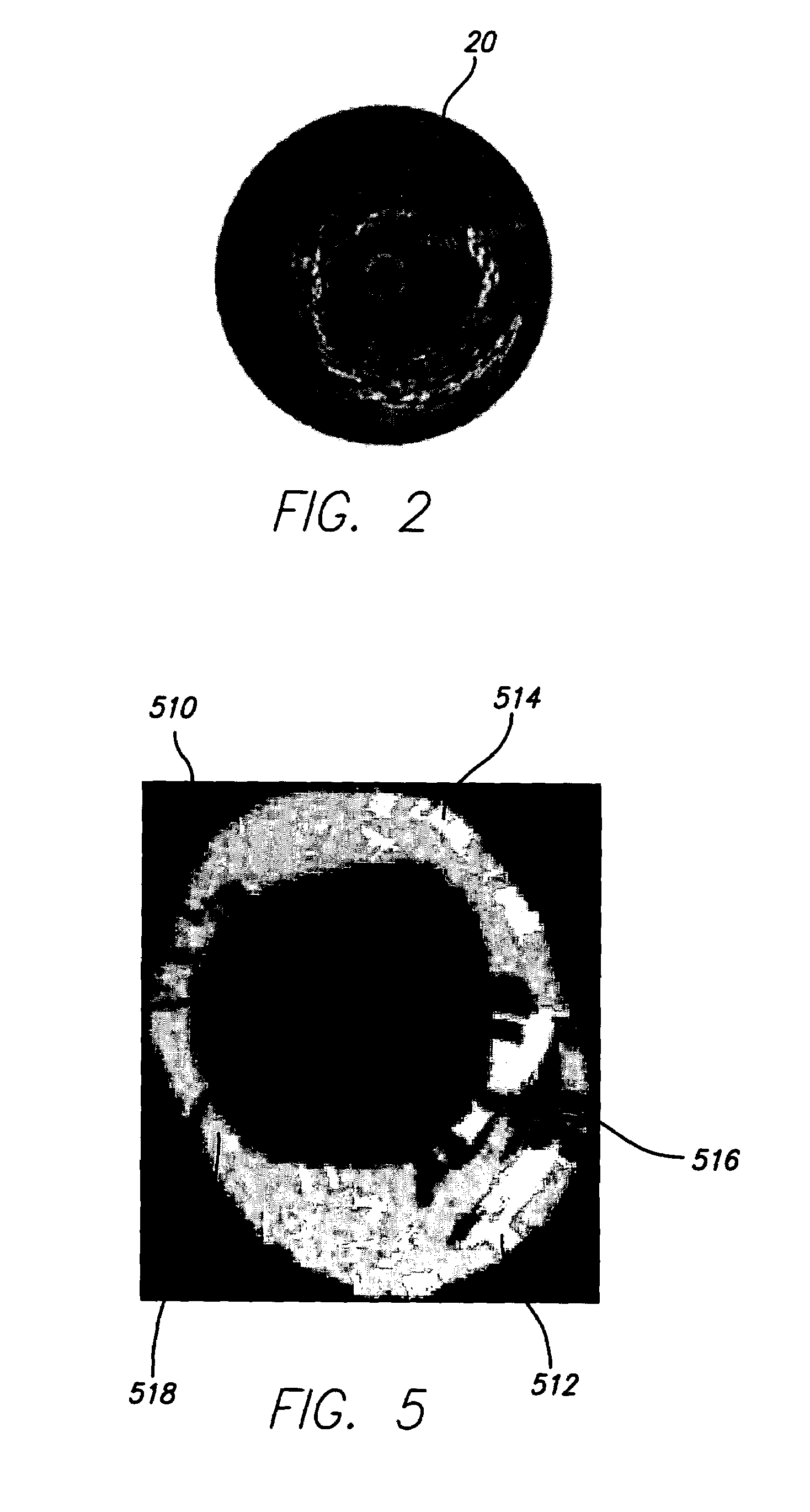
[0022]FIG. 1 illustrates a tissue-characterization system 10 operating in accordance with the present invention. In this embodiment, an intra-vascular ultrasound (IVUS) console 110 is electrically connected to an IVUS catheter 120 and used to acquire RF backscattered data (i.e., IVUS data) from a blood vessel. Specifically, a transducer 122 is attached to the end of the catheter 120 and is carefully maneuvered through a patient's body to a point of interest. The transducer is then pulsed to acquire echoes or backscattered signals reflected from the tissue of the vascular object. Because different types and densities of tissue absorb and reflect the ultrasound pulse differently, the reflected data (i.e., IVUS data) can be used to image the vascular object. In other words, the IVUS data can be used (e.g., by the IVUS console 110) to create an IVUS image. An exemplary IVUS image 20 is provided in FIG. 2, where the light and dark regions indicate different tissue types and / or densities....

second embodiment

[0034]Accordingly, in the present invention, the characterization application is adapted to receive IVUS data, determine parameters related thereto, and use the parameters stored in the database (i.e., histology data) to identify tissue type(s) or characterization(s) thereof. Specifically, with reference to FIG. 1, the characterization application 132 is adapted to receive IVUS data from the IVUS console 110. The characterization application 132 is then adapted to identify at least one parameter associated (either directly or indirectly) with the IVUS data. It should be appreciated that the IVUS data may either be received in real-time (e.g., while the patient is in the operating room) or after a period of delay (e.g., via CD-ROM, etc.). It should further be appreciated that the identified parameters should be related (generally) to the stored parameters. Thus, for example, an estimated Y intercept parameter should be identified if data related to a signal's estimated Y intercept is...


PUM


Abstract

A system and method is provided for using backscattered data and known parameters to characterize vascular tissue. Specifically, in one embodiment of the present invention, an ultrasonic device is used to acquire RF backscattered data (i.e., IVUS data) from a blood vessel. The IVUS data is then transmitted to a computing device and used to create an IVUS image. The blood vessel is then cross-sectioned and used to identify its tissue type and to create a corresponding image (i.e., histology image). A region of interest (ROI), preferably corresponding to the identified tissue type, is then identified on the histology image. The computing device, or more particularly, a characterization application operating thereon, is then adapted to identify a corresponding region on the IVUS image. To accurately match the ROI, however, it may be necessary to warp or morph the histology image to substantially fit the contour of the IVUS image. After the corresponding region is identified, the IVUS data that corresponds to this region is identified. Signal processing is then performed and at least one parameter is identified. The identified parameter and the tissue type (e.g., characterization data) is stored in a database. In another embodiment of the present invention, the characterization application is adapted to receive IVUS data, determine parameters related thereto (either directly or indirectly), and use the parameters stored in the database to identify a tissue type or a characterization thereof.

Description

CROSS-REFERENCE TO RELATED APPLICATIONS[0001]This application claims the benefit pursuant to 35 U.S.C. § 119(e) of U.S. Provisional Patent Application Nos. 60 / 406,183, filed Aug. 26, 2002, 60 / 406,254, filed Aug. 26, 2002, 60 / 406,148, filed Aug. 26, 2002, 60 / 406,184, filed Aug. 26, 2002, 60 / 406,185, filed Aug. 26, 2002, and 60 / 406,234, filed Aug. 26, 2002, all of which are incorporated herein, in their entirety, by reference.BACKGROUND OF THE INVENTION[0002]1. Field of the Invention[0003]The present invention relates to vascular tissue, or more particularly, to a system and method of using backscattered data and known parameters to characterize vascular tissue.[0004]2. Description of Related Art[0005]The present invention relates to the intra-vascular ultrasound (IVUS) analysis arts. It finds particular application to a system and method for quantitative component identification within a vascular object including characterization of tissue. It should be appreciated that while the pre...

Claims


Application Information

Patent Timeline
Patent Type & Authority Patents(United States)
IPC IPC(8): A61B8/00A61B8/12A61B8/08G06T7/00
CPCA61B5/02007A61B8/12G06T7/0012A61B5/7257A61B5/726G06T2207/30101A61B8/0833
Inventor NAIR, ANUJAVINCE, D. GEOFFREYKLINGENSMITH, JON D.KUBAN, BARRY D.
Owner THE CLEVELAND CLINIC FOUND
Why Eureka
Try Eureka