Internal biochemical sensing device

a biochemical and sensing device technology, applied in the field of implanted devices, can solve the problems of repeated painful finger pricks, difficult to achieve, and serious complications, and achieve the effect of convenient patient transpor

Inactive Publication Date: 2006-08-22
UNIV OF SOUTHERN CALIFORNIA
View PDF49 Cites 140 Cited by
  • Summary
  • Abstract
  • Description
  • Claims
  • Application Information

AI Technical Summary

Benefits of technology

The present invention provides a small and easy-to-carry bio-sensing device that can detect substances inside a person's body. It consists of a special sensor made up of materials that change color when they come into contact with specific molecules (analytes). A thin light guide connects this sensor to a larger machine called an information analyzer which processes the data collected by the sensors. This analysis allows healthcare professionals or patients themselves to monitor various biological markers such as blood glucose levels without needing to draw any fluid samples.

Problems solved by technology

The technical problem addressed in this patent text is the difficulty of repeatedly measuring specific biomolecules (also called analytes) inside a person's body without causing discomfort or requiring complicated equipment. Current methods require multiple fingersprikes and specialized devices, leading to poor patient adherence. This patent seeks to develop a less invasive and more efficient way to measure various analytes including glucose, cholesterol, and proteins.

Method used


Image

  • Internal biochemical sensing device
  • Internal biochemical sensing device
  • Internal biochemical sensing device

Examples


example 1

Material and Method of Forming a Biosensor Element

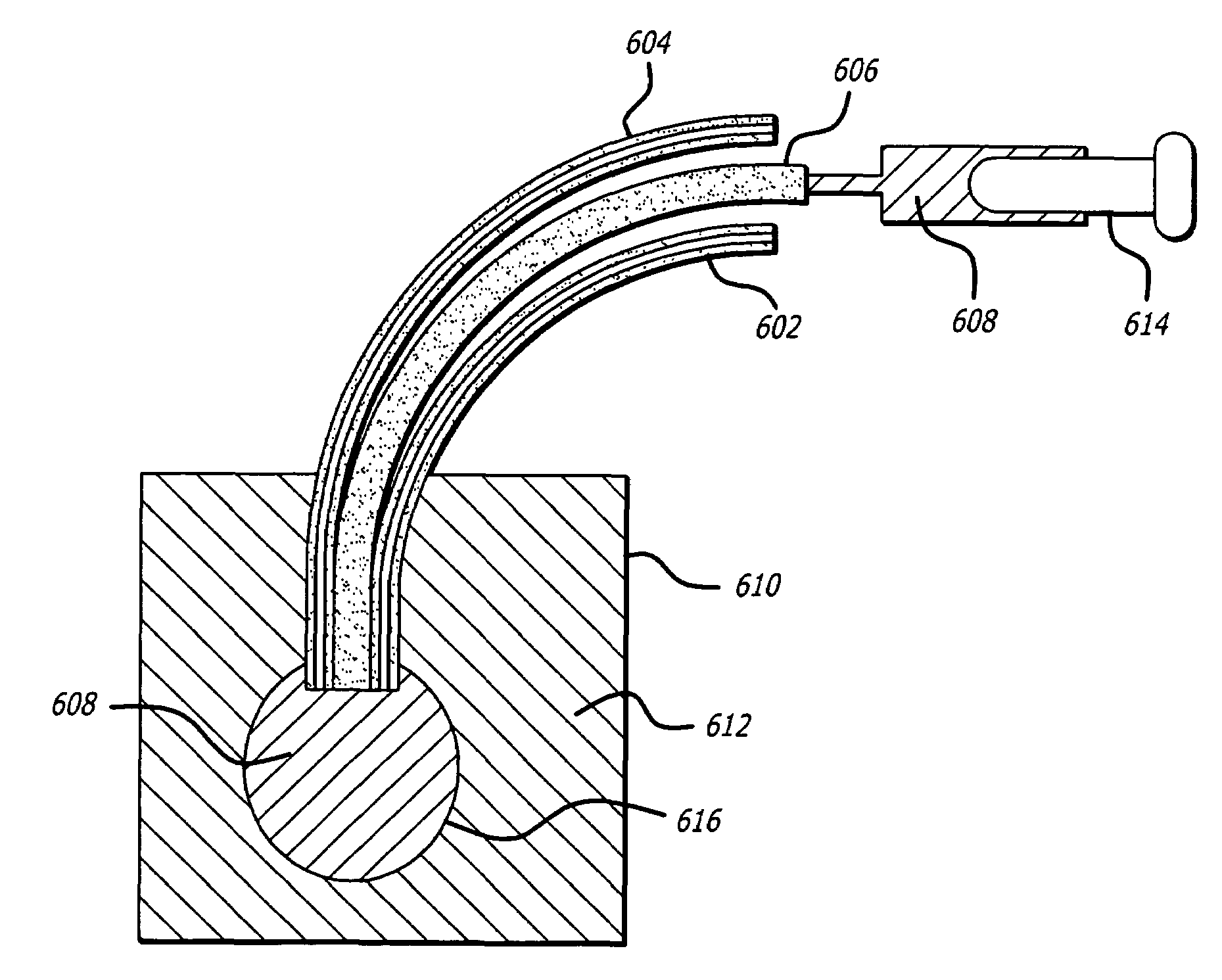
[0065]Note that the outer layer described and the method of preparing the outer layer described (in steps 3 and 5, for example) are optional. The outer layer may be desirable where it is desired to prevent the inner PEG layer (having reactants and fluorophores) from being directly exposed to the patient's tissue.

[0066]A. Composition of polyethylene glycol hydrogel inner and outer layer, including an exemplary sensing system for measurement of glucose by competitive binding of fluorophores.

[0067]

Inner PEGOuter PEGLayer (w / w)Layer (w / w)PEG-DA 575 (polyethylene glycol-32.06%32.15%diacrylate)PEG-NHS 3400 (a-acryloyl,ω-N-hydroxysuccimidyl ester of poly(ethylene 0.16%noneglycol)-propionic acid,Con A immobilized)TRITC-Con A / FITC-dextran (100 / 1) 0.13%noneDMPA (2,2-dimethoxy-2-phenylacetophenone, radical initiator) 0.32% 0.32%TPT (trimethylol propane 3.2% 3.3%triacrylate, Cross-linking agent to formcopolymer hydrogel for the sake ofstability ...

example 2

Fluorescence Resonance Energy Transfer Phenomenon as Applied to an Affinity Binding Assay System for Glucose Sensing

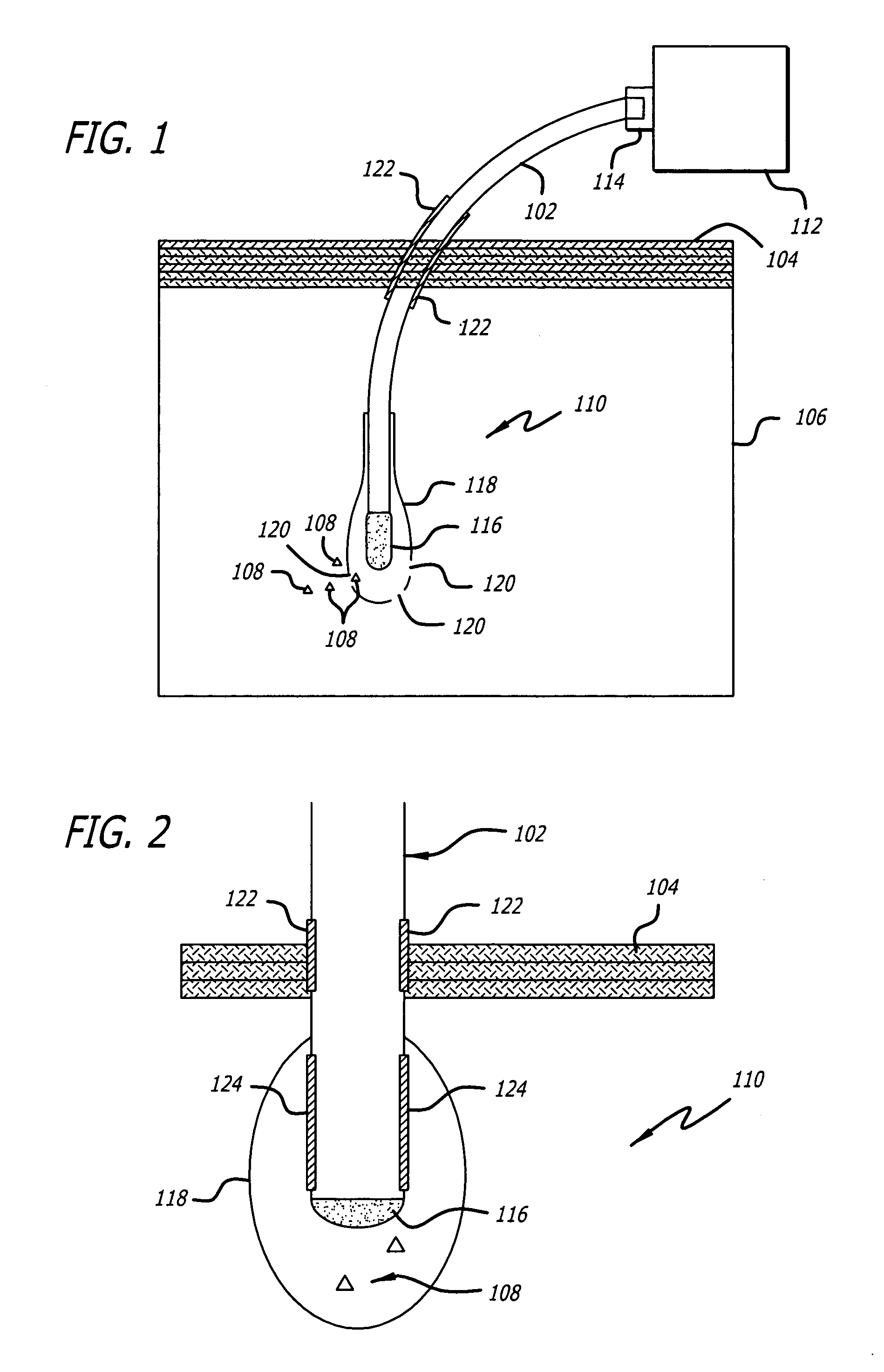
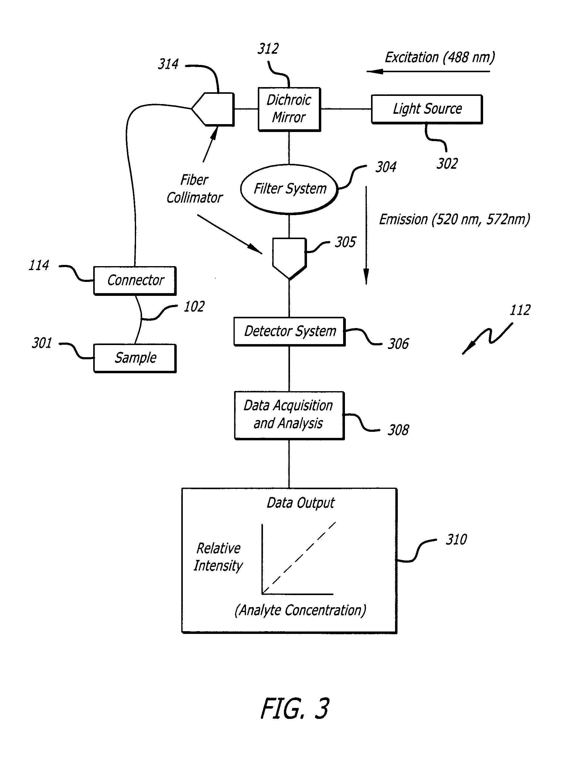
[0074]This example describes the fluorescence transduction techniques based the affinity binding assay system for polysaccharides based on the jack bean lectin concanavalin A (ConA). Mansouri S, Schultz J. “A Miniature Optical Glucose Sensor Based on Affinity Binding.”Biotechnology 1984, 885–90. This system uses a biosensor based on competitive binding between glucose and dextran. The assay aqueous solution is encapsulated within a hollow dialysis membrane permeable to glucose. In the assay, Con A is used as a receptor for competing species of fluorescein isothiocyanate (FITC) labeled dextran and glucose. FITC-dextran reversibly binds to Con A; as glucose concentration increases, FITC-dextran is displaced from ConA receptor sites, thus increasing the concentration and measurable fluorescence intensity of FITC-dextran.

[0075]Also, fluorescence resonance energy transfer (...

example 3

Clinical Analytes for which the Biosensing Device may be Configured to Detect, Include, but are not Limited to:

[0077]

Biological ElementsAnalytesEnzymes / Metabolitesglucose, cholesterol, amylase, urea, triglyceridespH, Na+, Ca2+, K+, H+, Cl−, HCO3, Fe, NH3Creatinine kinase, Creatinine, Aspartate amino-transferase, Phenylalanine, Lactate dehydrogen-ase, Akaline phosphotase, GOT, BilirubinGasesoxygen, carbon dioxide, ammoniaTherapeutic DrugsSalicylate, Acetaminophen (paracetamol),Ethanol, Theophylline, Dilantin, Gentamicin,HormonesTobramicin, Digoxin cortisol, estriol, pro-gesterone, aldosterone, cortisone, thyroxinebinding globulin, placental lactogen, prolactin,human chorionic gonadotropin, insulin,parathyroid hormone, growth hormone,angiotensin, oxytocin, vasopressinAntibodiesIgM (total), IgG (specific), Syphilis, RubellaAntigensA & B, Rh, Hepatitis, Alpha-fetoprotein,various cancer proteinsHall E. (1991) Biosensors. New Jersey: Prentice Hall.


PUM


Abstract

A biosensing device for detecting biological analytes, and methods of use and manufacture, are disclosed. The device includes a biosensing element that can remain implanted for extended periods of time. The biosensing element is connected to an optical fiber terminating outside of the body. The optical fiber is also connected to an information analyzer. The information analyzer directs light through the optical fiber into the biosensing element. The light excites fluorophores, created by a chemical reaction between analytes and biosensing material within the biosensing element. Emitted fluorescent light is redirected through the optical fiber to the information analyzer. Optical gratings within the filtering member of the information analyzer selectively deflect fluorescent emissions into a cladding layer. Detectors attached to the cladding layer detect the deflected fluorescent emissions and, according to their determined wavelength, report the presence or quantity of specific analytes to the patient on an external display.

Description


Claims


Application Information

Patent Timeline
Owner UNIV OF SOUTHERN CALIFORNIA
Why Eureka
Try Eureka