Latent human tuberculosis model, diagnostic antigens, and methods of use
a human tuberculosis and diagnostic antigen technology, applied in the field of mycobacterial latency, can solve the problems of inability to specifically identify latently infected individuals, difficult to contain tuberculosis outbreaks, and inability to detect latently infected individuals
- Summary
- Abstract
- Description
- Claims
- Application Information
AI Technical Summary
Benefits of technology
Problems solved by technology
Method used
Image
Examples
example 1
Preparation and Characterization of an In Vitro Granuloma Model
[0194]The principal defense of the human host against a mycobacterial infection is the formation of granulomas, which are compact organized collections of activated macrophages, including epithelioid and multinucleated giant cells, surrounded by T lymphocytes, and later by fibroblasts and collagen that aggregate around the macrophage-core. The granuloma may prevent active (non-latent) disease by sequestering the invading organisms. If the granuloma is maintained, these bacteria may remain latent for many years.
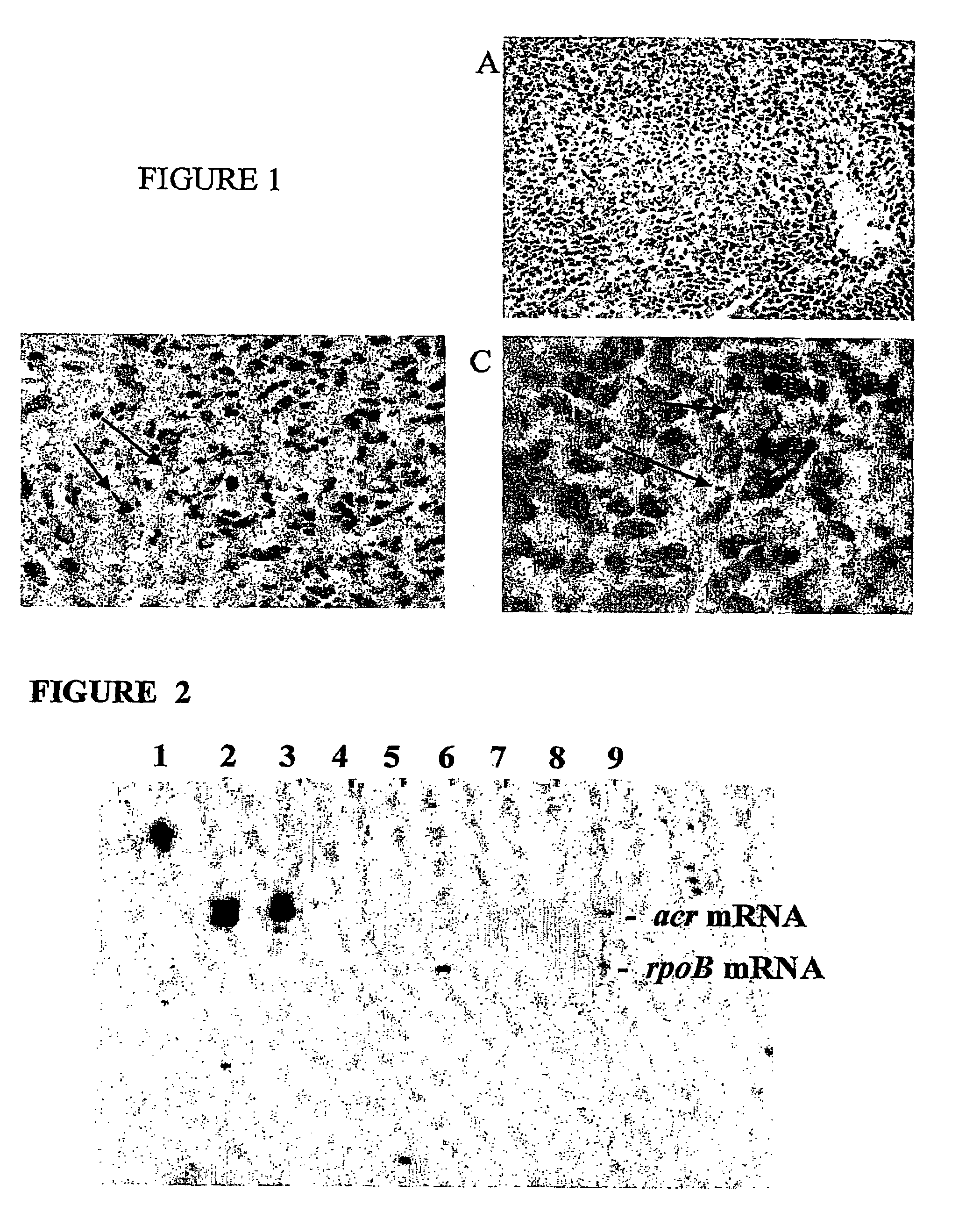
[0195]To study this process of granuloma formation and the granuloma's subsequent breakdown when host defenses are compromised, an in vitro model was developed. This example provides a description of one method for producing the in vitro granuloma that can be used as a model system, as well as several methods used to characterize the model. In overview, human peripheral blood mononuclear cells, autologous macrophag...
example 2
Detection of Differential Transcription of Acr mRNA in a Granuloma Model
[0200]Using bacteria cultured in an anoxic chamber, M. tuberculosis genes were identified that were differentially expressed after much of the available oxygen had been utilized. The genes that were differentially expressed were acr, sigF, oxyR and aphC. Of these four genes, acr encodes a protein (α-crystallin) that is secreted by the Mycobacterium.
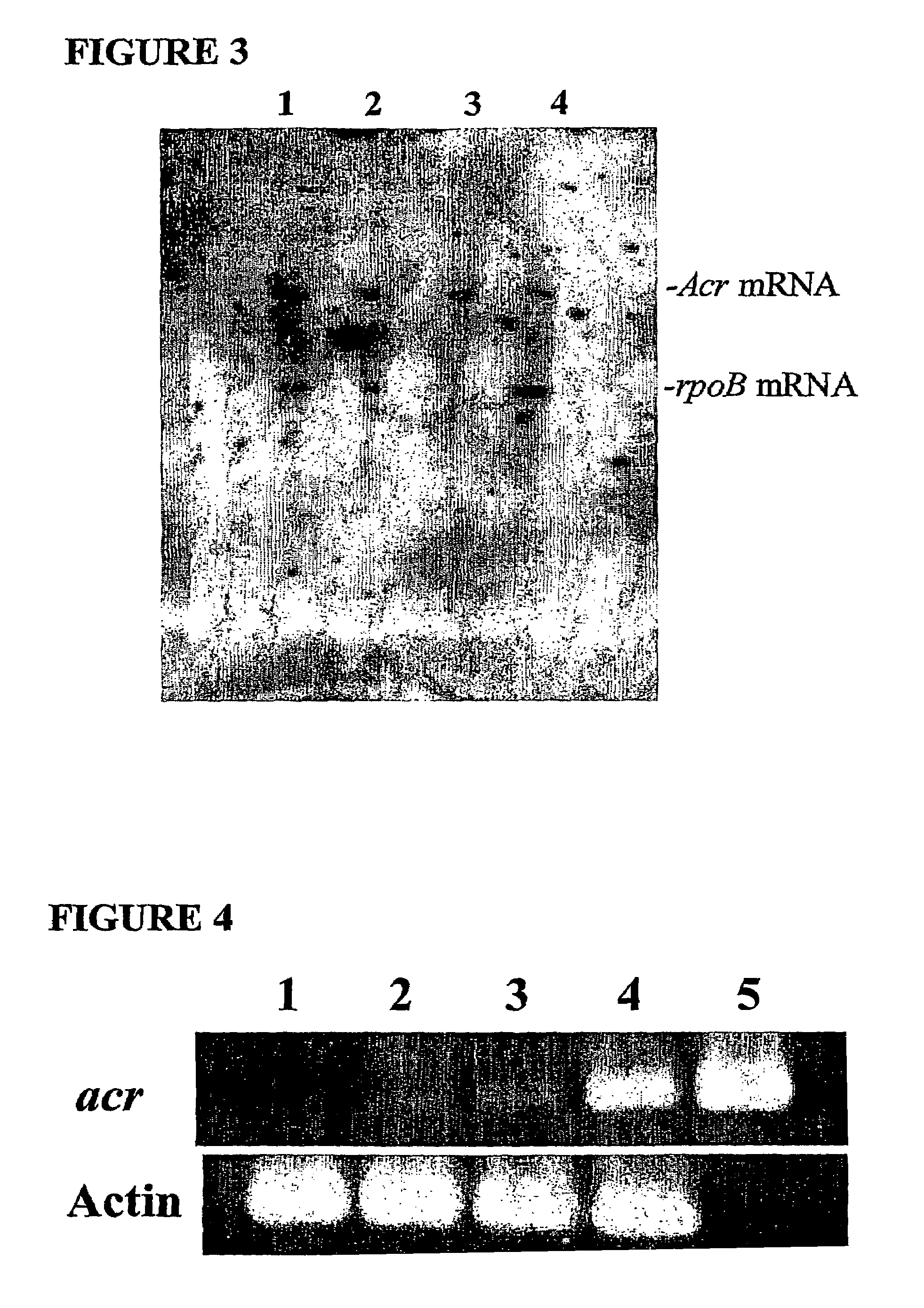
[0201]In order to confirm that these genes are expressed in the in vitro granuloma model, RT-PCR and RNA protection assays were performed. These assays showed that mRNA from mycobacterial genes acr, aphC, and sigF were transcribed. These transcripts were not found in uninfected in vitro granuloma controls.
[0202]Typical results from representative experiments are shown in FIGS. 2 and 3, which are ribonuclease protection assay (RPA) blots. In FIG. 2, acr mRNA was observed at all four time points while RpoB mRNA was only observed in aerobically grown cultures. In FIG. 3,...
example 3
Other In Vitro Latency Models
[0203]The following example provides other in vitro latency models, which can be used, for example, to confirm results obtained with the in vitro granuloma model (e.g., to screen drugs and immunostimulatory compounds and to identify latency-specific antigens).
Guinea Pig Aerosol Infection Model
[0204]When infected by aerosol inoculation using a small number of M. tuberculosis bacilli, it was observed that the bacteria cause formation of granulomas associated with the epithelial pneumocytes in the deep alveoli of the lung. Though these granulomas apparently are not able to completely contain the infection, and the bacteria eventually overwhelm this animal, there are a number of similarities with human granulomas. For example, these granulomas center on necrotic areas and contain predominantly macrophages, including epithelioid and multinucleated giant cells and T lymphocytes. Differential transcription of the bacterial Acr gene has been detected in these gr...
PUM
| Property | Measurement | Unit |
|---|---|---|
| Tm | aaaaa | aaaaa |
| diameter | aaaaa | aaaaa |
| time | aaaaa | aaaaa |
Abstract
Description
Claims
Application Information
 - R&D
- Intellectual Property
- Life Sciences
- Materials
- Tech Scout
- Unparalleled Data Quality
- Higher Quality Content
- 60% Fewer Hallucinations
Browse by: Latest US Patents, China's latest patents, Technical Efficacy Thesaurus, Application Domain, Technology Topic, Popular Technical Reports.
© 2025 PatSnap. All rights reserved.Legal|Privacy policy|Modern Slavery Act Transparency Statement|Sitemap|About US| Contact US: help@patsnap.com


