Direct selection of cells by secretion product
a technology of secretion product and cell, applied in the field of cell population analysis and cell separation, can solve the problems of inability to accurately identify and/or use the procedures that analyze and separate cells by markers naturally associated with the cell surface to separate secretor cells from non-secretor cells, and inability to identify quantitative differences in secretor cells. quantitative differences, the effect of reducing the number of secretor cells
- Summary
- Abstract
- Description
- Claims
- Application Information
AI Technical Summary
Benefits of technology
Problems solved by technology
Method used
Image
Examples
example 1
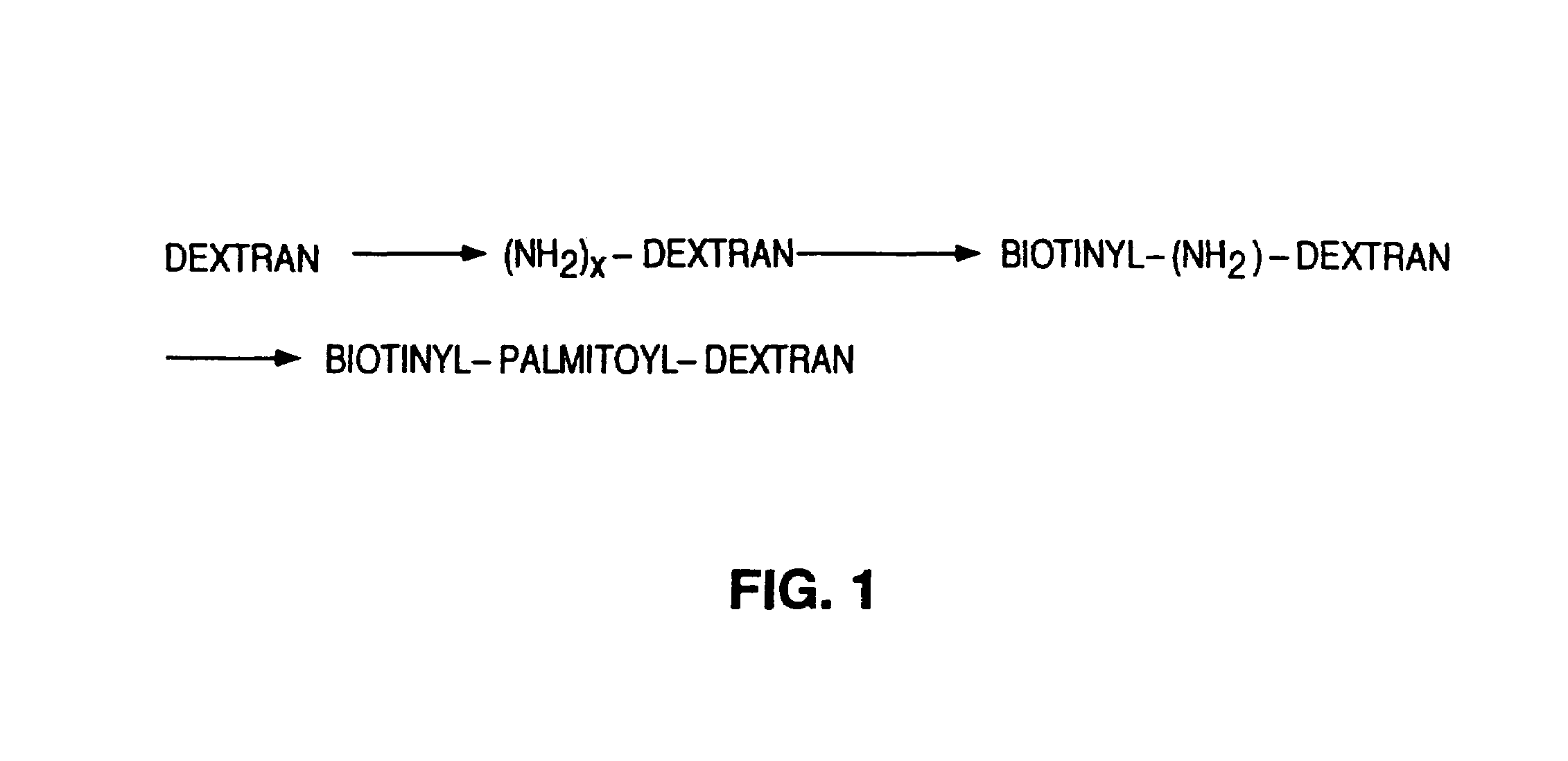
[0090]The purpose of this example was to separate living cells that secrete a given product from a mixture of externally identical cells. The B.1.8. hybridoma cell line and the X63. Ag 8 6.5.3. myeloma cell line were used as the test system. About 60% of B.1.8. cells secrete IgM; the myeloma line secretes no protein. The secreting cells were to be separated from B.1.8. and from mixtures of B.1.8. with Ag 8 6.5.3. cells. To achieve this, a procedure was developed to capture, on the cell surface, a product secreted by the cell, hold the product on the cell surface, and thus label the cell in question. The capture antibodies were attached in two steps: 1) biotinylation of the cells; and 2) attachment of the capture antibody through an avidin-biotin coupling reaction. The labeled cells were then separated from cell mixtures.
Biotinylation of the Cell Surface with Biotinylpalmitoyldextran
[0091]The objective was the synthesis of an anchor moiety which is a large macromolecule with biotin g...
example 2
[0135]This example demonstrates the effect of carrying out the secretion phase in a gelatinous medium as compared to a high viscosity medium on the capture of secreted product by cells containing a biotin anchor moiety linked via avidin to the capture moiety.
Chemical Biotinylation of Cells Using NHS-LC-Biotin
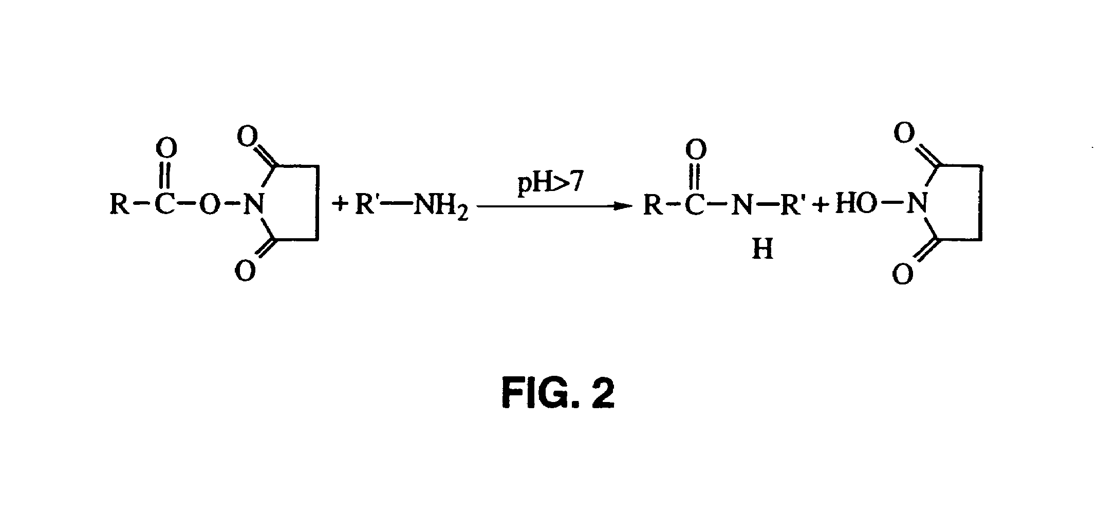
[0136]A mixture of B.1.8. and X63 cells was chemically biotinylated by the following procedure. Cell suspensions containing 107 to 108 cells were centrifuged, the supernatant removed, and the pellet resuspended in a solution of 200 μl PBS pH 8.5, containing 0.1 to 1 mg / ml NHS-LC-Biotin (Pierce, Rockford, Ill., U.S.A.). After incubation for 30 minutes at room temperature the cells were washed two times extensively with 50 ml PBS / BSA. Labeling with avidin conjugate was within 24 hours of the biotinylation.
Linkage of the Biotinylated Cells to Capture Antibodies with Avidin
[0137]The cells biotinylated by reaction with NHS-LC-Biotin were labeled with an avidin conjugate of LS136 (con...
example 3
[0144]The following describes a method to measure the absolute amount of secretion and to compensate for different amounts of capture moiety on the cell surfaces.
[0145]During the secretion phase the cells are exposed to a low concentration of tagged product supplied with the medium; the tagged product binds to but does not saturate the product binding sites on the cells. Incubation during this phase causes both the secreted product and the tagged product to bind to the cells. The cells are then subjected to labeling with the label is moiety specific for the product (both tagged and secreted). Measurement of the tag using one parameter, and the total product in the other parameter, the amount secreted by a cell is normalized, and the different amounts of capture antibody on the cells in the mixture is compensated for.
PUM
 Abstract
Description
Claims
Application Information
 - R&D
- Intellectual Property
- Life Sciences
- Materials
- Tech Scout
- Unparalleled Data Quality
- Higher Quality Content
- 60% Fewer Hallucinations
Browse by: Latest US Patents, China's latest patents, Technical Efficacy Thesaurus, Application Domain, Technology Topic, Popular Technical Reports.
© 2025 PatSnap. All rights reserved.Legal|Privacy policy|Modern Slavery Act Transparency Statement|Sitemap|About US| Contact US: help@patsnap.com


