Endoscopic mucosal resection device and method of use

a mucosal resection and endoscope technology, applied in the field of endoscope, to achieve the effect of reducing the risk of alimentary canal perforation, reducing patient burns, and ensuring the safety of resection

Active Publication Date: 2007-03-06
ETHICON ENDO SURGERY INC
View PDF34 Cites 71 Cited by
  • Summary
  • Abstract
  • Description
  • Claims
  • Application Information

AI Technical Summary

Benefits of technology

[0007]Once tissue is drawn into the resection chamber, an electrosurgical wire can be used for transection. In contrast to the flexible electrosurgical snares used in existing devices, the present invention can employ a relatively rigid wire positioned within the device to be drawn across or pushed across the chamber opeing to excise the entrapped tissue. The wire is only electrically active over the portion, which is exposed, non insulated, to the chamber opening. The present invention can also include a flexible, electrically conductive tissue stop, which can function to limit the depth of tissue that can enter the suction chamber for resection. Such a tissue stop can provide for greater safety of resection by reducing risk of alimentary canal perforation and reducing patient burns from monopolar ground pads. The tissue stop can also be perforated for communicating vacuum.

Problems solved by technology

Unfortunately, definitive therapy has historically involved invasive surgical resection of the lesion and adjacent bowel.

Method used


Image

  • Endoscopic mucosal resection device and method of use
  • Endoscopic mucosal resection device and method of use
  • Endoscopic mucosal resection device and method of use

Examples


Embodiment Construction

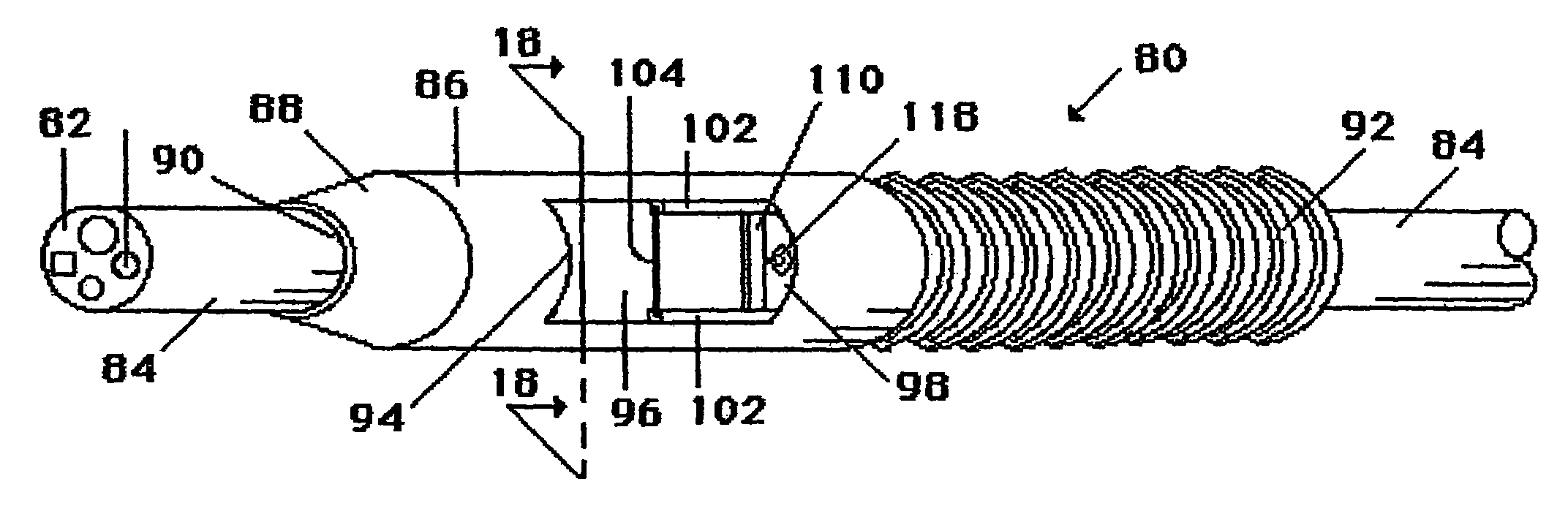
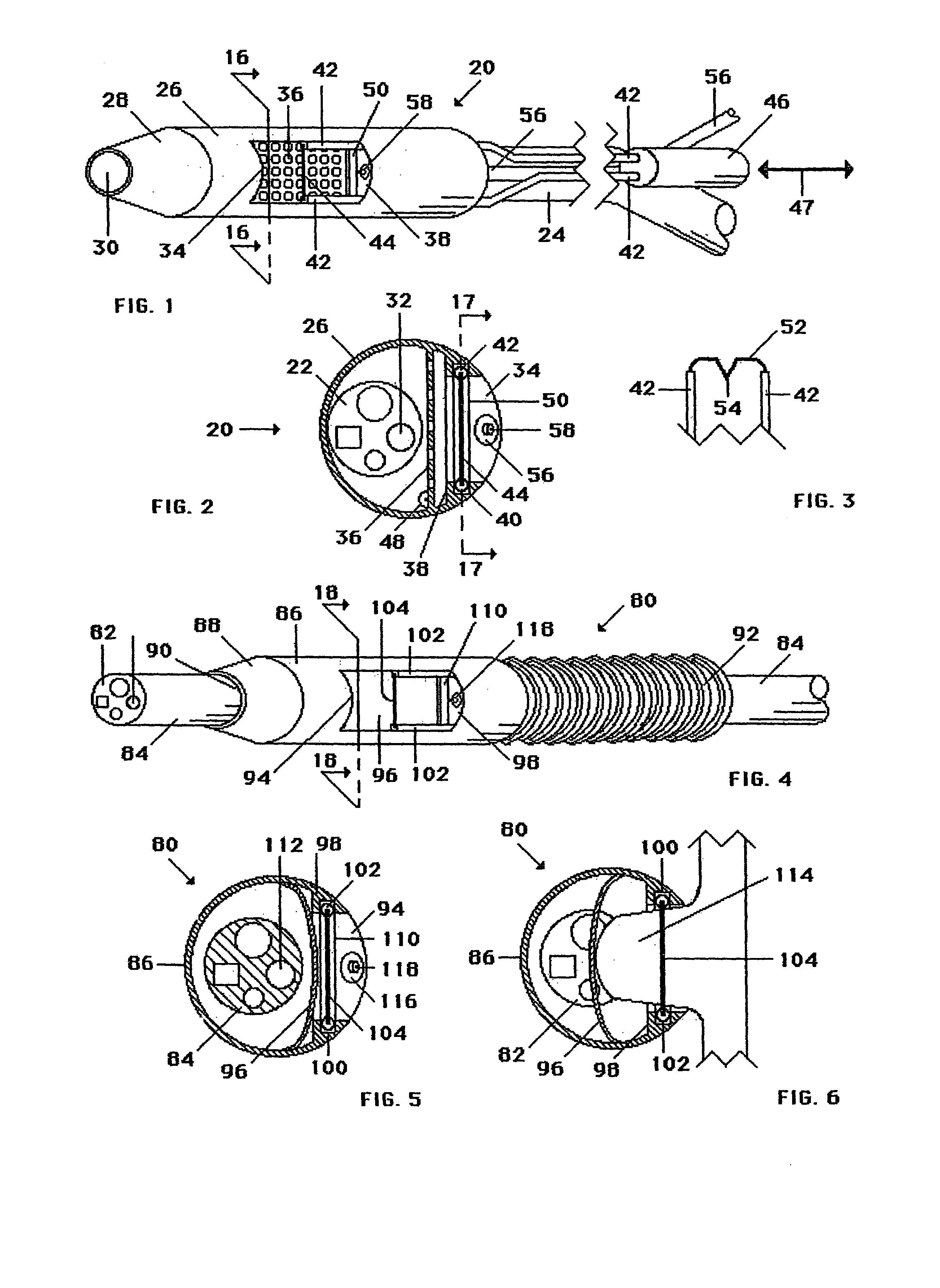
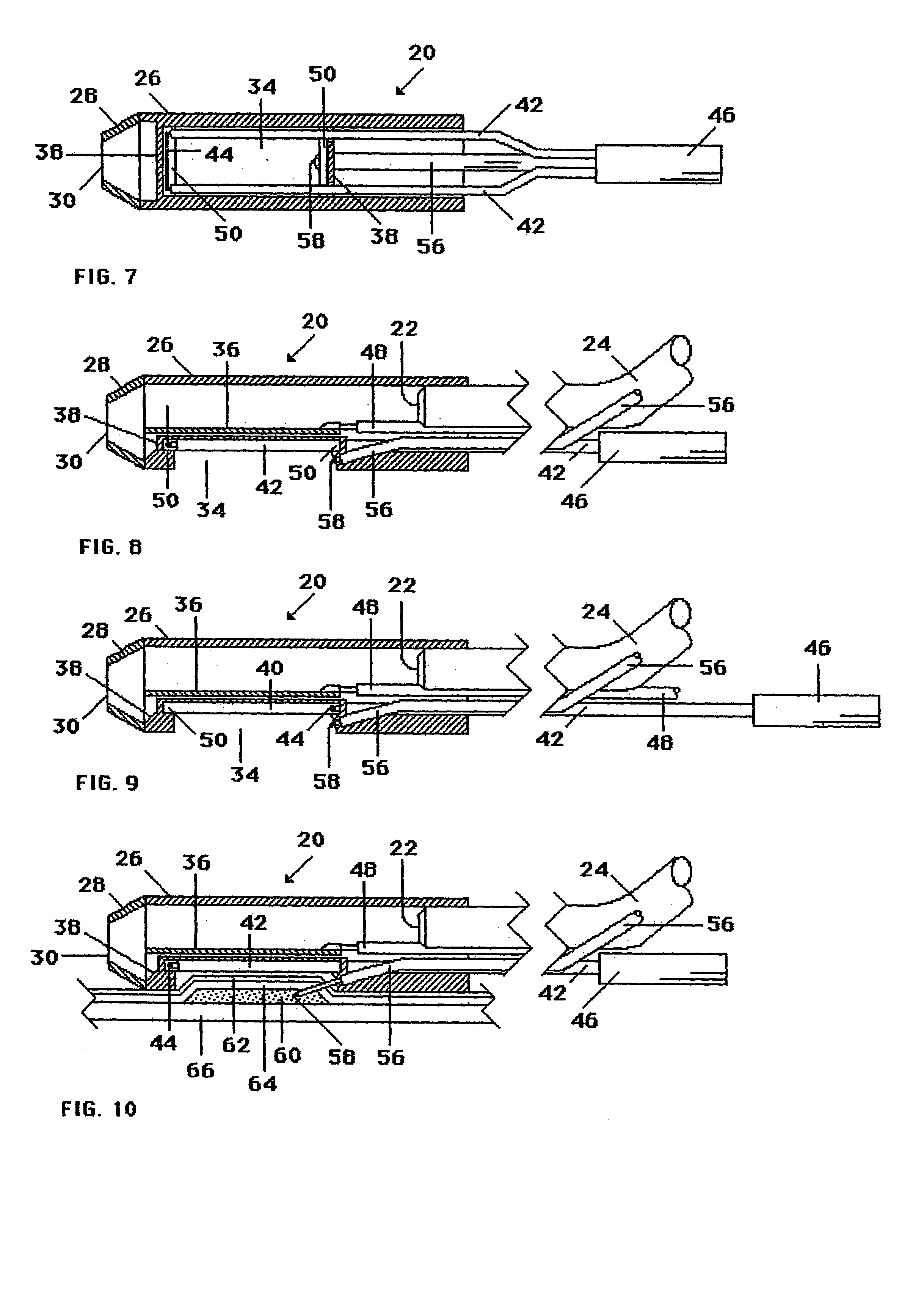
[0036]With reference to FIGS. 1, 2, 7&8, one embodiment of a cutting device 20 of the present invention is shown attached to a distal end 22 of a commercially available endoscope. Endoscope 24 may be made by Olympus Optical, having an outside diameter of about 0.2 to 0.7 inches. Cutting device 20 can have a rigid or semi-rigid cylindrical cutter support 26 which is attached to the endoscope perimeter by any suitable means, such as by shrink wrap, adhesive, snap fit, press fit, threaded engagement, or other suitable means known in the art for connecting one generally hollow member to another along parallel longitudinal axes.

[0037]Distal end 22 of endoscope 24 can be located at one end of cutter support 26. A flexible conical member 28 can be attached to the opposite, distal end of cutter support 26. Conical member 28 can be employed to provide for a smooth entry of cutting device 20 into the alimentary canal of a patient. Conical member 28 can have an open distal end 30 of about 0.3 ...


PUM


Abstract

A medical apparatus and method useful for resecting tissue from the gastrointestinal tract are disclosed. The apparatus can include an RF tissue cutting device disposed inward of a side opening in the device. A tissue stop can be used to control the depth of tissue resected, and the tissue stop can include holes for communicating vacuum for drawing tissue into the side opening. The tissue stop can be electrically grounded with respect to the RF tissue cutting device, and the tissue stop can provide one pole of an RF electrical circuit.

Description

FIELD OF THE INVENTION[0001]The present invention is related generally to endoscopy and more particularly to endoscopic mucosal resection.BACKGROUND OF THE INVENTION[0002]Cancerous or benign lesions of the GI tract often start in the mucosal layer of the stomach or intestines. With improved diagnostics and screening, such lesions are being identified prior to extension into the wall of the stomach or intestines. Unfortunately, definitive therapy has historically involved invasive surgical resection of the lesion and adjacent bowel. Treatment of such early lesions by local excision of the mucosal, with access via natural orifices, would represent a far less invasive approach.[0003]Existing approaches to local mucosal resection have utilized a variety of endoscopic instruments. Current methods can be described as “suck and cut” or “lift and cut”. In the suck and cut method, a chamber attached to the end of the endoscope is placed near the lesion, suction is applied to draw the lesion ...

Claims


Application Information

Patent Timeline
Patent Type & Authority Patents(United States)
IPC IPC(8): A61B18/14A61B10/00A61B10/02A61B1/00A61B17/22A61B17/30A61B17/32A61B17/34A61B18/00A61B18/12A61B19/00
CPCA61B17/320016A61B17/3478A61B10/0266A61B10/0283A61B17/320068A61B17/320783A61B2019/303A61B18/1492A61B2017/00269A61B2017/306A61B2017/32004A61B2018/00982A61B2090/033A61B2017/32007A61B1/00
Inventor NOBIS, RUDOLPHCLEM, MICHAELHESS, CHRISTOPHER J.
Owner ETHICON ENDO SURGERY INC
Why Eureka
Try Eureka