Traction trocar apparatus and method

a technology of trocar and obturator, which is applied in the field of trocar systems, can solve the problems of damage to internal organs and structures, inconvenient and patient injuries, and achieve the effect of facilitating the removal of trocar cannulas from the body wall and reducing the radial force of the sleeves

Inactive Publication Date: 2007-03-13
APPL MEDICAL RESOURCES CORP
View PDF18 Cites 42 Cited by
  • Summary
  • Abstract
  • Description
  • Claims
  • Application Information

AI Technical Summary

Benefits of technology

[0011]In a preferred embodiment a tubular mesh sleeve is initially disposed in the hollow shaft of the obturator. This sleeve is pulled out of a hole at the tip of the obturator shaft and drawn radially and proximally along the outer surface of the shaft. The mesh sleeve inverts at the distal tip facilitating its movement interiorly of the shaft and its traction with the parted tissue exteriorly of the shaft.
[0012]In one aspect of the invention, an apparatus is provided for creating an opening through body tissue. The apparatus includes a shaft having an axis and a channel extending axially between a proximal end and a distal end. The shaft has a distal tip and a hole in the tip communicating with the channel of the shaft. Portions of the tip define a leading surface of the tip. Means is disposed along this leading surface and is moveable relative to the tip for creating generally opposing forces on the body tissue which tend to part the body tissue and thereby create the opening through the body tissue.
[0017]In another method of operation, first and second cannulas are inserted through body tissue by providing an obturator having a shaft with an outer surface and a traction tread moveable relative to the outer surface. Placing the obturator in the first cannula, the body tissue is engaged with the tread and the tread is moved relative to the outer surface of the shaft to facilitate penetration of the body tissue by the shaft and the first cannula. The obturator is then removed from the first cannula and placed in the second cannula where again the traction thread engages the tissue and facilitates penetration of the body tissue by the shaft in the second cannula. Removing the obturator from the second cannula leaves both the first cannula and the second cannula operatively disposed across the body wall.
[0018]In another method associated with the invention, removal of a trocar cannula from a body wall is facilitated by placing a mesh sleeve between the cannula and the body wall. The sleeve is provided with properties which exert a radial force on the cannula tending to resist removal of the cannula from the body wall. However, an axial force can be applied to the sleeve to reduce the radial force of the sleeve on the cannula. During this step of applying the axial force, the cannula can be removed from the body wall.

Problems solved by technology

Tissue that is mechanically separated tends to heal better than tissue that is cut with tissue that is mechanically separated the healing process requires only that the affected tissues re-approximate each other with cut tissue, and in particular muscle fibers, the healing process must reconstruct the damaged tissue, often with resultant scaring and incomplete reconstruction.
Even with this separation, however, there is a risk that the patient will be injured by the sharp cutting tip of the obturator as it breaks through the abdominal wall.
It has been noted and observed that even with these mechanical shield mechanisms that the risk is not completely eliminated and that the rigid shields themselves can cause damage to internal organs and structures.
These plastic tips, however, are not as sharp as the metal bladed variety and therefore require a higher insertion force which in turn increase abdominal wall distortion.
This distortion or tenting brings the obturator tip into closer proximity with the internal organs and increases the chances for potential damage.
The wound created by such a device is still a cut and not a mechanical separation, as it still suffers from the above-mentioned disadvantages.
However, the needle still presents a risk to internal organs, and the system is more expensive and complex than those associated with the cutting obturator devices.
The use of sharp cutting elements common to all systems presents an unavoidable risk to the patient.

Method used


Image

  • Traction trocar apparatus and method
  • Traction trocar apparatus and method
  • Traction trocar apparatus and method

Examples


Embodiment Construction

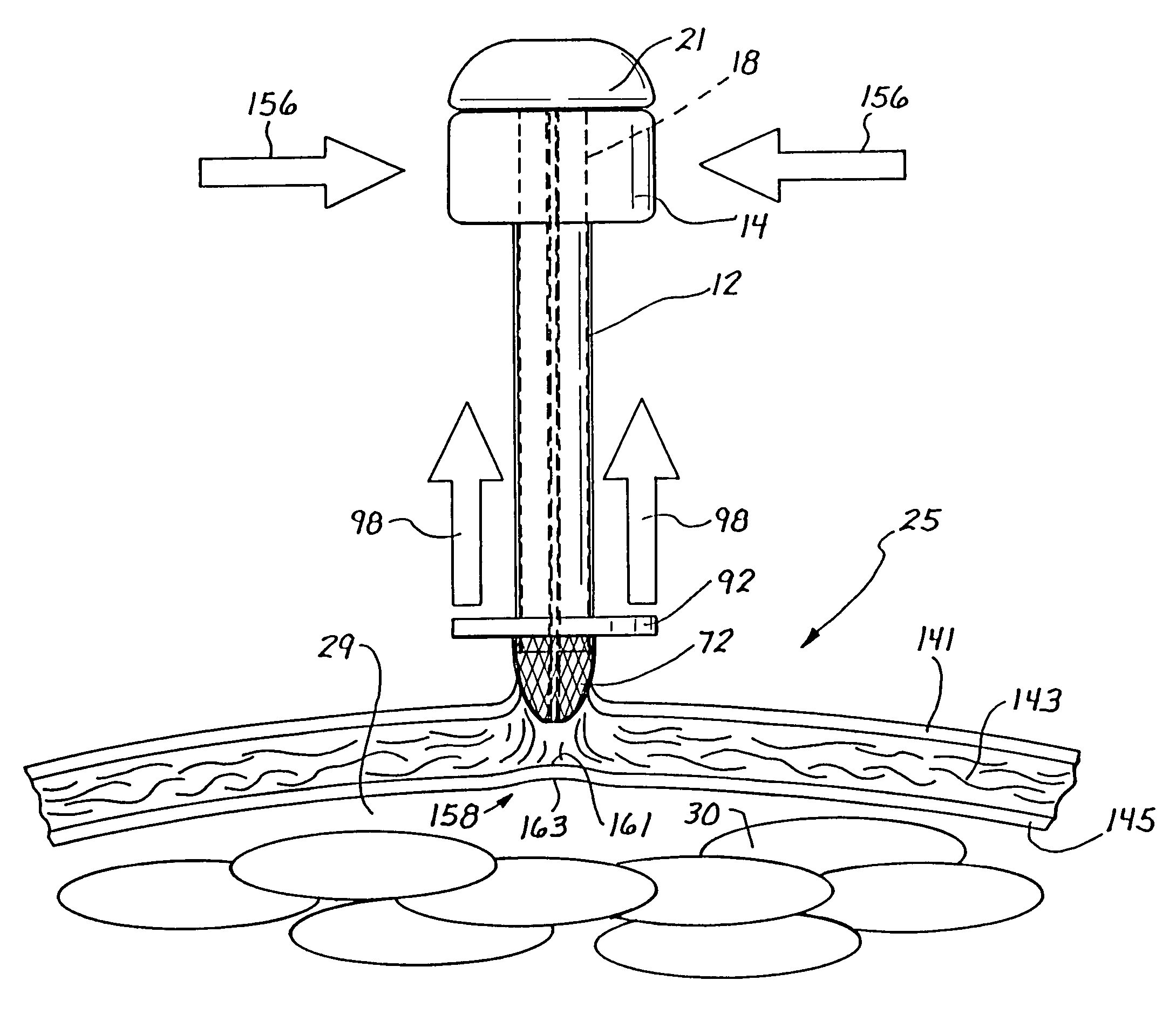
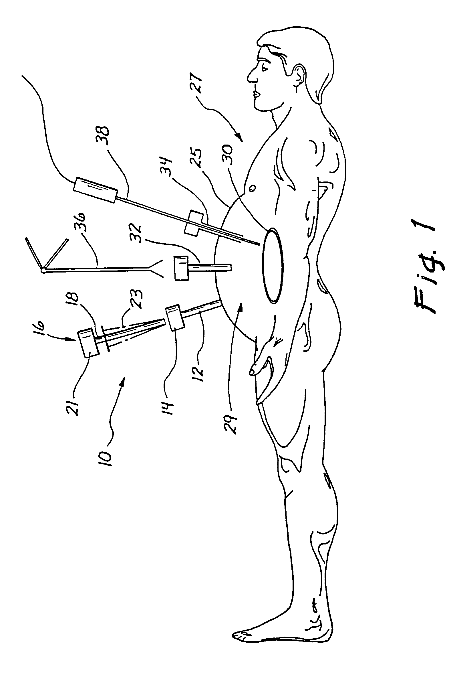
[0068]A trocar system of the present invention is illustrated generally in FIG. 1 and designated by the reference numeral 10. The system 10 includes a trocar cannula 12 having a seal housing 14, and an obturator 16 with a shaft 18 and handle 21, and including a traction mechanism 23 of particular interest to the present invention. The obturator 16 is used in placing the cannula 12 across a body wall such as an abdominal wall 25, associated with a patient 27. In the case of the abdomen, the wall 25 defines an abdominal cavity 29 which includes many organs such as that designated by the reference numeral 30.

[0069]In less evasive laparoscopic procedures, multiple cannulas 32 and 34 are used to provide access across the abdominal wall 25 to facilitate surgical procedures within the abdominal cavity 29. By way of example, the removal of a gallbladder is typically accomplished with such a laparoscopic procedure. Initially, cannulas 12, 32 and 34 are placed across the abdominal wall 25, ea...


PUM


Abstract

The trocar system includes a cannula insertable through a body wall using an obturator having a distal tip. A traction tread disposed interiorly of the obturator inverts at the distal tip and extends proximally along the outer surface of the obturator or cannula. At the distal tip the tread can facilitate parting rather than cutting the tissue. Along the outer surface, the tread can engage the tissue to pull it proximally along the advancing obturator. This produces counter forces which can result in a net proximal force facilitating distention of the abdominal wall and separation of the abdominal wall from internal organs. The traction tread can be axially and / or radially continuous. An associated method of operation includes the steps of contacting the body wall with the traction tread at the distal tip, and engaging the body wall with the traction tread along wall portions facing the outer surface.

Description

BACKGROUND OF THE INVENTION[0001]1. Field of Invention[0002]This invention relates generally to trocar systems and more specifically to obturator apparatus and methods for placing a trocar cannula across a body wall.[0003]2. Discussion of the Prior Art and Related Technology[0004]It is generally well known that holes can be created through body tissue either by cutting the tissue or by mechanically parting the tissue along lines of weakness. Where tissue is cut, it is severed along a line, which is determined by the direction of the cutting implement. Where tissue is parted, it separates along natural tissue planes such as those defined by muscle fibers or differing layers of tissue such as skin and muscle. Tissue that is mechanically separated tends to heal better than tissue that is cut with tissue that is mechanically separated the healing process requires only that the affected tissues re-approximate each other with cut tissue, and in particular muscle fibers, the healing proces...

Claims


Application Information

Patent Timeline
Patent Type & Authority Patents(United States)
IPC IPC(8): A61B17/34A61B19/00
CPCA61B17/34A61B17/3421A61B17/3476A61B17/3496A61B17/3417A61B2019/481A61B2017/3454A61B17/3431A61B2017/32006A61B2090/08021
Inventor HART, CHARLES C.PINGLETON, EDWARD D.BRUSTAD, JOHN R.HILAL, NABILPINEDJIAN, RAFFI
Owner APPL MEDICAL RESOURCES CORP
Why Eureka
Try Eureka