Method and apparatus for improving the vessel/tissue contrast in time-of-flight angiography of a magnetic resonance tomography measurement

a technology of time-of-flight angiography and magnetic resonance tomography, which is applied in the field of magnetic resonance tomography, can solve the problems of insufficient time for the magnetization to build up completely, low blood flow velocity, and inability to achieve the ideal case in reality, etc., and achieve the effect of improving the contrast of time-of-flight angiography

Inactive Publication Date: 2007-04-17
SIEMENS AG
View PDF8 Cites 2 Cited by
  • Summary
  • Abstract
  • Description
  • Claims
  • Application Information

AI Technical Summary

Benefits of technology

[0014]It is an object of the present invention to improve the contrast of time-of-flight angiography.

Problems solved by technology

Given a flipping of the spins in immediate succession, there is not enough time for the magnetization to build up completely again in the longitudinal direction.
This ideal case often cannot be achieved in reality since the slice thicknesses in the exposures of vascular anatomy are very large (approximately 4–6 cm), and the blood has a low flow velocity, especially in smaller vessels.

Method used


Image

  • Method and apparatus for improving the vessel/tissue contrast in time-of-flight angiography of a magnetic resonance tomography measurement
  • Method and apparatus for improving the vessel/tissue contrast in time-of-flight angiography of a magnetic resonance tomography measurement
  • Method and apparatus for improving the vessel/tissue contrast in time-of-flight angiography of a magnetic resonance tomography measurement

Examples


Embodiment Construction

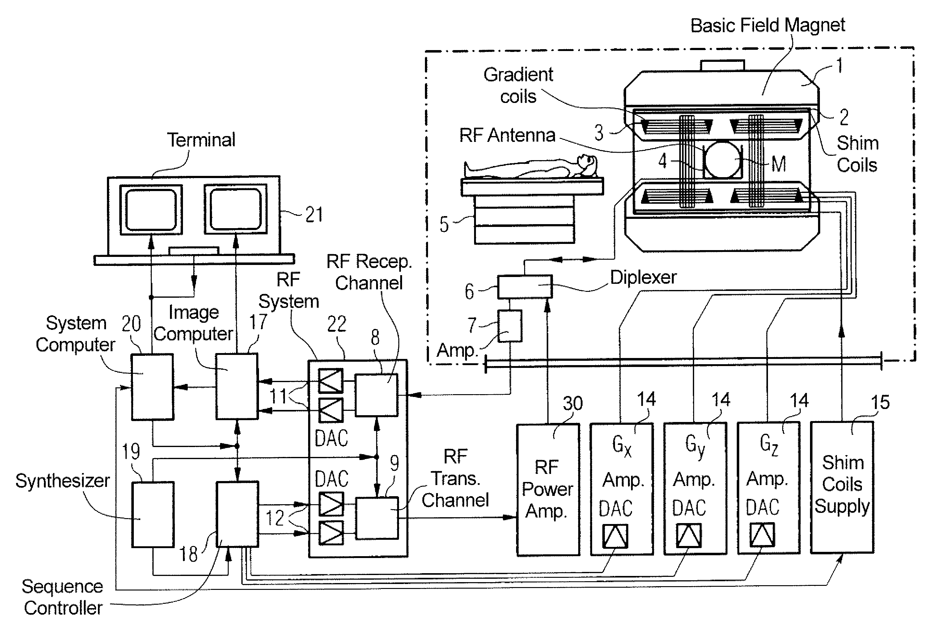
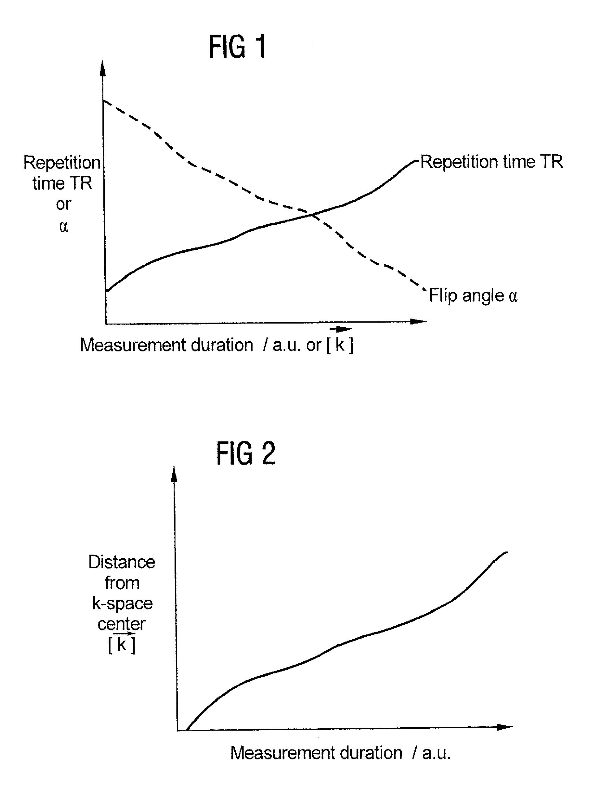
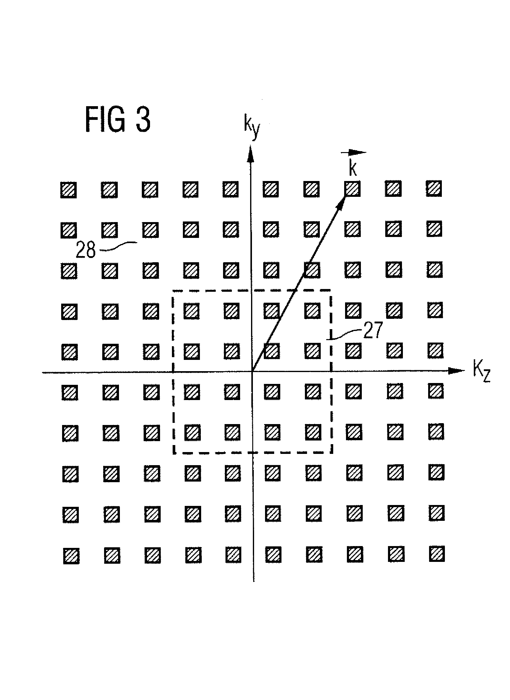
[0029]FIG. 5 is a schematic illustration of a nuclear magnetic resonance tomography apparatus with improved contrast behavior of MR time-of-flight angiography exposures according to the present invention. The basic components of the magnetic resonance tomography apparatus correspond to those of a conventional tomography apparatus, with the differences discussed below. A basic field magnet 1 generates a temporally constant, strong magnetic field for the polarization or alignment of the nuclear spins in the examination region of a subject such as, for example, a part of a human body to be examined. The high homogeneity of the basic magnetic field required for the magnetic resonance measurement is defined in a spherical measurement volume M into which the parts of the human body to be examined are introduced. For satisfying the homogeneity requirements and, in particular, for eliminating time-invariable influences, shim plates of ferromagnetic material are attached at suitable location...


PUM


Abstract

In a method and apparatus for improving the vessel / tissue contrast in time-of-flight angiography with a magnetic resonance tomography measurement, a sequence of radiofrequency excitation pulses is generated that each have a flip angle and a temporal spacing in the sequence defined by a repetition time, gradient pulses are generated with gradient coils such that, using an analog-to-digital converter, the magnetic resonance response signals are sampled in the frequency domain (k-space) in a slice referred to as a k-matrix, the k-matrix is sampled such that the respective distances of the measuring points from the center of the k-matrix are continuously increased (or decreased), and the repetition time is varied during the sampling, and / or the flip angle is varied during the sampling.

Description

BACKGROUND OF THE INVENTION[0001]1. Field of the Invention[0002]The present invention is directed in general to magnetic resonance tomography (MRT) as employed in medicine for examining patients. The present invention is particularly directed to a method for the optimization of k-space trajectories in the location encoding of a magnetic resonance tomography apparatus. An optimally fast sampling of k-matrix achieved as a result thereof leads to the utmost effectiveness of the sequence employed.[0003]2. Description of the Prior Art[0004]MRT is based on the physical phenomenon of nuclear magnetic resonance and has been successfully employed in medicine and biophysics for more than 15 years. In this examination modality, the subject is subjected to a strong, constant magnetic field. As a result thereof, the nuclear spins in the subject align, these having been previously irregularly oriented. Radiofrequency energy can then excite these “ordered” spins to a specific oscillation. This osc...

Claims


Application Information

Patent Timeline
Patent Type & Authority Patents(United States)
IPC IPC(8): A61B5/05G01R33/563
CPCG01R33/56308G01R33/5635
Inventor KUEHN, BERND
Owner SIEMENS AG
Why Eureka
Try Eureka