
Shunt for the treatment of glaucoma

a shunt and glaucoma technology, applied in the field of surgical treatment of glaucoma, can solve the problems of increased intraocular pressure, progressive damage to the optic nerve, and loss of central and peripheral visual field, and achieve the effect of reducing intraocular hypertension

Inactive Publication Date: 2007-04-24
SOLX
View PDF121 Cites 181 Cited by
  • Summary
  • Abstract
  • Description
  • Claims
  • Application Information

AI Technical Summary

Benefits of technology

"The present invention provides a system for reducing intraocular hypertension by implanting a shunt with a microchannel that allows aqueous flows to migrate from the anterior chamber through the microchannel. The shunt has an inflow port located at the first end of the microchannel and an outflow port located at the second end of the microchannel. The shunt can be implanted through a small incision in the eye and can be photo-ablated to increase the flow of aqueous flows through the shunt. The system can also include an anchor to hold the shunt in place. The invention has the advantage of reducing the risk of complications and providing a minimally invasive treatment for intraocular hypertension."

Problems solved by technology

Glaucoma is a major public health problem, affecting about two percent of the U.S. population and the third most common cause of blindness in the U.S. There are several forms of glaucoma however each results in elevated intraocular pressure (IOP) in the eye, which can cause progressive damage to the optic nerve, and both central and peripheral visual field loss.
If the IOP remains high for an extended period of time, total vision loss can occur.
The elevated IOP is caused by an imbalance in fluid inflows and outflows in the eye, and the pressure reduces the blood supply to the optic nerve.
It is believed that an abnormal trabecular cell metabolism results in compacted meshwork layers or a build up of extracellular materials within the meshwork that inhibits fluid flows.
The treatments generally require applications at least daily and are relatively expensive.
Furthermore, drugs may have occasional serious side effects, such as blurred vision, allergic reactions, headaches and potentially dangerous interactions with other drugs.
The benefits of the dissection procedures diminish over time due to the body's wound healing response and resulting fibrosis that repairs and closes the dissected opening in the meshwork.
Thus these expensive procedures do not provide a long-lasting cure.
Trabeculectomies and shunt surgeries and variations thereof have several disadvantages and moderate success rates.
Further, the surgeries cause substantial trauma to the eye.
Thus, the state of the art shunts and surgical techniques have yet to provide a cost-effective and long-lasting solution which has short recovery periods and low risk.

Method used


Image

  • Shunt for the treatment of glaucoma
  • Shunt for the treatment of glaucoma
  • Shunt for the treatment of glaucoma

Examples


Embodiment Construction

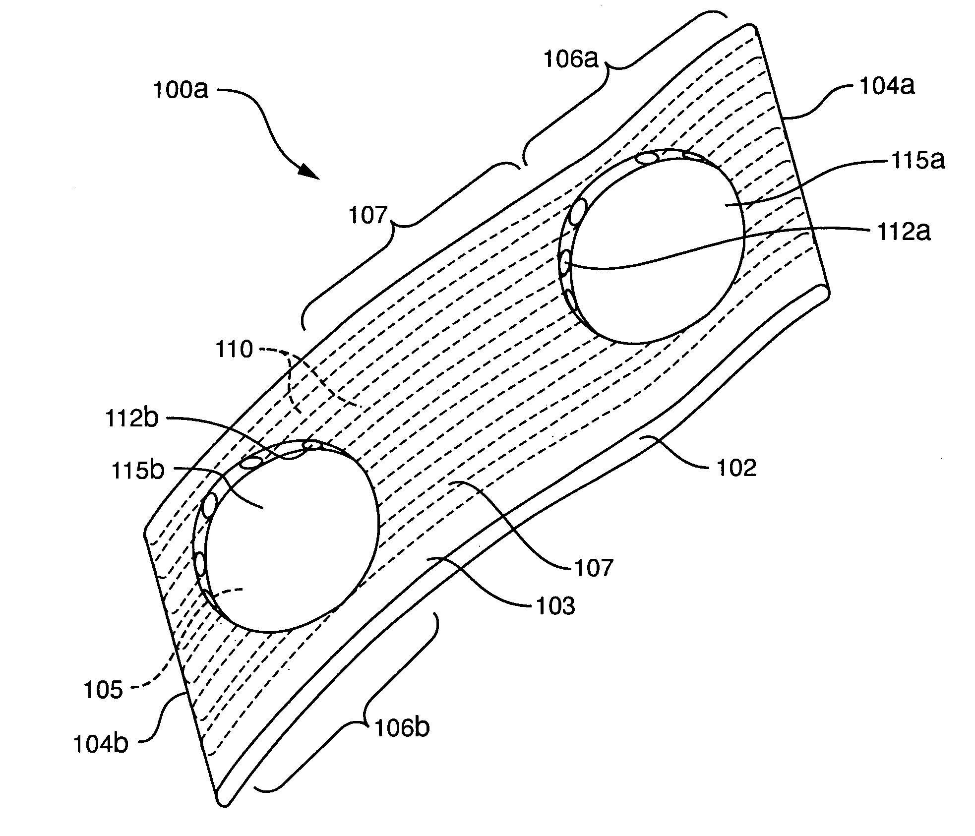
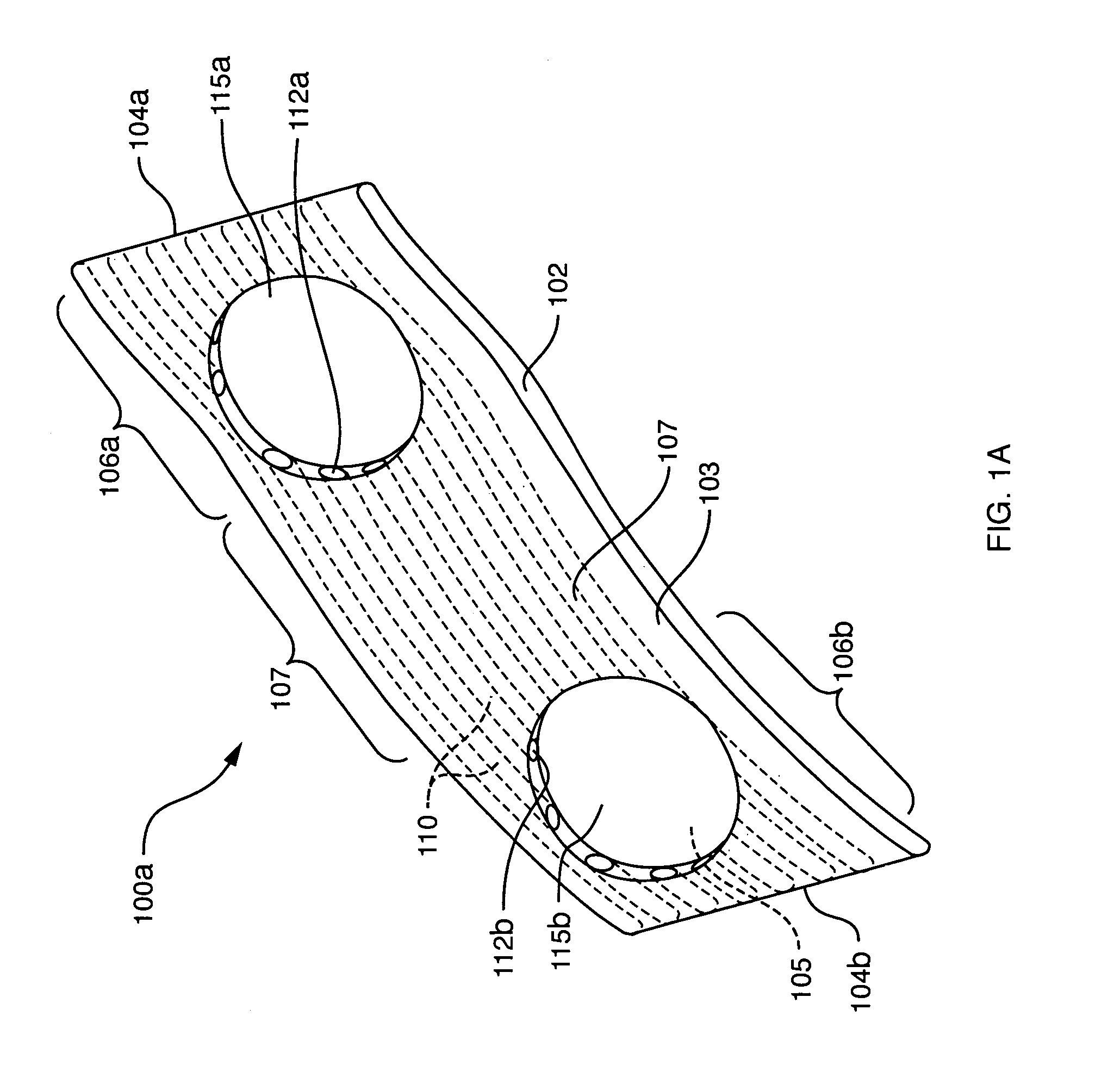
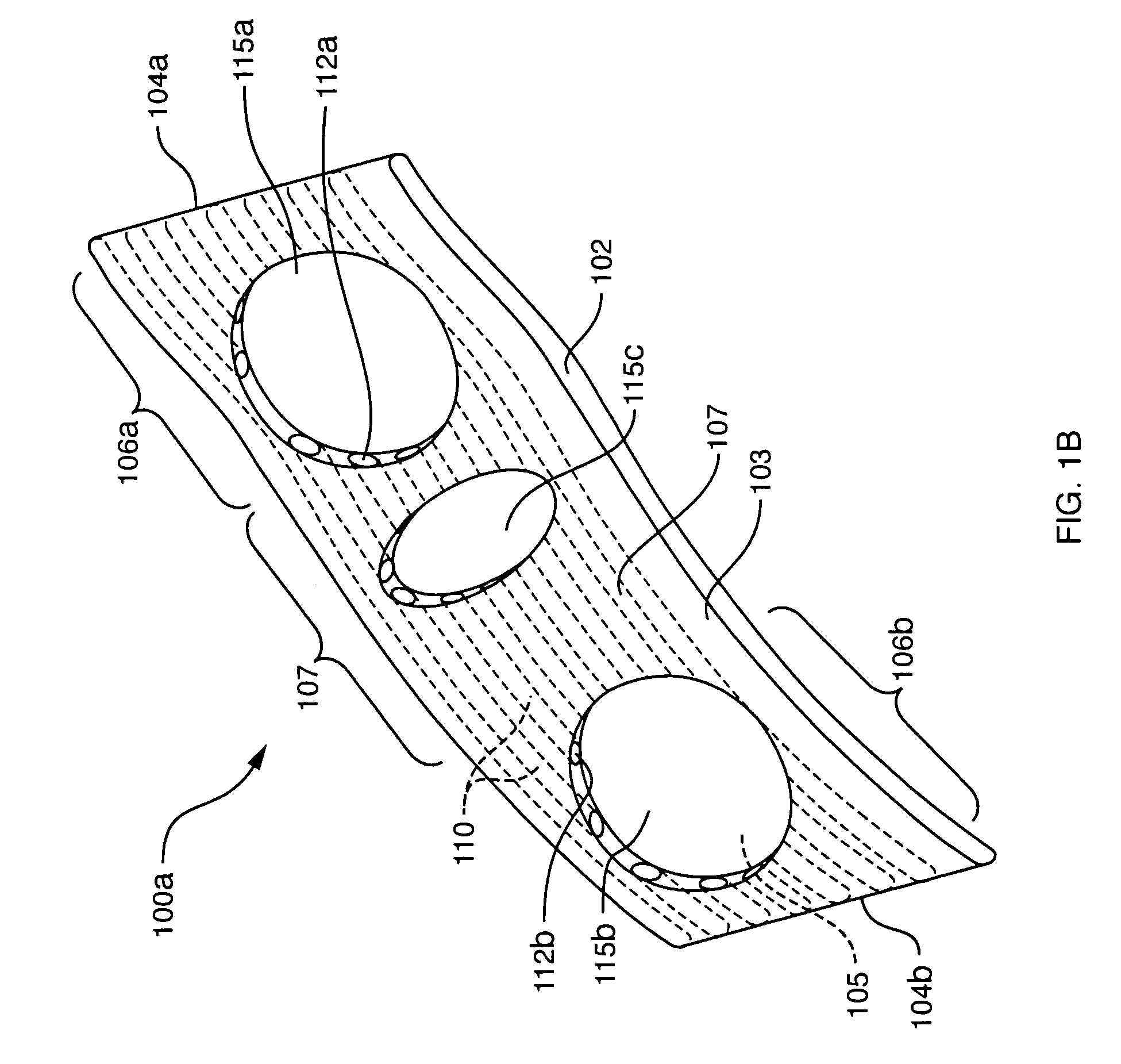
[0054]One embodiment of the present invention comprises a thin planar shunt device that can have various shapes and configurations as depicted in FIGS. 1 through 6. Various embodiments of the shunts are adapted to cooperate with the introducer or delivery apparatus used to facilitate placement of the shunt into the patient's eye.

[0055]Referring to FIG. 1, one embodiment of a shunt device 100A comprises a thin planar body 102 that extends along a longitudinal axis 105 from a first end section 106A to a second end section 106B with medial section 107 therebetween. The shape and configuration of the planar member, herein defined as its “planform”, is shown as being substantially rectangular with relatively straight side edges 103 and endwise edges 104A, 104B. However, other shapes and configuration are well within the scope of the invention as is readily appreciated by one skilled in the art. For example, the first end section 106A can be bulbous or pointed as illustrative examples. Li...


PUM


Abstract

A system is provided for reducing intraocular hypertension, the system having an implantable shunt, with a planar member having at least one microchannel disposed within the planar member. There is an inflow port disposed proximate to a first end of the microchannel and an outflow port disposed proximate to a second end of the microchannel. The inflow port is configured such that when the implantable shunt is implanted, the inflow port is located approximately within the region of an angle of an anterior chamber.

Description

RELATED APPLICATIONS[0001]This application claims the benefit of U.S. Provisional Application No. 60 / 478,895, filed Jun. 16, 2003. This application is herein incorporated in its entirety by reference.FIELD OF THE INVENTION[0002]The present invention pertains to surgical treatments for glaucoma and methods for reducing intraocular pressure (IOP), and more particularly relates to an implantable shunt device for allowing aqueous outflows from the eye's anterior chamber and associated methods thereof.BACKGROUND OF THE INVENTION[0003]Glaucoma is a major public health problem, affecting about two percent of the U.S. population and the third most common cause of blindness in the U.S. There are several forms of glaucoma however each results in elevated intraocular pressure (IOP) in the eye, which can cause progressive damage to the optic nerve, and both central and peripheral visual field loss. If the IOP remains high for an extended period of time, total vision loss can occur. The elevated...

Claims


Application Information

Patent Timeline
Patent Type & Authority Patents(United States)
IPC IPC(8): A61M5/00A61F9/007
CPCA61F9/00781
Inventor SIMON, GABRIEL
Owner SOLX
Learn More