
Method for mapping temperature rise using pulse-echo ultrasound

a pulse-echo ultrasound and temperature rise technology, applied in the field of ultrasound, can solve the problems of affecting the operation, affecting the accuracy of the temperature map, so as to achieve the effect of less artifacts and faster temperature maps

Inactive Publication Date: 2007-05-01
ETHICON ENDO SURGERY INC
View PDF197 Cites 38 Cited by
  • Summary
  • Abstract
  • Description
  • Claims
  • Application Information

AI Technical Summary

Benefits of technology

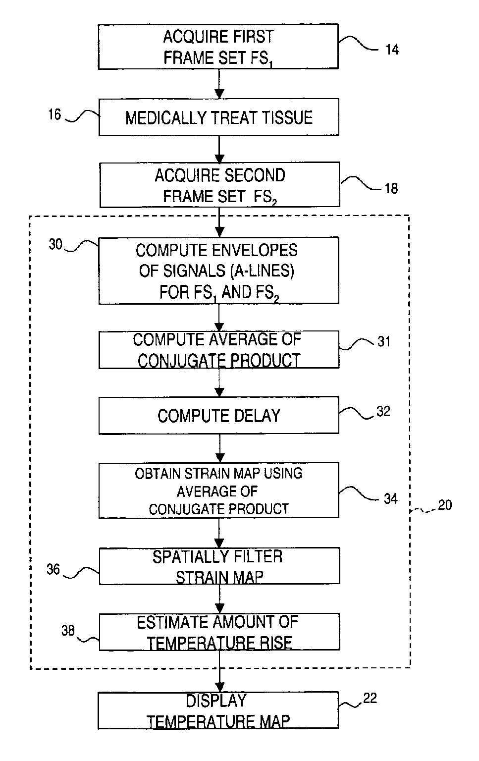
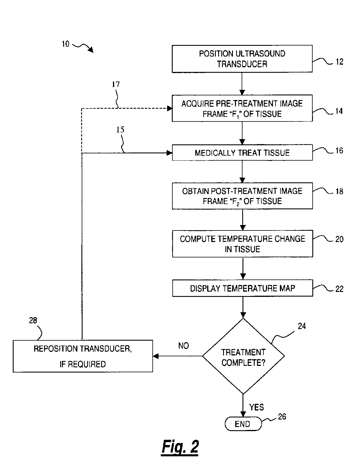
[0015]The present invention overcomes the limitations of the known art by estimating the time-dependent delay between two sets of narrow-band ultrasound signals. The travel time of a first signal is measured prior to treating anatomical tissue, then the travel time of a second signal is measured after the tissue is treated. The change in travel time between the first and second signals is used to generate a complete two-dimensional delay map from several two-dimensional multiplication and fast Fourier transform operations, rather than creating a map from a large quantity of individual time delay estimation steps. As a result, temperature maps can be generated more quickly as compared to prior methods. In addition, temperature maps generated using the present invention have fewer artifacts.

Problems solved by technology

However, this arrangement is not desirable for several reasons.
In addition, the thermocouple array may have a relatively slow response time, providing the operator with delayed feedback regarding the status of the treatment.
As a result, MRI-assisted ultrasound methods are expensive and cumbersome, making such treatment impractical for routine use in surgical treatments.
These approaches suffer from several drawbacks, including low spatial resolution and high artifactual content.
Since many time-delay estimation steps are needed to build up a temperature image, these approaches are also computationally inefficient.

Method used


Image

  • Method for mapping temperature rise using pulse-echo ultrasound
  • Method for mapping temperature rise using pulse-echo ultrasound
  • Method for mapping temperature rise using pulse-echo ultrasound

Examples


Embodiment Construction

[0028]It is well-known that the travel time of an ultrasound wave varies with the path length. The path length may vary in anatomic tissue due to thermal expansion of the tissue as a result of therapeutic ultrasound treatment, such as hyperthermia and ablation. Further, the travel time of an ultrasound wave varies with the speed of the ultrasound wave, which is a function of path temperature. Thus, changes in ultrasound wave travel times may be used to estimate temperature rise.

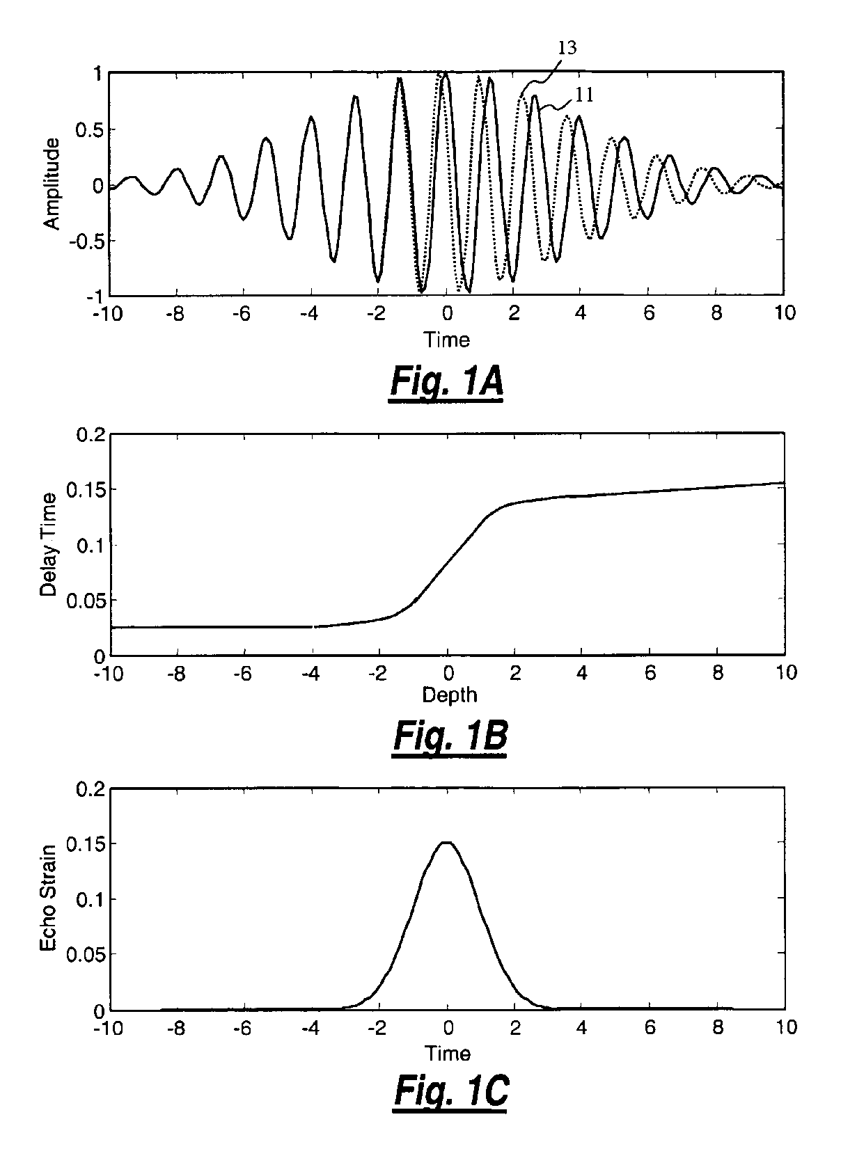
[0029]FIGS. 1A-1C provide an overview of a method for mapping temperature rise according to an embodiment of the present invention. FIG. 1A generally illustrates a first ultrasound signal 11 returning to an ultrasound transducer (not shown) before treatment of a region of tissue, while ultrasound signal 13 represents an ultrasound signal returning to the ultrasound transducer after treatment of the tissue. The relative delay between ultrasound echos returning to an ultrasound transducer, measured before and a...


PUM


Abstract

A method for measuring the temperature rise in anatomical tissue as a result of ultrasound treatment. A first ultrasound signal is obtained prior to treating the anatomical tissue, then a second ultrasound signal is obtained after the tissue is treated. Complex analytic signals are computed from the first and second ultrasound signals, then the depth-dependent delay is computed from the complex analytic signals. An echo strain map is generated from the slope of the depth-dependent delay. The echo strain map is used to estimate the amount of temperature rise from the first ultrasound signal to the second ultrasound signal. An image may then be created showing where temperature rise is occurring in the anatomical tissue.

Description

RELATED APPLICATIONS[0001]This is a continuation-in-part of application Ser. No. 10 / 153,241, filed May 22, 2002 now abandoned, which claims priority to U.S. Provisional Application Ser. No. 60 / 294,135 filed May 29, 2001. The entire disclosures are incorporated herein by reference.FIELD OF THE INVENTION[0002]The present invention relates generally to ultrasound, and more particularly, to a method for measuring temperature rise using pulse-echo ultrasound waves.BACKGROUND OF THE INVENTION[0003]Ultrasound medical systems and methods include ultrasound imaging of anatomical tissue to identify tissue for medical treatment. Ultrasound may also be used to medically treat and destroy unwanted tissue by heating the tissue. Imaging is done using low-intensity ultrasound waves, while medical treatment is performed with high-intensity ultrasound waves. High-intensity ultrasound waves, when focused at a focal zone a distance away from the ultrasound source, will substantially medically affect ti...

Claims


Application Information

Patent Timeline
IPC IPC(8): A61B8/00A61B18/00A61BA61B5/00A61B8/08A61B8/12A61B10/00A61B10/02A61B17/22A61B17/28A61B17/32A61B17/34A61B19/00A61N7/00A61N7/02H04R17/00
CPCA61B5/015A61B8/08A61B8/12A61B8/4488A61B8/5223A61B17/320092A61N7/022G01S7/52042A61B10/0233A61B17/22012A61B17/2202A61B17/29A61B2019/5276A61B2019/528A61B2019/5429A61B2019/5475A61N7/02A61N2007/0078A61B2090/378A61B2090/3784A61B2090/3929A61B2090/3975G16H50/30
Inventor MAST, T. DOUGLASFAIDI, WASEEM
Owner ETHICON ENDO SURGERY INC
Learn More