Optical computed tomography scanner for small laboratory animals

a computed tomography and laboratory animal technology, applied in the field of optical computed tomography scanners for scanning small laboratory animals, can solve the problems of inability to show volumetric data that can allow quantitative estimation of tissue volume, inability to generate mature t-lymphocytes, and inability to accurately estimate the quantity of tagged tissue within the sampl

Active Publication Date: 2007-05-01
IMAGING DIAGNOSTIC SYST
View PDF26 Cites 18 Cited by
  • Summary
  • Abstract
  • Description
  • Claims
  • Application Information

AI Technical Summary

Benefits of technology

[0011]It is another object of the present invention to provide an optical CT scanner that can image both attenuation and fluorescence distributions in small laboratory animals injected with a fluorophore, thereby to locate and quantify the volumetric size of tumors growing in these animals for research purposes.

Problems solved by technology

Although these two-dimensional images show the distribution of the fluorophore, accurate estimation of the quantity of tagged tissue within the sample (e.g. the volume of a tumor) is difficult without using actual three-dimensional fluorescent images of the sample.
None of these systems, however, show volumetric data that can allow quantitative estimation of tissue volumes.
The mice are unable to generate mature T-lymphocytes and therefore are unable to mount most types of immune responses, including antibody formation and rejection of transplanted tissues.

Method used


Image

  • Optical computed tomography scanner for small laboratory animals
  • Optical computed tomography scanner for small laboratory animals
  • Optical computed tomography scanner for small laboratory animals

Examples


Embodiment Construction

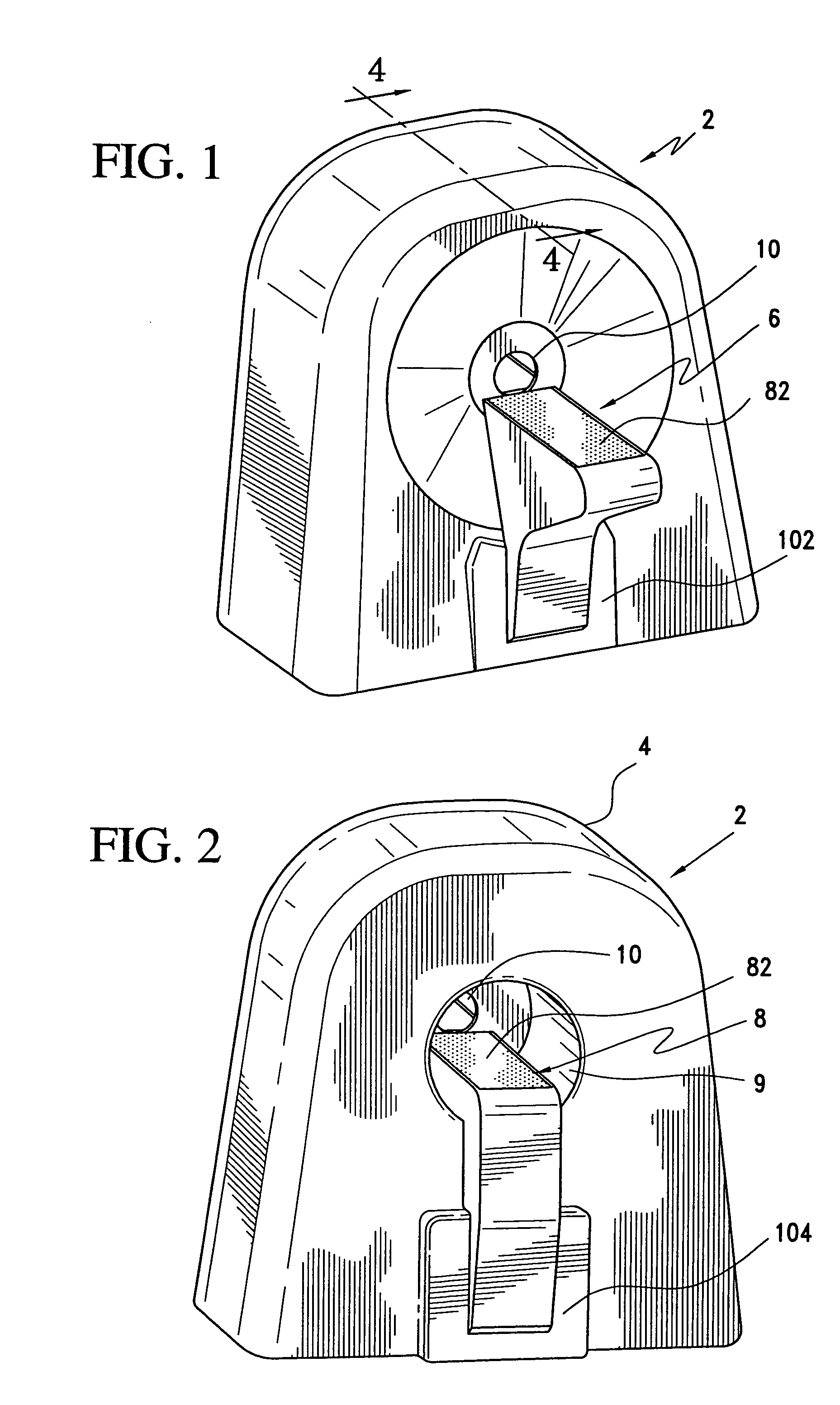
[0028]An optical computed tomography scanner 2 made in accordance with the present invention is disclosed in FIGS. 1 and 2. The scanner 2 includes a housing 4 and horizontal front and rear tables 6 and 8, respectively. The housing has a vertical opening 10 to allow the test subject to go from the front table 6 to the rear table 8 during scanning, as will be further described below. The housing 4 includes a well 9 having a bottom at which the opening 10 is disposed. The well 9 advantageously allows the user to observe the test subject as it progresses toward the rear table 8.

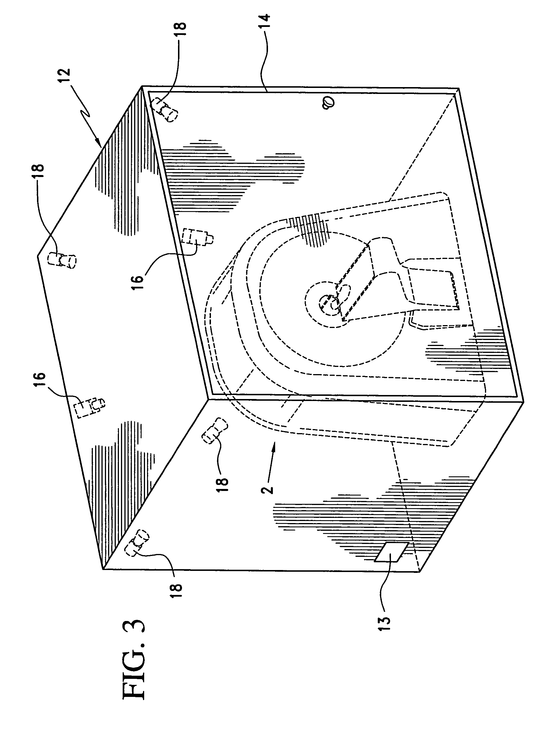
[0029]The scanner 2 is placed within a light-tight enclosure 12 to prevent room light from contaminating the light transmitted through the test subject by the scanner. The enclosure 12 is provided with light-tight door 14 to provide access to the scanner 2.

[0030]Video cameras 16 are provided within the enclosure 12 to provide remote monitoring of the test subject during the scanning period. Light fixtures 18 prov...


PUM


Abstract

An optical CT scanner for small laboratory animals comprises a housing having a vertical through opening through which a test subject is passed through during a scanning session, the housing including a peripheral slot disposed around the perimeter of the opening; a movable horizontal table disposed through the opening, the table being split with a horizontal slot aligned with the peripheral slot; a light beam directed toward the peripheral slot and orbitable around the opening; a plurality of collimators directed toward the peripheral slot and orbitable around the opening together with the light beam; a plurality of main photodetectors to detect the light beam after passing through the test subject and the collimators; a perimeter photodetector adapted to provide perimeter data of the test subject during a scanning session; and a first computer programmed to reconstruct an image of the test subject from the output of the perimeter and main photodetectors.

Description

RELATED APPLICATION[0001]This is a continuation-in-part application of application Ser. No. 10 / 717,969, filed Nov. 21, 2003, now U.S. Pat. No. 7,155,274 hereby incorporated by reference.FIELD OF THE INVENTION[0002]The present invention relates generally to an optical computed tomography (CT) scanner and specifically to an optical CT scanner for scanning small laboratory animals, such as nude mice carrying cancer cells with fluorophore, such as green fluorescent protein (GFP).BACKGROUND OF THE INVENTION[0003]Fluorescence is often used in biology and particularly in optical microscopy for tissue identification. A fluorophore chosen for its abilities to bind to or ‘tag’ a specific type of tissue is introduced into the sample being interrogated. When the sample is illuminated by light at the excitation wavelength of the fluorophore, the tissue tagged by the fluorophore will emit light at the fluorophore's emission wavelength, thereby allowing optical detection of that tissue's presence ...

Claims


Application Information

Patent Timeline
Patent Type & Authority Patents(United States)
IPC IPC(8): A61B6/00
CPCA61B5/0062A61B5/0073A61B5/415A61B2503/40B33Y80/00
Inventor WAKE, ROBERT H.HUMMER, BRIANBECKER, GARY M.PONDER, STEVEN L.MAGRANER, VINCE
Owner IMAGING DIAGNOSTIC SYST
Why Eureka
Try Eureka