Sensor for measuring tissue perfusion

Inactive Publication Date: 2007-05-08
DAMGAARD LARS RIIS +3
View PDF9 Cites 1 Cited by
  • Summary
  • Abstract
  • Description
  • Claims
  • Application Information

AI Technical Summary

Benefits of technology

[0009]It is a further object of the present invention to provide at least one embodiment of the general inventive idea which makes it possible to carry out non-invasive measurements of skin perfusion or measurements of prefusion in the surface layers of an organ, for instance for assessment of insufficient blood circulation.
[0013]According to the present invention the dimensions of the contact region between said tracer-permeable barrier and the surrounding tissue can be varied and thereby the region over which the tissue perfusion measurement is being carried out. It is also possible to vary the second area providing access to the detection cavity. By varying the geometry of the sensor, i.e. the relative layout of the reservoir, barrier and detection cavity, it is possible to vary the sensitivity and the radial resolution of the measurements being performed. It is furthermore possible to utilise a mixture of at least two tracers which might be supplied and removed substantially momentarily. A time-based measurement after instantaneous supply / removal to / from the tracer reservoir of two tracers with different diffusion coefficients will make it possible to distinguish between how much of the diffusion of the tracers away from the tracer reservoir is due to the concentration gradient within the tissue and how much is due to the transportation of the tracers away from the tissue by the blood. Thus, independent measures of perfusion and of diffusion coefficients within the tissue can be obtained.

Problems solved by technology

66, pp 337–345), but this technique suffers from a number of drawbacks in that its temporal resolution only amounts to approximately half an hour which is insufficient in many situations.
Furthermore the location of the injection of the radioactive matter into the tissue relative to the location where the radioactivity is being measured is not particularly well-defined and finally, the application of radioactive matter per se involves potential hazards.
This method also suffers from an insufficient temporal resolution.
In connection with monitoring tissue perfusion for instance during surgical operations, the above-mentioned prior art suffers from the drawbacks of either insufficient temporal resolution or a very limited measurement space.

Method used


Image

  • Sensor for measuring tissue perfusion
  • Sensor for measuring tissue perfusion
  • Sensor for measuring tissue perfusion

Examples


Embodiment Construction

[0028]In the following detailed part of the present description a number of different embodiments of the present invention will be described with reference to the accompanying drawings, but it is understood that these embodiments only constitute examples of the general inventive idea, and that other embodiments may be conceivable by a person skilled in the art.

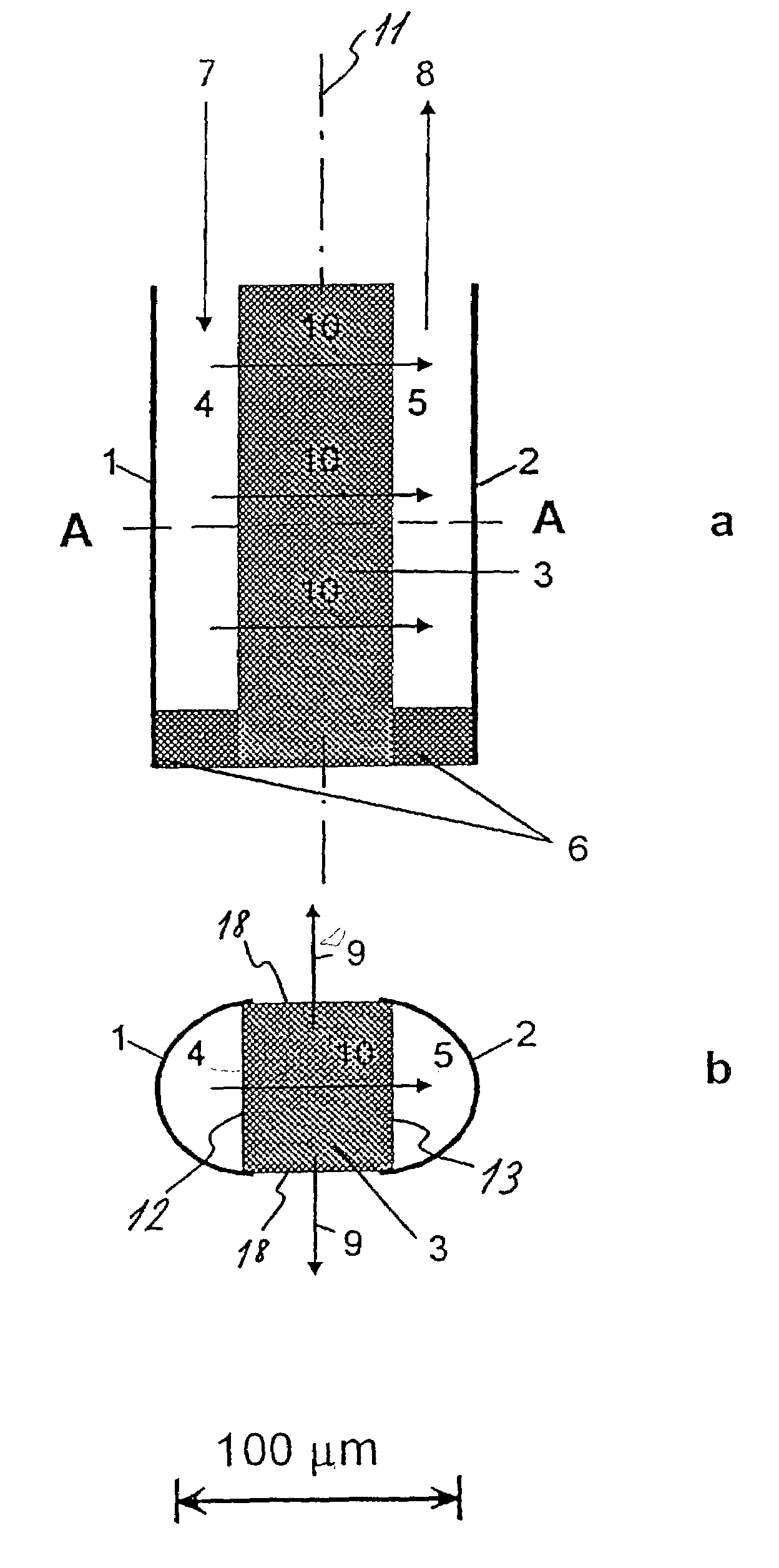
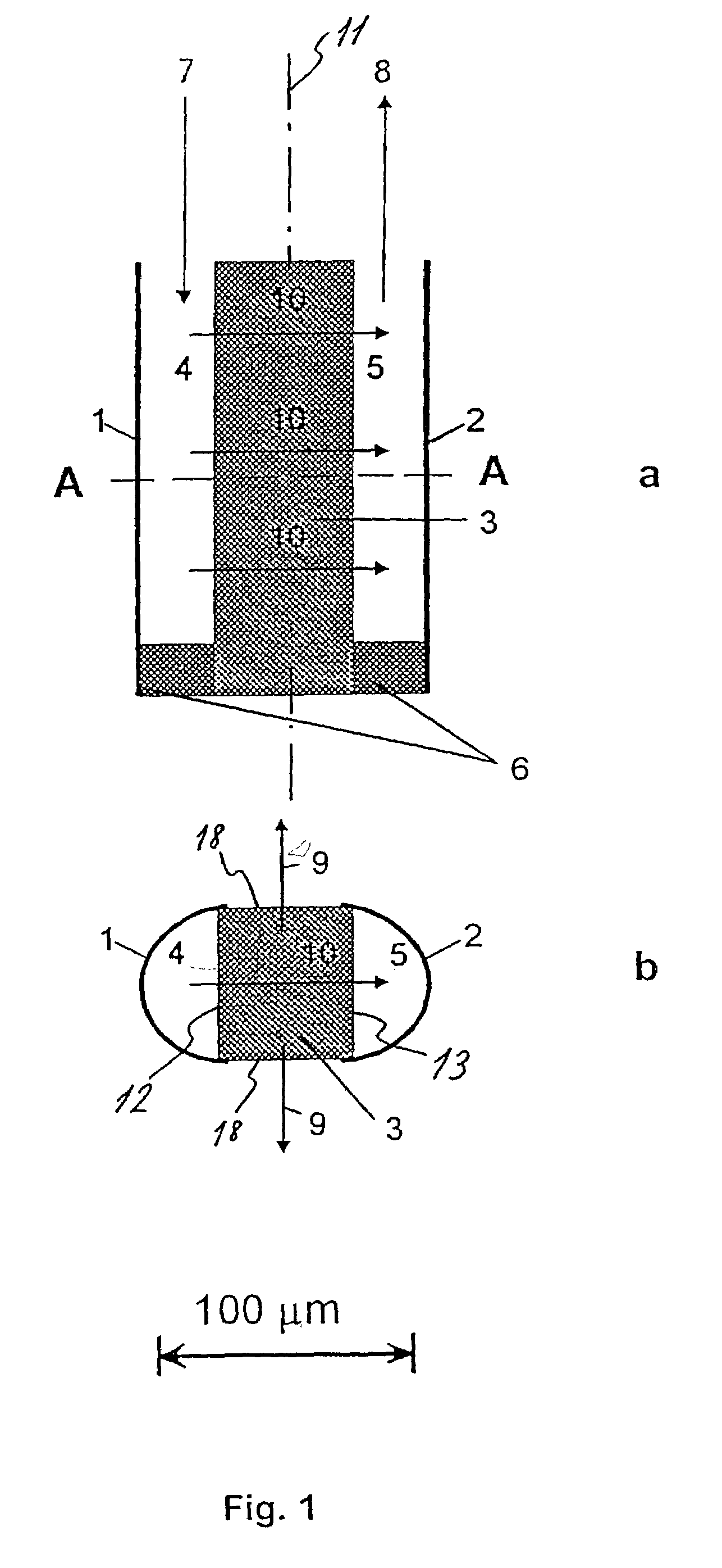
[0029]The first embodiment of the sensor is shown in FIG. 1a and FIG. 1b. The sensor is substantially symmetrical about a vertical plane 11 and comprises two U-shaped profiles 1, 2, the reservoir profile 1 and the detection profile 2 made of a gas-impermeable material such as metal or a suitable plastic material. The open sides 12, 13 of these two profiles 1, 2 are both in sealing abutment with a barrier 3 disposed between the reservoir 4 and the detection cavity 5 and extending throughout the vertical length of the sensor. The barrier 3 is made from a gas-permeable material, such as silicon or Teflon, such that two cavities, ...


PUM


Abstract

The invention relates to a method and a sensor for measurement of tissue perfusion. The sensor is provided with a reservoir (4) for a fluid or gaseous tracer and a tracer-permeable barrier (3), a sub-surface of which is in contact with the surrounding tissue and another sub-surface of which is in contact with a detection cavity (5) which is connected to a suitable apparatus for the measurement of tracer concentration in the detection cavity. The concentration of the tracer in the detection cavity is a measure of perfusion in the surrounding tissue. According to another embodiment of the invention it is also possible to carry out measurements of perfusion in the surface layers of the skin or of an organ.

Description

TECHNICAL FIELD OF THE INVENTION[0001]The present invention relates generally to devices for measurements of tissue perfusion according to the preamble of independent claim 1 and more particularly to a sensor for measurement of tissue perfusion over a given variable region and having a short response time.BACKGROUND ART[0002]Tissue perfusion is a measure of the amount (volume) of blood passing through a unit quantity of the tissue and is often measured with the unit ml blood / 100 g tissue. Since all blood tissues are at the same time being supplied with nutrients and excrete waste products through diffusion between tissue cells and the blood, tissue perfusion is a very important factor indicating the state of health of a tissue. A method for the measurement of tissue perfusion is therefore highly pertinent, for instance for monitoring tissue during and after surgical operations and transplantations. Monitoring of potentially threatened tissue, e.g. muscular tissue, whose blood supply...

Claims


Application Information

Patent Timeline
IPC IPC(8): A61B5/00A61B5/05A61B5/0275A61B5/145G01F1/704
CPCA61B5/0275G01F1/704
Inventor DAMGAARD, LARS RIISGUNDERSEN, JENS KRISTIANLARSEN, LARS HAUERKJAER, THOMAS
Owner DAMGAARD LARS RIIS
Why Eureka
Try Eureka