Method and apparatus for correcting motion in image reconstruction

a motion correction and image reconstruction technology, applied in the field of medical imaging, can solve problems such as blurring, streaking, or discontinuities, and various motion-related image artifacts, and achieve the effects of improving image quality, reducing image quality, and improving image quality

Active Publication Date: 2007-05-22
GENERAL ELECTRIC CO
View PDF73 Cites 56 Cited by
  • Summary
  • Abstract
  • Description
  • Claims
  • Application Information

AI Technical Summary

Problems solved by technology

CT imaging techniques, however, may present certain challenges when imaging dynamic internal tissues, such as the heart.
For example, in cardiac imaging, the motion of the heart causes inconsistencies in the projection data, which, after reconstruction, may result in various motion-related image artifacts such as blurring, streaking, or discontinuities.
The increasing centripetal force and the tolerances of the gantry components may comprise, therefore, a mechanical limitation to increases in gantry velocity.
Both mechanical and X-ray flux considerations, therefore, are obstacles to increasing the gantry rotation speed sufficiently to achieve a temporal resolution of 20 ms or better in CT reconstructions.

Method used


Image

  • Method and apparatus for correcting motion in image reconstruction
  • Method and apparatus for correcting motion in image reconstruction
  • Method and apparatus for correcting motion in image reconstruction

Examples


Embodiment Construction

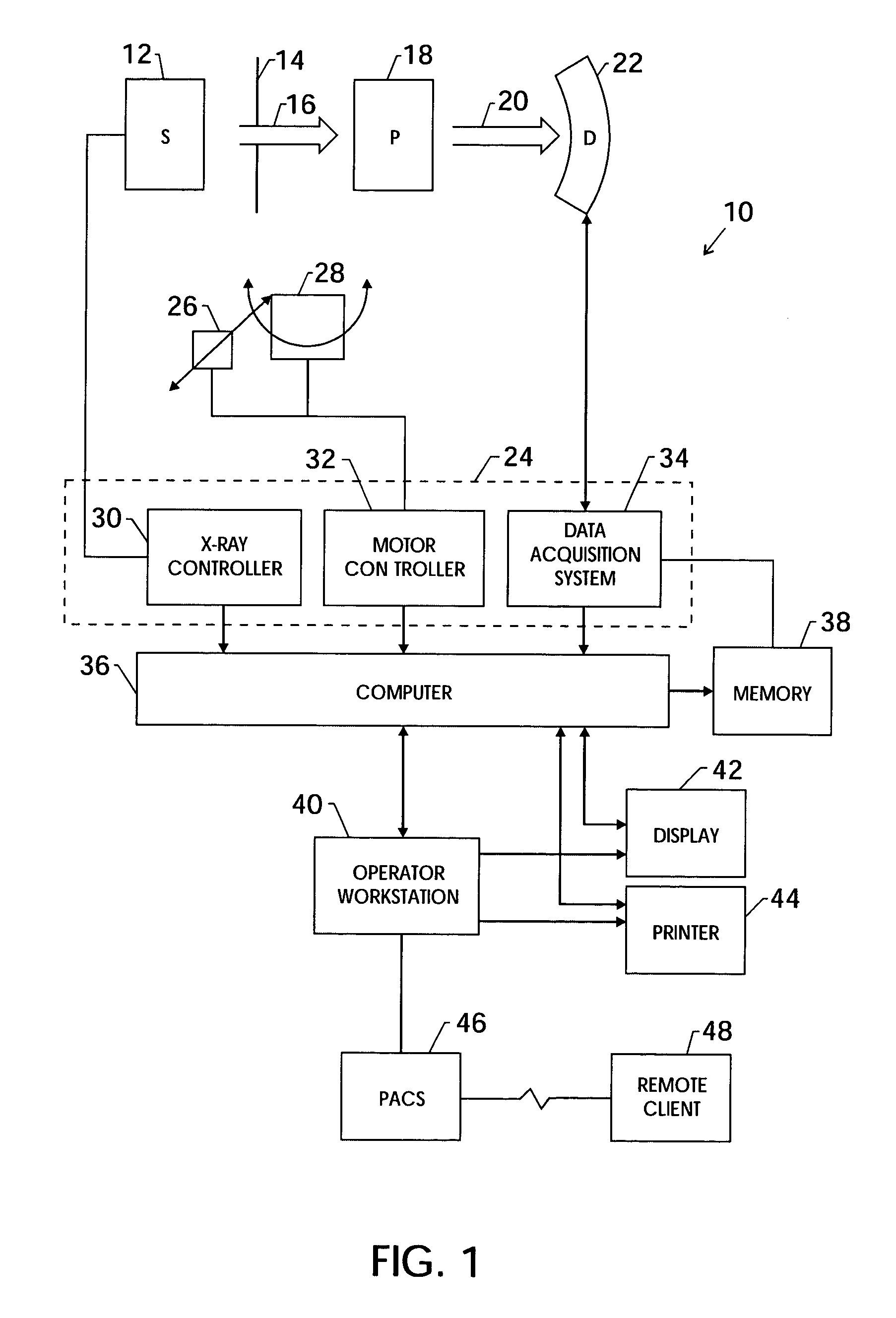
[0022]FIG. 1 illustrates diagrammatically an imaging system 10 for acquiring and processing image data. In the illustrated embodiment, system 10 is a computed tomography (CT) system designed to acquire X-ray projection data, to reconstruct the projection data into an image, and to process the image data for display and analysis in accordance with the present technique. In the embodiment illustrated in FIG. 1, imaging system 10 includes a source of X-ray radiation 12 positioned adjacent to a collimator 14. In this exemplary embodiment, the source of X-ray radiation source 12 is typically an X-ray tube.

[0023]Collimator 14 permits a stream of radiation 16 to pass into a region in which a subject, such as a human patient 18 is positioned. The stream of radiation 16 may be generally fan or cone shaped, depending on the configuration of the detector array, discussed below, as well as the desired method of data acquisition. A portion of the radiation 20 passes through or around the subject...


PUM

PropertyMeasurementUnit
fan angleaaaaaaaaaa
correlation thresholdaaaaaaaaaa
CTaaaaaaaaaa

Abstract

One or more techniques are provided for adapting a reconstruction process to account for the motion of an imaged object or organ, such as the heart. In particular, projection data of the moving object or organ is acquired using a slowly rotating CT gantry. Motion data may be determined from the projection data or from images reconstructed from the projection data. The motion data may be used to reconstruct motion-corrected images from the projection data. The motion-corrected images may be associated to form motion-corrected volume renderings.

Description

CROSS-REFERENCE TO RELATED APPLICATIONS[0001]This application claims the benefit of U.S. Provisional Application No. 60 / 397,658 filed on Jul. 23, 2002 and U.S. Provisional Application 60 / 398,463 filed on Jul. 25, 2002.BACKGROUND OF THE INVENTION[0002]The present invention relates generally to the field of medical imaging and more specifically to the field of imaging dynamic, internal tissue, such as cardiac tissue, by computed tomography. In particular, the present invention relates to the characterization of internal motion and to the reconstruction of images that account for the characterized motion.[0003]Computed tomography (CT) imaging systems measure the attenuation of X-ray beams passed through a patient from numerous angles. Based upon these measurements, a computer is able to reconstruct images of the portions of a patient's body responsible for the radiation attenuation. As will be appreciated by those skilled in the art, these images are based upon separate examination of ...

Claims


Application Information

Patent Timeline
Patent Type & Authority Patents(United States)
IPC IPC(8): G01N23/04A61B6/00A61B6/03G06T5/00G06T5/50G06T7/20G06T11/00
CPCA61B6/032A61B6/405A61B6/4241A61B6/482A61B6/541G01N23/046G06T5/50G06T7/2033G06T11/005G06T5/003A61B6/4035G01N2223/612A61B6/503G06T2211/408G06T2211/412Y10S378/901G06T2207/10081G06T2207/30004G01N2223/419A61B6/563G06T7/246
Inventor EDIC, PETER MICHAELIATROU, MARIACESMELI, ERDOGANDE MAN, BRUNOBASU, SAMIT
Owner GENERAL ELECTRIC CO
Why Eureka
Try Eureka